Dorsalis pedis artery

Last updated
Dorsalis pedis artery
Gray553.png
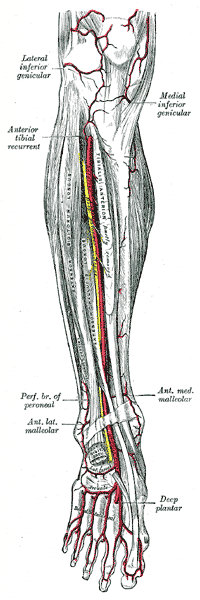
Anterior tibial artery, dorsalis pedis artery and the muscles and bones of the leg (anterior view).
Details
Source anterior tibial artery
Branches first dorsal metatarsal artery, deep plantar artery
Supplies dorsal surface of the foot
Identifiers
Latin arteria dorsalis pedis
TA98 A12.2.16.048
TA2 4714
FMA 43915
Anatomical terminology

In human anatomy, the dorsalis pedis artery (dorsal artery of foot) is a blood vessel of the lower limb. It arises from the anterior tibial artery, and ends at the first intermetatarsal space (as the first dorsal metatarsal artery and the deep plantar artery). It carries oxygenated blood to the dorsal side of the foot. It is useful for taking a pulse. It is also at risk during anaesthesia of the deep peroneal nerve.

Contents

Structure

The dorsalis pedis artery is located 1/3 from medial malleolus of the ankle. It arises at the anterior aspect of the ankle joint and is a continuation of the anterior tibial artery. [1] [2] It ends at the proximal part of the first intermetatarsal space. Here, it divides into two branches, the first dorsal metatarsal artery, and the deep plantar artery. [2] It is covered by skin and fascia, but is fairly superficial. [2]

The dorsalis pedis communicates with the plantar blood supply of the foot through the deep plantar artery. Along its course, it is accompanied by a deep vein, the dorsalis pedis vein.

Function

The dorsalis pedis artery supplies oxygenated blood to the dorsal side of the foot. [1]

Clinical significance

Pulse

The dorsalis pedis artery pulse can be palpated readily lateral to the extensor hallucis longus tendon (or medially to the extensor digitorum longus tendon) on the dorsal surface of the foot, distal to the dorsal most prominence of the navicular bone which serves as a reliable landmark for palpation. [3] It is often examined, by physicians, when assessing whether a given patient has peripheral vascular disease. It is absent, unilaterally or bilaterally, in 2–3% of young healthy individuals. [4]

Ultrasound

The dorsalis pedis artery may be studied using ultrasound. [2] Doppler ultrasound can be used to investigate blood flow. [2]

Local anaesthesia

The dorsalis pedis artery is at risk when injecting anaesthetic into the deep peroneal nerve. [5] Ultrasound can be used to help to avoid the artery. [5] The injection site is just lateral to the artery. [5]

Related Research Articles

Human leg Lower extremity or limb of the human body (foot, lower leg, thigh and hip)

The human leg, in the general word sense, is the entire lower limb of the human body, including the foot, thigh and even the hip or gluteal region. However, the definition in human anatomy refers only to the section of the lower limb extending from the knee to the ankle, also known as the crus or, especially in non-technical use, the shank. Legs are used for standing, and all forms of locomotion including recreational such as dancing, and constitute a significant portion of a person's mass. Female legs generally have greater hip anteversion and tibiofemoral angles, but shorter femur and tibial lengths than those in males.

Fibularis longus Superficial muscle in the lateral compartment of the leg

In human anatomy, the fibularis longus is a superficial muscle in the lateral compartment of the leg. It acts to tilt the sole of the foot away from the midline of the body (eversion) and to extend the foot downward away from the body at the ankle.

Popliteal artery Continuation of the femoral artery that supplies the lower leg

The popliteal artery is a deeply placed continuation of the femoral artery opening in the distal portion of the adductor magnus muscle. It courses through the popliteal fossa and ends at the lower border of the popliteus muscle, where it branches into the anterior and posterior tibial arteries.

Tibialis anterior muscle Flexor muscle in humans that dorsiflexes the foot

The tibialis anterior muscle is a muscle in humans that originates along the upper two-thirds of the lateral (outside) surface of the tibia and inserts into the medial cuneiform and first metatarsal bones of the foot. It acts to dorsiflex and invert the foot. This muscle is mostly located near the shin.

The posterior tibial artery of the lower limb is an artery that carries blood to the posterior compartment of the leg and plantar surface of the foot. It branches from the popliteal artery via the tibial-fibular trunk.

The anterior tibial artery is an artery of the leg. It carries blood to the anterior compartment of the leg and dorsal surface of the foot, from the popliteal artery.

Dorsal interossei of the foot Four muscles situated between the metatarsal bones

In human anatomy, the dorsal interossei of the foot are four muscles situated between the metatarsal bones.

Extensor digitorum longus muscle Pennate muscle, situated at the lateral part of the front of the leg

The extensor digitorum longus is a pennate muscle, situated at the lateral part of the front of the leg.

Extensor digitorum brevis muscle Muscle on the upper surface of the foot that helps extend digits 2 through 4

The extensor digitorum brevis muscle is a muscle on the upper surface of the foot that helps extend digits 2 through 4.

Fibularis brevis Shorter and smaller of the fibularis (peroneus) muscles

In human anatomy, the fibularis brevis is a muscle that lies underneath the fibularis longus within the lateral compartment of the leg. It acts to tilt the sole of the foot away from the midline of the body (eversion) and to extend the foot downward away from the body at the ankle.

The common fibular nerve is a nerve in the lower leg that provides sensation over the posterolateral part of the leg and the knee joint. It divides at the knee into two terminal branches: the superficial fibular nerve and deep fibular nerve, which innervate the muscles of the lateral and anterior compartments of the leg respectively. When the common fibular nerve is damaged or compressed, foot drop can ensue.

Superficial fibular nerve Nerve in the human leg and foot

The superficial fibular nerve innervates the fibularis longus and fibularis brevis muscles and the skin over the antero-lateral aspect of the leg along with the greater part of the dorsum of the foot.

Deep fibular nerve

The deep fibular nerve begins at the bifurcation of the common fibular nerve between the fibula and upper part of the fibularis longus, passes infero-medially, deep to the extensor digitorum longus, to the anterior surface of the interosseous membrane, and comes into relation with the anterior tibial artery above the middle of the leg; it then descends with the artery to the front of the ankle-joint, where it divides into a lateral and a medial terminal branch.

Fibularis muscles Group of muscles in the leg

The fibularis muscles are a group of muscles in the lower leg.

The lateral tarsal artery arises from the dorsalis pedis, as that vessel crosses the navicular bone; it passes in an arched direction lateralward, lying upon the tarsal bones, and covered by extensor hallucis brevis and extensor digitorum brevis; it supplies these muscles and the articulations of the tarsus, and receives the arcuate over the base of the fifth metatarsal. It may receive contributions from branches of the anterior lateral malleolar and the perforating branch of the peroneal artery directed towards the joint capsule, and from the lateral plantar arteries through perforating arteries of the foot.

The arcuate artery of the foot arises from dorsalis pedis slightly anterior to the lateral tarsal artery, specifically over the naviculocuneiform joint; it passes lateralward, over the bases of the lateral four metatarsal bones, beneath the tendons of the extensor digitorum brevis, its direction being influenced by its point of origin; and it terminates in the lateral tarsal artery. It communicates with the plantar arteries through the perforating arteries of the foot.

Plantar arch

The plantar arch is a circulatory anastomosis formed from:

Anterior compartment of leg Part of the Fascial compartments of leg

The anterior compartment of the leg is a fascial compartment of the lower [leg]. It contains muscles that produce dorsiflexion and participate in inversion and eversion of the foot, as well as vascular and nervous elements, including the anterior tibial artery and veins and the deep fibular nerve.

A neurovascular bundle is a structure that binds nerves and veins with connective tissue so that they travel in tandem through the body.

Outline of human anatomy Overview of and topical guide to human anatomy

The following outline is provided as an overview of and topical guide to human anatomy:

References

  1. 1 2 Meyr, Andrew J.; Steinberg, John S.; Attinger, Christopher E. (2012). "3 - Vascular anatomy and its surgical implications". Lower Extremity Soft Tissue & Cutaneous Plastic Surgery (2nd ed.). Saunders. pp. 13–21. doi:10.1016/B978-0-7020-3136-6.00003-5. ISBN   978-0-7020-3136-6.
  2. 1 2 3 4 5 Gaggl Sr., Alexander Johann; Borumandi, Farzad; Bürger, Heinz (2017). "42 - Other Free Flaps Used in Head and Neck Reconstruction". Maxillofacial Surgery (3rd ed.). Churchill Livingstone. pp. 584–615. doi:10.1016/B978-0-7020-6056-4.00043-5. ISBN   978-0-7020-6056-4.
  3. Mowlavi, A; Whiteman, J; Wilhelmi, BJ; Neumeister, MW; McLafferty, R (2002). "Dorsalis pedis arterial pulse: palpation using a bony landmark". Postgraduate Medical Journal. 78 (926): 746–7. doi:10.1136/pmj.78.926.746. PMC   1757948 . PMID   12509693.
  4. Robertson, GS; Ristic, CD; Bullen, BR (1990). "The incidence of congenitally absent foot pulses". Annals of the Royal College of Surgeons of England. 72 (2): 99–100. PMC   2499134 . PMID   2185683.
  5. 1 2 3 Shastri, Uma; Kwofie, Kwesi; Salviz, Emine Aysu; Xu, Daquan; Hadzic, Admir (2014). "54 - Lower Extremity Nerve Blocks". Practical Management of Pain (5th ed.). Mosby. pp. 732–744. doi:10.1016/B978-0-323-08340-9.00054-2. ISBN   978-0-323-08340-9.