This article needs additional citations for verification .(April 2021) |
Extensor hallucis longus muscle | |
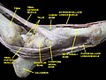
---|---|
Details | |
Origin | Arises from the middle portion of the fibula on the anterior surface and the interosseous membrane |
Insertion | Inserts on the dorsal side of the base of the distal phalanx of the big toe |
Artery | Anterior tibial artery |
Nerve | Deep fibular nerve, L5 (L4-S1) |
Actions | Extends (raises) the big toe and assists in dorsiflexion of the foot at the ankle. Also is a weak evertor/invertor |
Antagonist | Flexor hallucis longus, flexor hallucis brevis |
Identifiers | |
Latin | musculus extensor hallucis longus |
TA98 | A04.7.02.040 |
TA2 | 2650 |
FMA | 22533 |
Anatomical terms of muscle |
The extensor hallucis longus muscle is a thin skeletal muscle, situated between the tibialis anterior and the extensor digitorum longus. It extends the big toe and dorsiflects the foot. It also assists with foot eversion and inversion.
The muscle ends as a tendon of insertion. The tendon passes through a distinct compartment in the inferior extensor retinaculum of foot. It crosses anterior tibial vessels lateromedially near the bend of the ankle.[ citation needed ] In the foot, its tendon is situated at along the medial side of the dorsum of the foot. [1] Opposite the metatarsophalangeal articulation, the tendon gives off a thin prolongation on either side, to cover the surface of the joint. An expansion from the medial side of the tendon is usually inserted into the base of the proximal phalanx.[ citation needed ]
The extensor hallucis longus muscle arises from the middle portion of [2] the anterior surface[ citation needed ] of the fibula and adjacent interosseous membrane of the leg. [3] Its origin is medial to the origin of the extensor digitorum longus muscle.[ citation needed ]
The muscle inserts at the base of the distal phalanx of the great toe. [4]
The muscle receives motor innervation from the deep fibular nerve (L5) [3] (a branch of common fibular nerve).
The anterior tibial vessels and deep fibular nerve pass between this muscle and the tibialis anterior muscle.
Occasionally united at its origin with the extensor digitorum longus.
The extensor ossis metatarsi hallucis, a small muscle, sometimes found as a slip from the extensor hallucis longus, or from the tibialis anterior, or from the extensor digitorum longus, or as a distinct muscle; it traverses the same compartment of the transverse ligament with the extensor hallucis longus.
The muscle extends/dorsiflects the big toe (primary action), and dorsiflects the foot (secondary action). [3]
The foot is an anatomical structure found in many vertebrates. It is the terminal portion of a limb which bears weight and allows locomotion. In many animals with feet, the foot is a separate organ at the terminal part of the leg made up of one or more segments or bones, generally including claws and/or nails.
The leg is the entire lower limb of the human body, including the foot, thigh or sometimes even the hip or buttock region. The major bones of the leg are the femur, tibia, and adjacent fibula. The thigh is between the hip and knee, while the calf (rear) and shin (front) are between the knee and foot.
The tibia, also known as the shinbone or shankbone, is the larger, stronger, and anterior (frontal) of the two bones in the leg below the knee in vertebrates ; it connects the knee with the ankle. The tibia is found on the medial side of the leg next to the fibula and closer to the median plane. The tibia is connected to the fibula by the interosseous membrane of leg, forming a type of fibrous joint called a syndesmosis with very little movement. The tibia is named for the flute tibia. It is the second largest bone in the human body, after the femur. The leg bones are the strongest long bones as they support the rest of the body.
The fibula or calf bone is a leg bone on the lateral side of the tibia, to which it is connected above and below. It is the smaller of the two bones and, in proportion to its length, the most slender of all the long bones. Its upper extremity is small, placed toward the back of the head of the tibia, below the knee joint and excluded from the formation of this joint. Its lower extremity inclines a little forward, so as to be on a plane anterior to that of the upper end; it projects below the tibia and forms the lateral part of the ankle joint.
The ankle, the talocrural region or the jumping bone (informal) is the area where the foot and the leg meet. The ankle includes three joints: the ankle joint proper or talocrural joint, the subtalar joint, and the inferior tibiofibular joint. The movements produced at this joint are dorsiflexion and plantarflexion of the foot. In common usage, the term ankle refers exclusively to the ankle region. In medical terminology, "ankle" can refer broadly to the region or specifically to the talocrural joint.
The tibialis anterior muscle is a muscle of the anterior compartment of the lower leg. It originates from the upper portion of the tibia; it inserts into the medial cuneiform and first metatarsal bones of the foot. It acts to dorsiflex and invert the foot. This muscle is mostly located near the shin.
The tibial nerve is a branch of the sciatic nerve. The tibial nerve passes through the popliteal fossa to pass below the arch of soleus.
The flexor hallucis longus muscle (FHL) attaches to the plantar surface of phalanx of the great toe and is responsible for flexing that toe. The FHL is one of the three deep muscles of the posterior compartment of the leg, the others being the flexor digitorum longus and the tibialis posterior. The tibialis posterior is the most powerful of these deep muscles. All three muscles are innervated by the tibial nerve which comprises half of the sciatic nerve.
The flexor digitorum longus muscle is situated on the tibial side of the leg. At its origin it is thin and pointed, but it gradually increases in size as it descends. It serves to flex the second, third, fourth, and fifth toes.
The extensor digitorum longus is a pennate muscle, situated at the lateral part of the front of the leg.
The extensor digitorum brevis muscle is a muscle on the upper surface of the foot that helps extend digits 2 through 4.
In human anatomy, the fibularis brevis is a muscle that lies underneath the fibularis longus within the lateral compartment of the leg. It acts to tilt the sole of the foot away from the midline of the body (eversion) and to extend the foot downward away from the body at the ankle.
In human anatomy, the fibularis tertius is a muscle in the anterior compartment of the leg. It acts to tilt the sole of the foot away from the midline of the body (eversion) and to pull the foot upward toward the body (dorsiflexion).
The extensor hallucis brevis is a muscle on the top of the foot that helps to extend the big toe.
The superficial fibular nerve is a mixed nerve that provides motor innervation to the fibularis longus and fibularis brevis muscles, and sensory innervation to skin over the antero-lateral aspect of the leg along with the greater part of the dorsum of the foot.
The deep fibular nerve begins at the bifurcation of the common fibular nerve between the fibula and upper part of the fibularis longus, passes infero-medially, deep to the extensor digitorum longus, to the anterior surface of the interosseous membrane, and comes into relation with the anterior tibial artery above the middle of the leg; it then descends with the artery to the front of the ankle-joint, where it divides into a lateral and a medial terminal branch.
In humans, the sole of the foot is anatomically referred to as the plantar aspect.
The inferior extensor retinaculum of the foot is a Y-shaped band placed in front of the ankle-joint, the stem of the Y being attached laterally to the upper surface of the calcaneus, in front of the depression for the interosseous talocalcaneal ligament; it is directed medialward as a double layer, one lamina passing in front of, and the other behind, the tendons of the peroneus tertius and extensor digitorum longus.
The superior extensor retinaculum of the foot is the upper part of the extensor retinaculum of foot which extends from the ankle to the heelbone.
The anterior compartment of the leg is a fascial compartment of the lower leg. It contains muscles that produce dorsiflexion and participate in inversion and eversion of the foot, as well as vascular and nervous elements, including the anterior tibial artery and veins and the deep fibular nerve.
This article incorporates text in the public domain from page 481 of the 20th edition of Gray's Anatomy (1918)