Abductor hallucis muscle

Last updated
Abductor hallucis muscle
Sobo 1909 310.png
First layer of muscles of the sole of the foot (abductor hallucis visible at lower right)
Details
Origin Medial process of calcaneal tuberosity, Plantar aponeurosis, Flexor retinaculum
Insertion Medial aspect of base of 1st phalanx of hallux
Artery Medial plantar artery
Nerve Medial plantar nerve
Actions Abducts hallux
Antagonist Adductor hallucis muscle
Identifiers
Latin musculus abductor hallucis
TA98 A04.7.02.056
TA2 2672
FMA 37448
Anatomical terms of muscle

The abductor hallucis muscle is an intrinsic muscle of the foot. It participates in the abduction and flexion of the great toe.

Contents

Structure

The abductor hallucis as seen from an inferior projection of the foot. This diagram shows the bottom-most layer of muscles, just under the plantar skin of the foot. 1124 Intrinsic Muscles of the Foot b.png
The abductor hallucis as seen from an inferior projection of the foot. This diagram shows the bottom-most layer of muscles, just under the plantar skin of the foot.

The abductor hallucis muscle is located in the medial border of the foot and contributes to form the prominence that is observed on the region. It is inserted behind on the tuberosity of the calcaneus, the flexor retinaculum, and the plantar aponeurosis. [1] Its muscle body, relatively thick behind, flattens as it goes forward. It ends in a common tendon with the medial head of the flexor hallucis brevis that inserts on the medial surface of the base of the first proximal phalanx and its related sesamoid bone. Its medial surface is superficial and covered with the muscle's fascia and the skin. [2]

Nerve supply

Abductor hallucis is supplied by the medial plantar nerve. The nerves that supply it enter the muscle from its upper border.

Additional images

See also

Related Research Articles

<span class="mw-page-title-main">Foot</span> Anatomical structure found in vertebrates

The foot is an anatomical structure found in many vertebrates. It is the terminal portion of a limb which bears weight and allows locomotion. In many animals with feet, the foot is a separate organ at the terminal part of the leg made up of one or more segments or bones, generally including claws and/or nails.

<span class="mw-page-title-main">Human leg</span> Lower extremity or limb of the human body (foot, lower leg, thigh and hip)

The leg is the entire lower limb of the human body, including the foot, thigh or sometimes even the hip or buttock region. The major bones of the leg are the femur, tibia, and adjacent fibula. The thigh is between the hip and knee, while the calf (rear) and shin (front) are between the knee and foot.

<span class="mw-page-title-main">Toe</span> Digit of a foot

Toes are the digits of the foot of a tetrapod. Animal species such as cats that walk on their toes are described as being digitigrade. Humans, and other animals that walk on the soles of their feet, are described as being plantigrade; unguligrade animals are those that walk on hooves at the tips of their toes.

<span class="mw-page-title-main">Fibularis longus</span> Superficial muscle in the lateral compartment of the leg

In human anatomy, the fibularis longus is a superficial muscle in the lateral compartment of the leg. It acts to tilt the sole of the foot away from the midline of the body (eversion) and to extend the foot downward away from the body at the ankle.

<span class="mw-page-title-main">Cuboid bone</span> Bone of the ankle

In the human body, the cuboid bone is one of the seven tarsal bones of the foot.
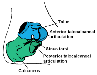
<span class="mw-page-title-main">Calcaneus</span> Bone of the tarsus of the foot

In humans and many other primates, the calcaneus or heel bone is a bone of the tarsus of the foot which constitutes the heel. In some other animals, it is the point of the hock.

<span class="mw-page-title-main">Tibial nerve</span> Branch of the sciatic nerve

The tibial nerve is a branch of the sciatic nerve. The tibial nerve passes through the popliteal fossa to pass below the arch of soleus.

<span class="mw-page-title-main">Dorsal interossei of the foot</span> Four muscles situated between the metatarsal bones

In human anatomy, the dorsal interossei of the foot are four muscles situated between the metatarsal bones.

<span class="mw-page-title-main">Flexor hallucis longus muscle</span> One of the three deep muscles in the lower leg

The flexor hallucis longus muscle (FHL) attaches to the plantar surface of phalanx of the great toe and is responsible for flexing that toe. The FHL is one of the three deep muscles of the posterior compartment of the leg, the others being the flexor digitorum longus and the tibialis posterior. The tibialis posterior is the most powerful of these deep muscles. All three muscles are innervated by the tibial nerve which comprises half of the sciatic nerve.

<span class="mw-page-title-main">Flexor digitorum longus muscle</span> Muscle located on the tibial side of the leg

The flexor digitorum longus muscle is situated on the tibial side of the leg. At its origin it is thin and pointed, but it gradually increases in size as it descends. It serves to flex the second, third, fourth, and fifth toes.
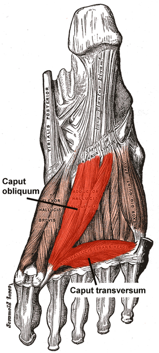
<span class="mw-page-title-main">Adductor hallucis muscle</span> Muscle responsible for adducting the big toe

The Adductor hallucis arises by two heads—oblique and transverse and is responsible for adducting the big toe. It has two heads, both are innervated by the lateral plantar nerve.
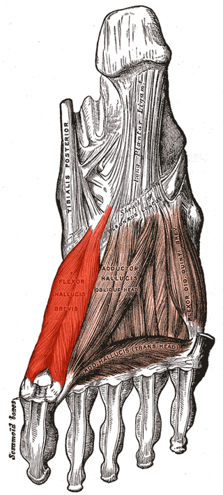
<span class="mw-page-title-main">Flexor hallucis brevis muscle</span> Muscle in sole of the foot that leads to the big toe

Flexor hallucis brevis muscle is a muscle of the foot that flexes the big toe.
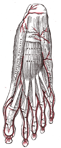
<span class="mw-page-title-main">Flexor digitorum brevis muscle</span> Lies in the middle of the sole of the foot

The flexor digitorum brevis is a muscle which lies in the middle of the sole of the foot, immediately above the central part of the plantar aponeurosis, with which it is firmly united.
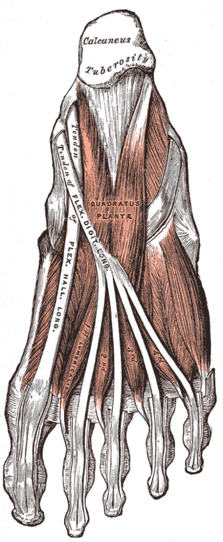
<span class="mw-page-title-main">Quadratus plantae muscle</span> Muscles of the sole of the foot

The quadratus plantae is separated from the muscles of the first layer by the lateral plantar vessels and nerve. It acts to aid in flexing the 2nd to 5th toes and is one of the few muscles in the foot with no homolog in the hand.
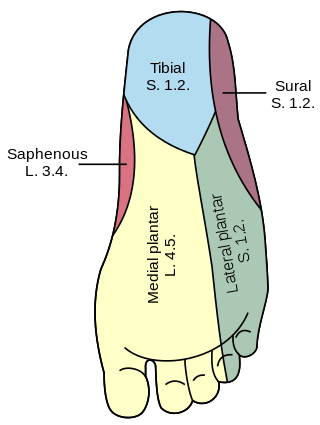
<span class="mw-page-title-main">Plantar nerve</span> Pair of nerves innervating the sole of the foot

The plantar nerves are a pair of nerves innervating the sole of the foot. They arise from the posterior branch of the tibial nerve.

<span class="mw-page-title-main">Sole (foot)</span> Bottom part of foot

The sole is the bottom of the foot.

<span class="mw-page-title-main">Medial plantar nerve</span> Larger of the two terminal divisions of the tibial nerve

The medial plantar nerve is the larger of the two terminal divisions of the tibial nerve, which accompanies the medial plantar artery.

<span class="mw-page-title-main">Medial plantar artery</span>

The medial plantar artery, much smaller than the lateral plantar artery, passes forward along the medial side of the foot.

The flexor retinaculum of foot is a strong fibrous band in the foot.

<span class="mw-page-title-main">Fifth metatarsal bone</span>

The fifth metatarsal bone is a long bone in the foot, and is palpable along the distal outer edges of the feet. It is the second smallest of the five metatarsal bones. The fifth metatarsal is analogous to the fifth metacarpal bone in the hand.

References

PD-icon.svgThis article incorporates text in the public domain from page 491 of the 20th edition of Gray's Anatomy (1918)

  1. "Abductor Hallucis". Loyola University Medical Education Network. Retrieved 7 January 2015.
  2. Latarjet, Michel; Ruiz Liard, Alfredo (2005). Human Anatomy (Spanish ed.). Editorial Médica Panamericana. ISBN   978-950-06-1368-2.[ page needed ]