Flexor digitorum longus muscle

Last updated
Flexor digitorum longus muscle
Flechisseur commun des orteils.png
Inferior view of flexor digitorum longus muscle - animation.gif
View from below
Details
Origin Posterior surface of the body of the tibia
Insertion Plantar surface; base of the distal phalanges of the four lesser toes
Artery Posterior tibial artery
Nerve Tibial nerve
Actions Flexion of the four smaller digits
Antagonist Extensor digitorum longus, extensor digitorum brevis
Identifiers
Latin musculus flexor digitorum longus
TA98 A04.7.02.052
TA2 2667
FMA 51071
Anatomical terms of muscle

The flexor digitorum longus muscle is situated on the tibial side of the leg. At its origin it is thin and pointed, but it gradually increases in size as it descends. It serves to flex the second, third, fourth, and fifth toes.

Contents

Structure

The flexor digitorum longus runs along the medial posterior side of the lower leg and aids in flexions of the toes (apart from the big toe). 1123 Muscles of the Leg that Move the Foot and Toes c.png
The flexor digitorum longus runs along the medial posterior side of the lower leg and aids in flexions of the toes (apart from the big toe).

The flexor digitorum longus muscle arises from the posterior surface of the body of the tibia, from immediately below the soleal line to within 7 or 8 cm of its lower extremity, medial to the tibial origin of the tibialis posterior muscle. It also arises from the fascia covering the tibialis posterior muscle.

The fibers end in a tendon, which runs nearly the whole length of the posterior surface of the muscle. This tendon passes behind the medial malleolus, in a groove, common to it and the tibialis posterior, but separated from the latter by a fibrous septum, each tendon being contained in a special compartment lined by a separate mucous sheath. The tendon of the tibialis posterior and the tendon of the flexor digitorum longus cross each other, in a spot above the medial malleolus, the crural tendinous chiasm. [1] [2] [3] It passes through the tarsal tunnel. [4]

It passes obliquely forward and lateralward, superficial to the deltoid ligament of the ankle-joint, into the sole of the foot, where it crosses over the tendon of the flexor hallucis longus at the level of the navicular bone at a location known as the knot of henry [5] (also referred to as plantar tendinous chiasm), [1] [2] [3] and receives from it a strong tendinous slip.

It then expands and is joined by the quadratus plantæ muscle, and finally divides into four tendons, which are inserted into the bases of the last phalanges of the second, third, fourth, and fifth toes, each tendon passing through an opening in the corresponding tendon of the flexor digitorum brevis muscle opposite the base of the first interphalangeal joint.

Variation

Flexor accessorius longus digitorum, not infrequent, origin from fibula, or tibia, or the deep fascia and ending in a tendon which, after passing beneath the laciniate ligament, joins the tendon of the long flexor or the quadratus plantæ.

Function

Similar to the flexor hallucis longus and tibialis posterior muscles, the flexor digitorum longus muscle functions to plantar flex and invert the foot. The flexor digitorum longus muscle is responsible for the movement and curling of the second, third, fourth and fifth toes. This muscle makes it possible for the toes to grip the surface of floors, which is important when it comes to maintaining postural balance on surfaces that are rough or uneven. The other deep muscles are the flexor hallucis longus and tibialis posterior; the tibialis posterior is the most powerful of these deep muscles. All three muscles are innervated by the tibial nerve which comprises half of the sciatic nerve.

Clinical significance

After passing through the tarsal tunnel, the flexor digitorum longus tendon must curve around a bony landmark called the sustentaculum tali. Flexor digitorum longus pain can occur with a trip and fall on uneven surface when the toes are not able to grip the surface totally. One can also injure the flexor digitorum longus muscle while running on a beach in the sand without any footwear, making the muscle vulnerable at the calcaneus attachment for injuries. In case of flexor digitorum longus pain or strain, the patient will find it tough to walk and will have excruciating pain in the feet and ankles. Support braces along with warm compresses are the most preferred way of treating flexor digitorum longus pain or strain.

Related Research Articles

<span class="mw-page-title-main">Foot</span> Anatomical structure found in vertebrates

The foot is an anatomical structure found in many vertebrates. It is the terminal portion of a limb which bears weight and allows locomotion. In many animals with feet, the foot is a separate organ at the terminal part of the leg made up of one or more segments or bones, generally including claws and/or nails.

<span class="mw-page-title-main">Human leg</span> Lower extremity or limb of the human body (foot, lower leg, thigh and hip)

The leg is the entire lower limb of the human body, including the foot, thigh or sometimes even the hip or buttock region. The major bones of the leg are the femur, tibia, and adjacent fibula. The thigh is between the hip and knee, while the calf (rear) and shin (front) are between the knee and foot.

<span class="mw-page-title-main">Cuboid bone</span> Bone of the ankle

In the human body, the cuboid bone is one of the seven tarsal bones of the foot.

<span class="mw-page-title-main">Tibia</span> Leg bone in vertebrates

The tibia, also known as the shinbone or shankbone, is the larger, stronger, and anterior (frontal) of the two bones in the leg below the knee in vertebrates ; it connects the knee with the ankle. The tibia is found on the medial side of the leg next to the fibula and closer to the median plane. The tibia is connected to the fibula by the interosseous membrane of leg, forming a type of fibrous joint called a syndesmosis with very little movement. The tibia is named for the flute tibia. It is the second largest bone in the human body, after the femur. The leg bones are the strongest long bones as they support the rest of the body.

<span class="mw-page-title-main">Fibula</span> Leg bone in vertebrates

The fibula or calf bone is a leg bone on the lateral side of the tibia, to which it is connected above and below. It is the smaller of the two bones and, in proportion to its length, the most slender of all the long bones. Its upper extremity is small, placed toward the back of the head of the tibia, below the knee joint and excluded from the formation of this joint. Its lower extremity inclines a little forward, so as to be on a plane anterior to that of the upper end; it projects below the tibia and forms the lateral part of the ankle joint.

<span class="mw-page-title-main">Ankle</span> Region where the foot and the leg meet

The ankle, the talocrural region or the jumping bone (informal) is the area where the foot and the leg meet. The ankle includes three joints: the ankle joint proper or talocrural joint, the subtalar joint, and the inferior tibiofibular joint. The movements produced at this joint are dorsiflexion and plantarflexion of the foot. In common usage, the term ankle refers exclusively to the ankle region. In medical terminology, "ankle" can refer broadly to the region or specifically to the talocrural joint.

<span class="mw-page-title-main">Extensor hallucis longus muscle</span> Thin muscle, situated between the tibialis anterior and the extensor digitorum longus

The extensor hallucis longus muscle is a thin skeletal muscle, situated between the tibialis anterior and the extensor digitorum longus. It extends the big toe and dorsiflects the foot. It also assists with foot eversion and inversion.

Chiasm may refer to:

<span class="mw-page-title-main">Tibialis anterior muscle</span> Flexor muscle in humans that dorsiflexes the foot

The tibialis anterior muscle is a muscle of the anterior compartment of the lower leg. It originates from the upper portion of the tibia; it inserts into the medial cuneiform and first metatarsal bones of the foot. It acts to dorsiflex and invert the foot. This muscle is mostly located near the shin.

<span class="mw-page-title-main">Tibial nerve</span> Branch of the sciatic nerve

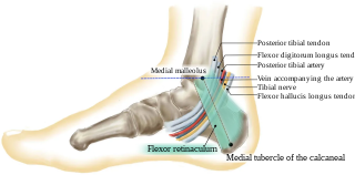
The tibial nerve is a branch of the sciatic nerve. The tibial nerve passes through the popliteal fossa to pass below the arch of soleus.

<span class="mw-page-title-main">Flexor hallucis longus muscle</span> One of the three deep muscles in the lower leg

The flexor hallucis longus muscle (FHL) attaches to the plantar surface of phalanx of the great toe and is responsible for flexing that toe. The FHL is one of the three deep muscles of the posterior compartment of the leg, the others being the flexor digitorum longus and the tibialis posterior. The tibialis posterior is the most powerful of these deep muscles. All three muscles are innervated by the tibial nerve which comprises half of the sciatic nerve.

<span class="mw-page-title-main">Flexor hallucis brevis muscle</span> Muscle in sole of the foot that leads to the big toe

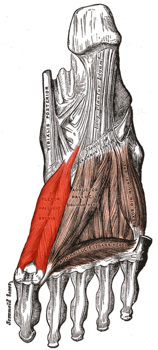
Flexor hallucis brevis muscle is a muscle of the foot that flexes the big toe.
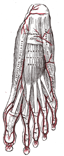
<span class="mw-page-title-main">Quadratus plantae muscle</span> Muscles of the sole of the foot

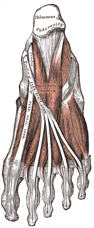
The quadratus plantae is separated from the muscles of the first layer by the lateral plantar vessels and nerve. It acts to aid in flexing the 2nd to 5th toes and is one of the few muscles in the foot with no homolog in the hand.

<span class="mw-page-title-main">Plantar nerve</span> Pair of nerves innervating the sole of the foot

The plantar nerves are a pair of nerves innervating the sole of the foot. They arise from the posterior branch of the tibial nerve.

<span class="mw-page-title-main">Sole (foot)</span> Bottom part of foot

In humans, the sole of the foot is anatomically referred to as the plantar aspect.

The flexor retinaculum of foot is a strong fibrous band in the foot.

<span class="mw-page-title-main">Malleolus</span> Ankle bone protrusion

A malleolus is the bony prominence on each side of the human ankle.

<span class="mw-page-title-main">Tarsal tunnel</span> Canal in the ankle area

The tarsal tunnel is a passage found along the inner leg underneath the medial malleolus of the ankle.

<span class="mw-page-title-main">Outline of human anatomy</span> Overview of and topical guide to human anatomy

The following outline is provided as an overview of and topical guide to human anatomy:

References

PD-icon.svgThis article incorporates text in the public domain from page 485 of the 20th edition of Gray's Anatomy (1918)

  1. 1 2 Walter Thiel (1997) Photographic atlas of practical anatomy - Volume 1, Part 1 - p.348
  2. 1 2 Johannes Sobotta, Reinhard Putz, Reinhard Pabst (1997) Sobotta Atlas of Human Anatomy: Thorax, abdomen, pelvis, lower limb, p.331
  3. 1 2 Jan Langman, Martinus Willem Woerdeman (1982) Atlas of medical anatomy p.323
  4. Gujar, Bansari; Flores, Raymond H. (2015-01-01), Hochberg, Marc C.; Silman, Alan J.; Smolen, Josef S.; Weinblatt, Michael E. (eds.), "81 - Entrapment neuropathies and compartment syndromes", Rheumatology (Sixth Edition), Philadelphia: Mosby, pp. 671–682, doi: 10.1016/b978-0-323-09138-1.00081-4 , ISBN   978-0-323-09138-1
  5. Knipe, Henry. "Knot of Henry | Radiology Reference Article | Radiopaedia.org". Radiopaedia. Retrieved 20 October 2020.