The quadratus femoris is a flat, quadrilateral skeletal muscle. Located on the posterior side of the hip joint, it is a strong external rotator and adductor of the thigh,[2] but also acts to stabilize the femoral head in the acetabulum. The quadratus femoris is used in Meyer's muscle pedicle grafting to prevent avascular necrosis of femur head.
It originates on the lateral border of the ischial tuberosity of the ischium of the pelvis.[1] From there, it passes laterally to its insertion on the posterior side of the head of the femur: the quadrate tubercle on the intertrochanteric crest and along the quadrate line, the vertical line which runs downward to bisect the lesser trochanter on the medial side of the femur. Along its course, quadratus is aligned edge to edge with the inferior gemellus above and the adductor magnus below, so that its upper and lower borders run horizontal and parallel.[3]
Groin pain can be a disabling ailment with many potential root causes: one such cause, often overlooked, is quadratus femoris tendinitis. Magnetic resonance imaging can show abnormal signal intensity at the insertion of the right quadratus femoris tendon, which suggests inflammation of the area.[4] Since the muscle works to laterally rotate and adduct the femur, actions involving the lower body can strain the muscle. In addition, patients present with hip pain and an increased signal intensity of the MRI of the quadratus femoris have been shown to also have a significantly narrower ischiofemoral space compared to the general populace. The ischiofemoral impingement may be a cause of the hip pain associated with quadratus femoris tendinitis.[5]
Quadratus femoris muscle
Additional images
Right femur. Posterior surface.
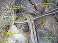
Structures surrounding right hip-joint.
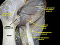
Nerves of the right lower extremity Posterior view.
↑ Platzer, Werner (2003). Color atlas and textbook of human anatomy (5th rev. and enlarged [English]ed.). Stuttgart: Thieme. p.238. ISBN978-1-58890-159-0. OCLC54767617.
↑ Abrahams, Peter H.; Marks, S. C.; Hutchings, R. T. (2003). McMinn's color atlas of human anatomy (5thed.). Edinburgh: Mosby. p.166. ISBN0-7234-3212-0. OCLC50477357.
Platzer, Werner (2004). Color Atlas of Human Anatomy, Vol 1: Locomotor system (5thed.). Thieme. ISBN3-13-533305-1. (ISBN for the Americas 1-58890-159-9.)
This page is based on this Wikipedia article Text is available under the CC BY-SA 4.0 license; additional terms may apply. Images, videos and audio are available under their respective licenses.