Structure
The soleus is located in the superficial posterior compartment of the leg.
The soleus exhibits significant morphological differences across species. It is unipennate in many species. In some animals, such as the rabbit, it is fused for much of its length with the gastrocnemius muscle.
The soleus is a complex, multi-pennate muscle in humans, normally having a separate (posterior) aponeurosis from the gastrocnemius muscle. Most soleus muscle fibers originate from each side of the anterior aponeurosis, attached to the tibia and fibula. [1] [2] Other fibers originate from the posterior (back) surfaces of the head of the fibula and its upper quarter, as well as the middle third of the medial border of the tibia.
The fibers originating from the anterior surface of the anterior aponeurosis insert onto the median septum, and the fibers originating from the posterior surface of the anterior aponeurosis insert onto the posterior aponeurosis. [1] [2] The posterior aponeurosis and median septum join in the lower quarter of the muscle and then join with the anterior aponeuroses of the gastrocnemius muscles to form the calcaneal tendon or Achilles tendon and inserts onto the posterior surface of the calcaneus, or heel bone.
In contrast to some animals, the human soleus and gastrocnemius muscles are relatively separate, so shear can be detected between the soleus and gastrocnemius aponeuroses. [3]
The soleus is vestigial in the horse. [4]
Function
The action of the calf muscles, including the soleus, is plantar flexion of the foot (that is, they increase the angle between the foot and the leg). They are powerful muscles vital in walking, running, and keeping balance. The soleus plays an important role in maintaining standing posture; if not for its constant pull, the body would fall forward.
Also, in upright posture, the soleus is responsible for pumping venous blood back into the heart from the periphery, and is often called the skeletal muscle pump, peripheral heart or the sural (tricipital) pump. [5]
Soleus muscles have more slow muscle fibers than many other muscles. In some animals, such as the guinea pig and cat, soleus consists of 100% slow muscle fibers. [6] [7] Human soleus fiber composition is variable, containing between 60% and 100% slow fibers. [8]
The soleus is the most effective muscle for plantar flexion in a bent knee position. The gastrocnemius originates on the femur, so bending the leg limits its effective tension. During regular movement (i.e., walking) the soleus is the primary muscle utilized for plantar flexion due to the slow-twitch fibers resisting fatigue. [9]
This page is based on this
Wikipedia article Text is available under the
CC BY-SA 4.0 license; additional terms may apply.
Images, videos and audio are available under their respective licenses.
 Animation.
 Bones of the right leg. Posterior surface.
 Cross-section through middle of leg.
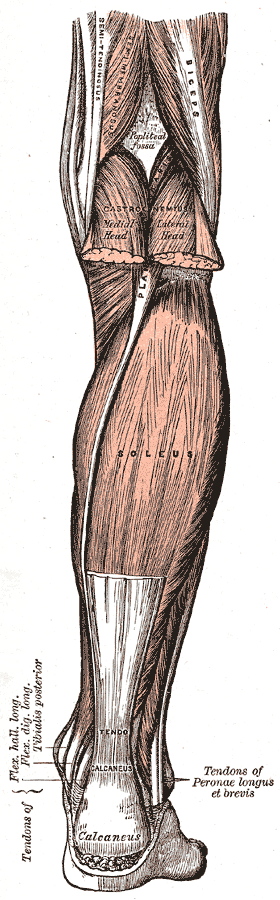
 Back of left lower extremity.
 Posterior view.