Adductor brevis muscle

Last updated
Adductor brevis muscle
Anterior Hip Muscles 2.PNG
The adductor brevis and nearby muscles
Gray344.png
Structures surrounding right hip-joint. (Adductor brevis at upper right.)
Details
Origin anterior surface of the inferior ramus and body of the pubis
Insertion the lesser trochanter and linea aspera of the femur
Artery deep femoral artery
Nerve obturator nerve
Actions adduction of hip
Identifiers
Latin musculus adductor brevis
TA98 A04.7.02.027
TA2 2629
FMA 22442
Anatomical terms of muscle

The adductor brevis is a muscle in the thigh situated immediately deep to the pectineus and adductor longus. It belongs to the adductor muscle group. The main function of the adductor brevis is to pull the thigh medially. [1] The adductor brevis and the rest of the adductor muscle group is also used to stabilize left to right movements of the trunk, when standing on both feet, or to balance when standing on a moving surface. The adductor muscle group is used pressing the thighs together to ride a horse, and kicking with the inside of the foot in soccer or swimming. Last, they contribute to flexion of the thigh when running or against resistance (squats, jumping, etc.). [2]

Contents

Structure

It is somewhat triangular in form, and arises by a narrow origin from the outer surfaces of the body of the pubis and inferior ramus of the pubis, between the gracilis and obturator externus.

The Adductor brevis muscle widens in triangular fashion to be inserted into the upper part of the linea aspera immediately lateral to the insertion of pectineus and above that of adductor longus.

Relations

By its anterior surface, the adductor brevis is in relation with the pectineus, adductor longus, and anterior branches of the obturator artery, the obturator vein, and the obturator nerve.

By its posterior surface with the adductor magnus and the posterior branches of the obturator artery, the obturator vein, and the obturator nerve.

By its outer border with the obturator externus, and the iliopsoas. By its inner border with the gracilis and adductor magnus.

It is pierced near its insertion by the middle perforating artery. [3]

Innervation

The adductor brevis is innervated dually by the anterior and posterior branches of the obturator nerve. [4]

Function

The muscle is primarily known as a hip adductor. It also functions as a hip flexor. Whether it acts to rotate the femur laterally or medially is dependent on position.

Related Research Articles

<span class="mw-page-title-main">Human leg</span> Lower extremity or limb of the human body (foot, lower leg, thigh and hip)

The human leg is the entire lower limb of the human body, including the foot, thigh or sometimes even the hip or buttock region. The major bones of the leg are the femur, tibia, and adjacent fibula. The thigh is between the hip and knee, while the calf (rear) and shin (front) are between the knee and foot.

<span class="mw-page-title-main">Thigh</span> The part of the body between the pelvis and the knee; upper leg

In anatomy, the thigh is the area between the hip (pelvis) and the knee. Anatomically, it is part of the lower limb.
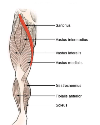
<span class="mw-page-title-main">Deep femoral artery</span> Large branch of the femoral artery that supplies the thigh

The deep femoral artery also known as the deep artery of the thigh, or profunda femoris artery, is a large branch of the femoral artery. It travels more deeply ("profoundly") than the rest of the femoral artery. It gives rise to the lateral circumflex femoral artery and medial circumflex femoral artery, and the perforating arteries, terminating within the thigh.

<span class="mw-page-title-main">Pectineus muscle</span> Adductor of the thigh

The pectineus muscle is a flat, quadrangular muscle, situated at the anterior (front) part of the upper and medial (inner) aspect of the thigh. The pectineus muscle is the most anterior adductor of the hip. The muscle's primary action is hip flexion; it also produces adduction and internal rotation of the hip.

<span class="mw-page-title-main">Groin</span> The two creases at the junction of the torso with the thighs, on either side of the pubic area

In human anatomy, the groin also known as the inguinal region or iliac region, is the junctional area between the torso and the thigh. The groin is at the front of the body on either side of the pubic tubercle, where the lower part of the abdominal wall meets the thigh. A fold or crease is formed at this junction known as the inguinal groove, or crease. This is also the area of the medial compartment of the thigh that contains attachments of the adductor muscles of the hip or the groin muscles. The groin is the common site for a hernia.

<span class="mw-page-title-main">Hip</span> Anatomical region between the torso and the legs, holding the buttocks and genital region

In vertebrate anatomy, hip refers to either an anatomical region or a joint.

<span class="mw-page-title-main">External obturator muscle</span> One of six small hip muscles in the lateral rotator group

The external obturator muscle, obturator externus muscle is a flat, triangular muscle, which covers the outer surface of the anterior wall of the pelvis.

<span class="mw-page-title-main">Adductor longus muscle</span> Skeletal muscle located in the thigh

In the human body, the adductor longus is a skeletal muscle located in the thigh. One of the adductor muscles of the hip, its main function is to adduct the thigh and it is innervated by the obturator nerve. It forms the medial wall of the femoral triangle.

<span class="mw-page-title-main">Adductor magnus muscle</span> Muscle in the thigh

The adductor magnus is a large triangular muscle, situated on the medial side of the thigh.

<span class="mw-page-title-main">Gracilis muscle</span> Most superficial muscle on the medial side of the thigh

The gracilis muscle is the most superficial muscle on the medial side of the thigh. It is thin and flattened, broad above, narrow and tapering below.

<span class="mw-page-title-main">Iliopsoas</span> Joined psoas and the iliacus muscles

The iliopsoas muscle refers to the joined psoas major and the iliacus muscles. The two muscles are separate in the abdomen, but usually merge in the thigh. They are usually given the common name iliopsoas. The iliopsoas muscle joins to the femur at the lesser trochanter. It acts as the strongest flexor of the hip.

<span class="mw-page-title-main">Muscles of the hip</span> Causes movement in the hip

In human anatomy, the muscles of the hip joint are those muscles that cause movement in the hip. Most modern anatomists define 17 of these muscles, although some additional muscles may sometimes be considered. These are often divided into four groups according to their orientation around the hip joint: the gluteal group; the lateral rotator group; the adductor group; and the iliopsoas group.

<span class="mw-page-title-main">Adductor muscles of the hip</span> Group of muscles

The adductor muscles of the hip are a group of muscles in the medial compartment of the thigh mostly used for bringing the thighs together.

<span class="mw-page-title-main">Lumbar plexus</span> Web of nerves in the lower spine

The lumbar plexus is a web of nerves in the lumbar region of the body which forms part of the larger lumbosacral plexus. It is formed by the divisions of the first four lumbar nerves (L1-L4) and from contributions of the subcostal nerve (T12), which is the last thoracic nerve. Additionally, the ventral rami of the fourth lumbar nerve pass communicating branches, the lumbosacral trunk, to the sacral plexus. The nerves of the lumbar plexus pass in front of the hip joint and mainly support the anterior part of the thigh.

<span class="mw-page-title-main">Obturator nerve</span>

The obturator nerve in human anatomy arises from the ventral divisions of the second, third, and fourth lumbar nerves in the lumbar plexus; the branch from the third is the largest, while that from the second is often very small.

<span class="mw-page-title-main">Pubis (bone)</span> Most forward-facing of the three main regions making up the os coxa

In vertebrates, the pubis or pubic bone forms the lower and anterior part of each side of the hip bone. The pubis is the most forward-facing of the three bones that make up the hip bone. The left and right pubic bones are each made up of three sections, a superior ramus, inferior ramus, and a body.

<span class="mw-page-title-main">Medial circumflex femoral artery</span>

The medial circumflex femoral artery is an artery in the upper thigh that arises from the profunda femoris artery. It supplies arterial blood to several muscles in the region, as well as the femoral head and neck.

<span class="mw-page-title-main">Medial compartment of thigh</span> One of the fascial compartments of the thigh

The medial compartment of thigh is one of the fascial compartments of the thigh and contains the hip adductor muscles and the gracilis muscle.

The anterior branch of the obturator nerve is a branch of the obturator nerve found in the pelvis and leg.

<span class="mw-page-title-main">Adductor minimus muscle</span> Small and flat skeletal muscle in the thigh

In human anatomy, the adductor minimus is a small and flat skeletal muscle in the thigh which constitutes the upper, lateral part of the adductor magnus muscle. It adducts and laterally rotates the femur.

References

PD-icon.svgThis article incorporates text in the public domain from page 473 of the 20th edition of Gray's Anatomy (1918)

  1. Saladin, Kenneth (2010). Anatomy & Physiology: The Unity of Form and Function (6th ed.). McGraw-Hill. ISBN   978-0-07-337825-1.[ page needed ]
  2. Moore, Keith. Clinically Oriented Anatomy (5th ed.). Lippincott Williams & Wilkins.[ page needed ]
  3. Wilson, Erasmus (1851). The anatomist's vade mecum: a system of human anatomy. John Churchill. pp.  260–1.
  4. Miura M, Nakamura E, Kato S, Usui T, Miyauchi R (August 1994). "The true nature of the adductor brevis dually innervated by the anterior and posterior branches of the obturator nerve in humans". Okajimas Folia Anatomica Japonica. 71 (2–3): 67–82. doi: 10.2535/ofaj1936.71.2-3_67 . PMID   7808725.

Additional images