Tibialis posterior muscle

Last updated
Tibialis posterior muscle
Gray442.png
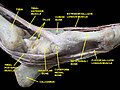
The mucous sheaths of the tendons around the ankle. Medial aspect. (Tibialis posterior labeled at top center.)
Details
Origin Tibia and fibula
Insertion Navicular and medial cuneiform bone
Artery Posterior tibial artery
Nerve Tibial nerve
Actions Inversion of the foot and plantar flexion of the foot at the ankle
Antagonist Fibularis brevis and longus, antagonist to the inversion.
Identifiers
Latin musculus tibialis posterior
TA98 A04.7.02.051
TA2 2666
FMA 51099
Anatomical terms of muscle

The tibialis posterior muscle is the most central of all the leg muscles, and is located in the deep posterior compartment of the leg. It is the key stabilizing muscle of the lower leg.

Contents

Posterior tibial tendonitis

Posterior tibial tendonitis is a condition that predominantly affects runners and active individuals. It involves inflammation or tearing of the posterior tibial tendon, which connects the calf muscle to the bones on the inside of the foot. It plays a vital role in supporting the arch and assisting in foot movement. This condition can cause pain, swelling, and potentially lead to flatfoot if left untreated. [1]

Structure

The tibialis posterior muscle originates on the inner posterior border of the fibula laterally. [2] It is also attached to the interosseous membrane medially, which attaches to the tibia and fibula. [2]

The tendon of the tibialis posterior muscle (sometimes called the posterior tibial tendon) descends posterior to the medial malleolus. [2] It terminates by dividing into plantar, main, and recurrent components. The main portion inserts into the tuberosity of the navicular bone. [2] The smaller portion inserts into the plantar surface of the medial cuneiform. The plantar portion inserts into the bases of the second, third and fourth metatarsals, the intermediate and lateral cuneiforms and the cuboid. The recurrent portion inserts into the sustentaculum tali of the calcaneus.

Blood is supplied to the muscle by the posterior tibial artery.

Nerve supply

The tibialis posterior muscle is supplied by the tibial nerve.

Function

The tibialis posterior muscle is a key muscle for stabilization of the lower leg. It also contracts to produce inversion of the foot, and assists in the plantarflexion of the foot at the ankle. [3] The tibialis posterior has a major role in supporting the medial arch of the foot. Dysfunction of the tibialis posterior, including rupture of the tibialis posterior tendon, can lead to flat feet in adults, as well as a valgus deformity due to unopposed eversion when inversion is lost. [4] [5]

Clinical significance

Injury to the distal tendon of the tibialis posterior muscle is rare. [3] It may be caused during exercise. [3] It usually presents with pain on the medial side of the ankle. [3] Injuries including dislocations and tears often require surgery. [6]

Additional images

Related Research Articles

<span class="mw-page-title-main">Foot</span> Anatomical structure found in vertebrates

The foot is an anatomical structure found in many vertebrates. It is the terminal portion of a limb which bears weight and allows locomotion. In many animals with feet, the foot is a separate organ at the terminal part of the leg made up of one or more segments or bones, generally including claws and/or nails.

<span class="mw-page-title-main">Human leg</span> Lower extremity or limb of the human body (foot, lower leg, thigh and hip)

The leg is the entire lower limb of the human body, including the foot, thigh or sometimes even the hip or buttock region. The major bones of the leg are the femur, tibia, and adjacent fibula. The thigh is between the hip and knee, while the calf (rear) and shin (front) are between the knee and foot.

<span class="mw-page-title-main">Fibularis longus</span> Superficial muscle in the lateral compartment of the leg

In human anatomy, the fibularis longus is a superficial muscle in the lateral compartment of the leg. It acts to tilt the sole of the foot away from the midline of the body (eversion) and to extend the foot downward away from the body at the ankle.

<span class="mw-page-title-main">Tibia</span> Leg bone in vertebrates

The tibia, also known as the shinbone or shankbone, is the larger, stronger, and anterior (frontal) of the two bones in the leg below the knee in vertebrates ; it connects the knee with the ankle. The tibia is found on the medial side of the leg next to the fibula and closer to the median plane. The tibia is connected to the fibula by the interosseous membrane of leg, forming a type of fibrous joint called a syndesmosis with very little movement. The tibia is named for the flute tibia. It is the second largest bone in the human body, after the femur. The leg bones are the strongest long bones as they support the rest of the body.

<span class="mw-page-title-main">Ankle</span> Region where the foot and the leg meet

The ankle, the talocrural region or the jumping bone (informal) is the area where the foot and the leg meet. The ankle includes three joints: the ankle joint proper or talocrural joint, the subtalar joint, and the inferior tibiofibular joint. The movements produced at this joint are dorsiflexion and plantarflexion of the foot. In common usage, the term ankle refers exclusively to the ankle region. In medical terminology, "ankle" can refer broadly to the region or specifically to the talocrural joint.

<span class="mw-page-title-main">Extensor hallucis longus muscle</span> Thin muscle, situated between the tibialis anterior and the extensor digitorum longus

The extensor hallucis longus muscle is a thin skeletal muscle, situated between the tibialis anterior and the extensor digitorum longus. It extends the big toe and dorsiflects the foot. It also assists with foot eversion and inversion.

<span class="mw-page-title-main">Tibialis anterior muscle</span> Flexor muscle in humans that dorsiflexes the foot

The tibialis anterior muscle is a muscle of the anterior compartment of the lower leg. It originates from the upper portion of the tibia; it inserts into the medial cuneiform and first metatarsal bones of the foot. It acts to dorsiflex and invert the foot. This muscle is mostly located near the shin.

<span class="mw-page-title-main">Gastrocnemius muscle</span> Calf muscle

The gastrocnemius muscle is a superficial two-headed muscle that is in the back part of the lower leg of humans. It is located superficial to the soleus in the posterior (back) compartment of the leg. It runs from its two heads just above the knee to the heel, extending across a total of three joints.

<span class="mw-page-title-main">Tibial nerve</span> Branch of the sciatic nerve

The tibial nerve is a branch of the sciatic nerve. The tibial nerve passes through the popliteal fossa to pass below the arch of soleus.

<span class="mw-page-title-main">Flexor hallucis longus muscle</span> One of the three deep muscles in the lower leg

The flexor hallucis longus muscle (FHL) attaches to the plantar surface of phalanx of the great toe and is responsible for flexing that toe. The FHL is one of the three deep muscles of the posterior compartment of the leg, the others being the flexor digitorum longus and the tibialis posterior. The tibialis posterior is the most powerful of these deep muscles. All three muscles are innervated by the tibial nerve which comprises half of the sciatic nerve.

<span class="mw-page-title-main">Flexor digitorum longus muscle</span> Muscle located on the tibial side of the leg

The flexor digitorum longus muscle is situated on the tibial side of the leg. At its origin it is thin and pointed, but it gradually increases in size as it descends. It serves to flex the second, third, fourth, and fifth toes.

<span class="mw-page-title-main">Extensor digitorum longus muscle</span> Pennate muscle, situated at the lateral part of the front of the leg

The extensor digitorum longus is a pennate muscle, situated at the lateral part of the front of the leg.

<span class="mw-page-title-main">Plantaris muscle</span> One of the superficial muscles of the superficial posterior compartment of the leg

The plantaris is one of the superficial muscles of the superficial posterior compartment of the leg, one of the fascial compartments of the leg.

<span class="mw-page-title-main">Fibularis brevis</span> Shorter and smaller of the fibularis (peroneus) muscles

In human anatomy, the fibularis brevis is a muscle that lies underneath the fibularis longus within the lateral compartment of the leg. It acts to tilt the sole of the foot away from the midline of the body (eversion) and to extend the foot downward away from the body at the ankle.

<span class="mw-page-title-main">Flexor hallucis brevis muscle</span> Muscle in sole of the foot that leads to the big toe

Flexor hallucis brevis muscle is a muscle of the foot that flexes the big toe.

<span class="mw-page-title-main">Deep fibular nerve</span> Type of nerve

The deep fibular nerve begins at the bifurcation of the common fibular nerve between the fibula and upper part of the fibularis longus, passes infero-medially, deep to the extensor digitorum longus, to the anterior surface of the interosseous membrane, and comes into relation with the anterior tibial artery above the middle of the leg; it then descends with the artery to the front of the ankle-joint, where it divides into a lateral and a medial terminal branch.

The flexor retinaculum of foot is a strong fibrous band in the foot.

<span class="mw-page-title-main">Deltoid ligament</span> Anatomical detail in the ankle

The deltoid ligament is a strong, flat, triangular band, attached, above, to the apex and anterior and posterior borders of the medial malleolus. The deltoid ligament supports the ankle joint and also resists excessive eversion of the foot. The deltoid ligament is composed of 4 fibers:

  1. Anterior tibiotalar ligament
  2. Tibiocalcaneal ligament
  3. Posterior tibiotalar ligament
  4. Tibionavicular ligament.
<span class="mw-page-title-main">Malleolus</span> Ankle bone protrusion

A malleolus is the bony prominence on each side of the human ankle.

<span class="mw-page-title-main">Tarsal tunnel</span> Canal in the ankle area

The tarsal tunnel is a passage found along the inner leg underneath the medial malleolus of the ankle.

References

  1. "Unveiling the Mystery: What is Posterior Tibial Tendonitis? - Auto-Ness Physical Therapy- Physical Therapy Scripps Ranch". antherapies.com. 2023-09-12. Retrieved 2023-12-05.
  2. 1 2 3 4 Ma, Yun-tao (2011-01-01), Ma, Yun-tao (ed.), "CHAPTER 14 - General Principles of Treating Soft Tissue Dysfunction in Sports Injuries", Acupuncture for Sports and Trauma Rehabilitation, Saint Louis: Churchill Livingstone, pp. 212–233, doi:10.1016/b978-1-4377-0927-8.00014-2, ISBN   978-1-4377-0927-8 , retrieved 2021-02-21
  3. 1 2 3 4 Hunt, Kenneth J. (2020-01-01), Porter, David A.; Schon, Lew C. (eds.), "10 - Posterior Tibialis Tendon Injury in the Athlete", Baxter's the Foot and Ankle in Sport (Third Edition), Philadelphia: Elsevier, pp. 206–223, doi:10.1016/b978-0-323-54942-4.00010-5, ISBN   978-0-323-54942-4, S2CID   219807856 , retrieved 2021-02-21
  4. Durrant, B., Chockalingam, N. and Hashmi, F., 2011. Posterior tibial tendon dysfunction: a review. Journal of the American Podiatric Medical Association, 101(2), pp.176-186.https://doi.org/10.7547/1010176
  5. Bowring, B. and Chockalingam, N., 2010. Conservative treatment of tibialis posterior tendon dysfunction—A review. The Foot, 20(1), pp.18-26.https://doi.org/10.1016/j.foot.2009.11.001
  6. Lohrer, H.; Nauck, T. (1 May 2010). "Posterior tibial tendon dislocation: a systematic review of the literature and presentation of a case". British Journal of Sports Medicine. 44 (6): 398–406. doi:10.1136/bjsm.2007.040204. PMID   18199628. S2CID   24338413.