Muscles of the hip

Last updated
Posterior view of several hip muscles Posterior Hip Muscles 1.PNG
Posterior view of several hip muscles
Anterior view of several hip muscles Anterior Hip Muscles 2.PNG
Anterior view of several hip muscles
Posterior view of gluteus maximus and gluteus medius Posterior Hip Muscles 3.PNG
Posterior view of gluteus maximus and gluteus medius

In human anatomy, the muscles of the hip joint are those muscles that cause movement in the hip. Most modern anatomists define 17 of these muscles, although some additional muscles may sometimes be considered. These are often divided into four groups according to their orientation around the hip joint: the gluteal group; the lateral rotator group; the adductor group; and the iliopsoas group.

Contents

Structure

The muscles of the hip consist of four main groups

Gluteal group

The gluteal muscles include the gluteus maximus, gluteus medius, gluteus minimus, and tensor fasciae latae. They cover the lateral surface of the ilium. The gluteus maximus, which forms most of the muscle of the buttocks, originates primarily on the ilium and sacrum and inserts on the gluteal tuberosity of the femur as well as the iliotibial tract, a tract of strong fibrous tissue that runs along the lateral thigh to the tibia and fibula. The gluteus medius and gluteus minimus originate anterior to the gluteus maximus on the ilium and both insert on the greater trochanter of the femur. The tensor fasciae latae shares its origin with the gluteus maximus at the ilium and also shares the insertion at the iliotibial tract.

Adductor group

The adductor brevis, adductor longus, adductor magnus, pectineus, and gracilis make up the adductor group. The adductors all originate on the pubis and insert on the medial, posterior surface of the femur, with the exception of the gracilis which inserts just below the medial condyle of the tibia.

Iliopsoas group

The iliacus and psoas major comprise the iliopsoas group. The psoas major is a large muscle that runs from the bodies and disc of the L1 to L5 vertebrae, joins with the iliacus via its tendon, and connects to the lesser trochanter of the femur. The iliacus originates on the iliac fossa of the ilium. Together these muscles are commonly referred to as the "iliopsoas".

Lateral rotator group

This group consists of the externus and internus obturators, the piriformis, the superior and inferior gemelli, and the quadratus femoris. These six originate at or below the acetabulum of the ilium and insert on or near the greater trochanter of the femur.

Other hip muscles

Additional muscles, such as the rectus femoris and the sartorius, can cause some movement in the hip joint. However these muscles primarily move the knee, and are not generally classified as muscles of the hip.

The hamstring muscles, which originate mostly from the ischial tuberosity inserting on the tibia/fibula, have a large moment assisting with hip extension.

Function

Movements of the hip occur because multiple muscles activate at once. Most muscles are also responsible for more than one type of movement.

Movements of the hip are described in anatomical terminology using anatomical terms of motion. The movement that brings the thighs close to the abdomen is called "flexion". When the legs open, such as in the lotus posture of yoga, this is called "lateral rotation", with the opposite movement called "medial rotation". Hip abduction occurs when the femur moves outward to the side, as in taking the thighs apart. Hip adduction occurs when the femur moves back to the midline. Many muscles contribute to these movements:

Hip muscles also play a role in maintaining the standing posture. These muscles work in an integrated system with muscles of the shoulder, neck, core, lower leg, and supporting muscles of the spine, to provide the ability to stand with good posture. [1] These muscles include the gluteus medius and gluteus minimus which abduct the thigh, prevent swaying of hips, stabilize pelvic region while keeping hips level, and shift an individual's weight in order to adjust body placement to increase one's overall body stability. [2]

Related Research Articles

<span class="mw-page-title-main">Human leg</span> Lower extremity or limb of the human body (foot, lower leg, thigh and hip)

The human leg is the entire lower limb of the human body, including the foot, thigh or sometimes even the hip or buttock region. The major bones of the leg are the femur, tibia, and adjacent fibula. The thigh is between the hip and knee, while the calf (rear) and shin (front) are between the knee and foot.

<span class="mw-page-title-main">Gluteus maximus</span> Largest and most superficial of the three gluteal muscles

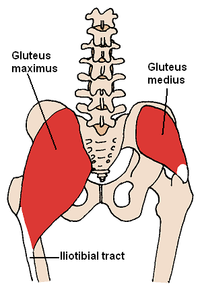
The gluteus maximus is the main extensor muscle of the hip in humans. It is the largest and outermost of the three gluteal muscles and makes up a large part of the shape and appearance of each side of the hips. It is the single largest muscle in the human body. Its thick fleshy mass, in a quadrilateral shape, forms the prominence of the buttocks. The other gluteal muscles are the medius and minimus, and sometimes informally these are collectively referred to as the glutes.

<span class="mw-page-title-main">Gluteus medius</span> One of the three gluteal muscles

The gluteus medius, one of the three gluteal muscles, is a broad, thick, radiating muscle. It is situated on the outer surface of the pelvis.

<span class="mw-page-title-main">Gluteus minimus</span> Smallest of the three gluteal muscles

The gluteus minimus, or glutæus minimus, the smallest of the three gluteal muscles, is situated immediately beneath the gluteus medius.

<span class="mw-page-title-main">Piriformis muscle</span> One of six small hip muscles in the lateral rotator group

The piriformis muscle is a flat, pyramidally-shaped muscle in the gluteal region of the lower limbs. It is one of the six muscles in the lateral rotator group.

<span class="mw-page-title-main">Hip</span> Anatomical region between the torso and the legs, holding the buttocks and genital region

In vertebrate anatomy, hip refers to either an anatomical region or a joint.

<span class="mw-page-title-main">Quadratus lumborum muscle</span> Muscle in the lower back

The quadratus lumborum muscle, informally called the QL, is a paired muscle of the left and right posterior abdominal wall. It is the deepest abdominal muscle, and commonly referred to as a back muscle. Each is irregular and quadrilateral in shape.

<span class="mw-page-title-main">Adductor magnus muscle</span> Muscle in the thigh

The adductor magnus is a large triangular muscle, situated on the medial side of the thigh.

<span class="mw-page-title-main">Iliopsoas</span> Joined psoas and the iliacus muscles.

The iliopsoas muscle refers to the joined psoas major and the iliacus muscles. The two muscles are separate in the abdomen, but usually merge in the thigh. They are usually given the common name iliopsoas. The iliopsoas muscle joins to the femur at the lesser trochanter. It acts as the strongest flexor of the hip.

<span class="mw-page-title-main">Tensor fasciae latae muscle</span> Muscle of the thigh

The tensor fasciae latae is a muscle of the thigh. Together with the gluteus maximus, it acts on the iliotibial band and is continuous with the iliotibial tract, which attaches to the tibia. The muscle assists in keeping the balance of the pelvis while standing, walking, or running.

<span class="mw-page-title-main">Gluteal muscles</span> Group of three muscles which make up the buttocks

The gluteal muscles, often called glutes, are a group of three muscles which make up the gluteal region commonly known as the buttocks: the gluteus maximus, gluteus medius and gluteus minimus. The three muscles originate from the ilium and sacrum and insert on the femur. The functions of the muscles include extension, abduction, external rotation, and internal rotation of the hip joint.

The linea aspera is a ridge of roughened surface on the posterior surface of the shaft of the femur. It is the site of attachments of muscles and the intermuscular septum.

<span class="mw-page-title-main">Adductor muscles of the hip</span> Group of muscles

The adductor muscles of the hip are a group of muscles in the medial compartment of the thigh mostly used for bringing the thighs together.

<span class="mw-page-title-main">Lateral rotator group</span> Group of six small muscles of the hip

The lateral rotator group is a group of six small muscles of the hip which all externally (laterally) rotate the femur in the hip joint. It consists of the following muscles: piriformis, gemellus superior, obturator internus, gemellus inferior, quadratus femoris and the obturator externus.

<span class="mw-page-title-main">Fascia lata</span> Deep fascia of the thigh

The fascia lata is the deep fascia of the thigh. It encloses the thigh muscles and forms the outer limit of the fascial compartments of thigh, which are internally separated by the medial intermuscular septum and the lateral intermuscular septum. The fascia lata is thickened at its lateral side where it forms the iliotibial tract, a structure that runs to the tibia and serves as a site of muscle attachment.

<span class="mw-page-title-main">Superior gluteal artery</span>

The superior gluteal artery is the terminal branch of the posterior division of the internal iliac artery. It exits the pelvis through the greater sciatic foramen before splitting into a superficial branch and a deep branch.

In human anatomy, the body of femur is the almost cylindrical, long part of the femur. It is a little broader above than in the center, broadest and somewhat flattened from before backward below. It is slightly arched, so as to be convex in front, and concave behind, where it is strengthened by a prominent longitudinal ridge, the linea aspera.

<span class="mw-page-title-main">Wing of ilium</span> Flat portion of the hip bone

The wing(ala)of ilium is the large expanded portion of the ilium, the bone which bounds the greater pelvis laterally. It presents for examination two surfaces—an external and an internal—a crest, and two borders—an anterior and a posterior.

<span class="mw-page-title-main">Pelvis</span> Lower part of the trunk of the human body between the abdomen and the thighs

The pelvis is the lower part of the trunk, between the abdomen and the thighs, together with its embedded skeleton.

References

  1. "The Muscles Used for Posture". Healthy Living. Retrieved 20 October 2020.
  2. "The Role of the Gluteus Medius and Minimus in Hip Stability". paramount-physiotherapy.com. Retrieved 20 October 2020.

Notes