Anterior compartment of thigh

Last updated
Anterior compartment of thigh
Gray432 color.png
Cross-section through the middle of the thigh. (Anterior compartment is at upper left, from the 7 o'clock position to the 2 o'clock position)
Details
Artery Femoral artery
Nerve Femoral nerve
Identifiers
Latin compartimentum femoris anterius
TA98 A04.7.01.002
TA2 2609
FMA 45151
Anatomical terminology

The anterior compartment of thigh contains muscles which extend the knee and flex the hip.

Contents

Structure

The anterior compartment is one of the fascial compartments of the thigh that contains groups of muscles together with their nerves and blood supply. The anterior compartment contains the sartorius muscle (the longest muscle in the body) and the quadriceps femoris group, which consists of the rectus femoris muscle and the three vasti muscles – the vastus lateralis, vastus intermedius, and the vastus medialis. [1]

The iliopsoas is sometimes considered a member of the anterior compartment muscles, [2] as is the articularis genus muscle. [3]

The anterior compartment is separated from the posterior compartment by the lateral intermuscular septum and from the medial compartment by the medial intermuscular septum.

Nerve supply

The nerve of the anterior compartment of thigh is the femoral nerve. [2] Innervation for the quadriceps muscles come from the posterior division of the femoral nerve, while the anterior division (which contains cutaneous as well as muscular components) gives a lateral and a medial branch, the second being responsible for the innervation of the sartorius muscle. [4] The iliacus and the psoas major and psoas minor muscles, sometimes considered part of the anterior compartment, do not share the same innervation. Whereas the iliacus is innervated by the femoral nerve, the psoas is innervated by ventral rami of L1-L3. [2]

Blood supply

When the external iliac artery crosses the inguinal ligament, it becomes the femoral artery, which supplies blood to the anterior compartment and is the largest blood vessel of the inferior member.

Function

Cross-section of the upper right thigh. In this diagram, the anterior surface is at the bottom (labelled "A"). The anterior compartment is separated by a fibrous septum which is visible from the femoral artery and vein at the bottom, surrounding the sartorius muscle, and travelling to the profunda femoris artery adjacent to the femur (not labelled), from here to the sciatic nerve, and then between the vastus lateralis and biceps femoris muscles. The compartments on the left-hand side of the image are contained within the compartment. Slide2EA.JPG
Cross-section of the upper right thigh. In this diagram, the anterior surface is at the bottom (labelled "A"). The anterior compartment is separated by a fibrous septum which is visible from the femoral artery and vein at the bottom, surrounding the sartorius muscle, and travelling to the profunda femoris artery adjacent to the femur (not labelled), from here to the sciatic nerve, and then between the vastus lateralis and biceps femoris muscles. The compartments on the left-hand side of the image are contained within the compartment.

The anterior compartment of thigh contains muscles which are extensors of the knee and flexors of the hip joints. [2]

Clinical significance

The anterior compartment may be affected as part of a compartment syndrome. [5]

Related Research Articles

<span class="mw-page-title-main">Human leg</span> Lower extremity or limb of the human body (foot, lower leg, thigh and hip)

The leg is the entire lower limb of the human body, including the foot, thigh or sometimes even the hip or buttock region. The major bones of the leg are the femur, tibia, and adjacent fibula. The thigh is between the hip and knee, while the calf (rear) and shin (front) are between the knee and foot.

<span class="mw-page-title-main">Thigh</span> The part of the body between the pelvis and the knee; upper leg

In anatomy, the thigh is the area between the hip (pelvis) and the knee. Anatomically, it is part of the lower limb.
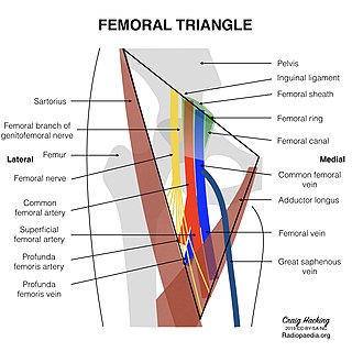
<span class="mw-page-title-main">Femoral triangle</span> Anatomical region of the thigh

The femoral triangle is an anatomical region of the upper third of the thigh. It is a subfascial space which appears as a triangular depression below the inguinal ligament when the thigh is flexed, abducted and laterally rotated.

<span class="mw-page-title-main">Pectineus muscle</span> Adductor of the thigh

The pectineus muscle is a flat, quadrangular muscle, situated at the anterior (front) part of the upper and medial (inner) aspect of the thigh. The pectineus muscle is the most anterior adductor of the hip. The muscle's primary action is hip flexion; it also produces adduction and internal rotation of the hip.

The biceps femoris is a muscle of the thigh located to the posterior, or back. As its name implies, it consists of two heads; the long head is considered part of the hamstring muscle group, while the short head is sometimes excluded from this characterization, as it only causes knee flexion and is activated by a separate nerve.

<span class="mw-page-title-main">Adductor magnus muscle</span> Muscle in the thigh

The adductor magnus is a large triangular muscle, situated on the medial side of the thigh.

<span class="mw-page-title-main">Vastus intermedius muscle</span> Body of the femur muscle

The vastus intermedius (Cruraeus) arises from the front and lateral surfaces of the body of the femur in its upper two-thirds, sitting under the rectus femoris muscle and from the lower part of the lateral intermuscular septum. Its fibers end in a superficial aponeurosis, which forms the deep part of the quadriceps femoris tendon.

The rectus femoris muscle is one of the four quadriceps muscles of the human body. The others are the vastus medialis, the vastus intermedius, and the vastus lateralis. All four parts of the quadriceps muscle attach to the patella by the quadriceps tendon.

<span class="mw-page-title-main">Iliopsoas</span> Joined psoas and the iliacus muscles

The iliopsoas muscle refers to the joined psoas major and the iliacus muscles. The two muscles are separate in the abdomen, but usually merge in the thigh. They are usually given the common name iliopsoas. The iliopsoas muscle joins to the femur at the lesser trochanter. It acts as the strongest flexor of the hip.

The linea aspera is a ridge of roughened surface on the posterior surface of the shaft of the femur. It is the site of attachments of muscles and the intermuscular septum.

<span class="mw-page-title-main">Femoral nerve</span> Long nerve down the thigh and inner leg

The femoral nerve is a nerve in the thigh that supplies skin on the upper thigh and inner leg, and the muscles that extend the knee. It is the largest branch of the lumbar plexus.

<span class="mw-page-title-main">Lumbar plexus</span> Web of nerves in the lower spine

The lumbar plexus is a web of nerves in the lumbar region of the body which forms part of the larger lumbosacral plexus. It is formed by the divisions of the first four lumbar nerves (L1-L4) and from contributions of the subcostal nerve (T12), which is the last thoracic nerve. Additionally, the ventral rami of the fourth lumbar nerve pass communicating branches, the lumbosacral trunk, to the sacral plexus. The nerves of the lumbar plexus pass in front of the hip joint and mainly support the anterior part of the thigh.
<span class="mw-page-title-main">Fascia lata</span> Deep fascia of the thigh

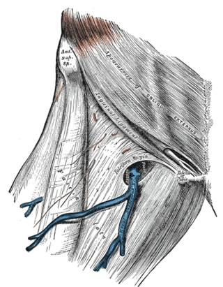
The fascia lata is the deep fascia of the thigh. It encloses the thigh muscles and forms the outer limit of the fascial compartments of thigh, which are internally separated by the medial intermuscular septum and the lateral intermuscular septum. The fascia lata is thickened at its lateral side where it forms the iliotibial tract, a structure that runs to the tibia and serves as a site of muscle attachment.

<span class="mw-page-title-main">Lateral circumflex femoral artery</span>

The lateral circumflex femoral artery is an artery in the upper thigh. It is usually a branch of the profunda femoris artery, and produces three branches. It is mostly distributed to the muscles of the lateral thigh, supplying arterial blood to muscles of the knee extensor group.

<span class="mw-page-title-main">Adductor canal</span> Aponeurotic tunnel in the middle third of the thigh

The adductor canal is an aponeurotic tunnel in the middle third of the thigh giving passage to parts of the femoral artery, vein, and nerve. It extends from the apex of the femoral triangle to the adductor hiatus.

The saphenous nerve is the largest cutaneous branch of the femoral nerve. It is derived from the lumbar plexus (L3-L4). It is a strictly sensory nerve, and has no motor function. It commences in the proximal (upper) thigh and travels along the adductor canal. Upon exiting the adductor canal, the saphenous nerve terminates by splitting into two terminal branches: the sartorial nerve, and the infrapatellar nerve. The saphenous nerve is responsible for providing sensory innervation to the skin of the anteromedial leg.

<span class="mw-page-title-main">Fascial compartments of arm</span> Anatomical compartments

The fascial compartments of arm refers to the specific anatomical term of the compartments within the upper segment of the upper limb of the body. The upper limb is divided into two segments, the arm and the forearm. Each of these segments is further divided into two compartments which are formed by deep fascia – tough connective tissue septa (walls). Each compartment encloses specific muscles and nerves.

<span class="mw-page-title-main">Posterior compartment of thigh</span> One of the fascial compartments that contains the knee flexors and hip extensors

The posterior compartment of the thigh is one of the fascial compartments that contains the knee flexors and hip extensors known as the hamstring muscles, as well as vascular and nervous elements, particularly the sciatic nerve.

<span class="mw-page-title-main">Vastus muscles</span> Human thigh muscles

The vastus muscles are three of the four muscles that make up the quadriceps femoris muscle of the thigh. The three muscles are the vastus intermedius, the vastus lateralis, and the vastus medialis located in the middle, on the outside, and inside of the thigh, respectively. The fourth muscle is the rectus femoris muscle a large fleshy muscle which covers the front and sides of the femur.

References

  1. Sauerland, Eberhardt K.; Tank, Patrick W. (2005). Grant's dissector . Hagerstown, MD: Lippincott Williams & Wilkins. pp.  125. ISBN   0-7817-5484-4.
  2. 1 2 3 4 Drake, Richard L.; Vogl, Wayne; Tibbitts, Adam W.M. Mitchell; illustrations by Richard; Richardson, Paul (2005). Gray's anatomy for students. Philadelphia: Elsevier/Churchill Livingstone. p. 518. ISBN   978-0-8089-2306-0.
  3. "Summary of Lower Limb". Archived from the original on 23 January 2008. Retrieved 2008-01-27.
  4. Latarjet, Michel; Ruiz Liard, Alfredo (2005). Human Anatomy (Spanish ed.). Editorial Médica Panamericana. pp. 846–847. ISBN   978-950-06-1368-2.
  5. Wadhawan, Himanshu; Upadhyay, Vishal; Sabboubeh, Adel; Al Hussainy, Haydar A. J.; Madan, Sanjeev (9 April 2007). "Acute Anterior Thigh Compartment Syndrome Revisited: A Case Report and Review of Literature". European Journal of Trauma and Emergency Surgery. 33 (4): 430–433. doi:10.1007/s00068-007-6952-5. PMID   26814739.