Human body

Last updated

Female (left) and male (right) adult human bodies photographed in ventral (above) and dorsal (below) perspectives. Naturally-occurring pubic, body, and facial hair have been deliberately removed to show anatomy. Human Body 02.png
Female (left) and male (right) adult human bodies photographed in ventral (above) and dorsal (below) perspectives. Naturally-occurring pubic, body, and facial hair have been deliberately removed to show anatomy.

The human body is the entire structure of a human being. It is composed of many different types of cells that together create tissues and subsequently organs and then organ systems. They ensure homeostasis and the viability of the human body.

Contents

It consists of head, hair, neck, torso (which includes the thorax and abdomen), arms and hands, legs and feet.

The study of the human body includes anatomy, physiology, histology and embryology. The body varies anatomically in known ways. Physiology focuses on the systems and organs of the human body and their functions. Many systems and mechanisms interact in order to maintain homeostasis, with safe levels of substances such as sugar and oxygen in the blood.

The body is studied by health professionals, physiologists, anatomists, and artists to assist them in their work.

Composition

Elements of the human body by mass. Trace elements are less than 1% combined (and each less than 0.1%).
201 Elements of the Human Body.02.svg ElementSymbolPercent massPercent atoms
OxygenO65.024.0
CarbonC18.512.0
HydrogenH9.562.0
NitrogenN3.21.1
CalciumCa1.50.22
PhosphorusP1.00.22
PotassiumK0.40.03
SulfurS0.30.038
SodiumNa0.20.037
ChlorineCl0.20.024
MagnesiumMg0.10.015
Trace elements< 0.1< 0.3

The human body is composed of elements including hydrogen, oxygen, carbon, calcium and phosphorus. These elements reside in trillions of cells and non-cellular components of the body.

The adult male body is about 60% water for a total water content of some 42 litres (9.2 imp gal; 11 US gal). This is made up of about 19 litres (4.2 imp gal; 5.0 US gal) of extracellular fluid including about 3.2 litres (0.70 imp gal; 0.85 US gal) of blood plasma and about 8.4 litres (1.8 imp gal; 2.2 US gal) of interstitial fluid, and about 23 litres (5.1 imp gal; 6.1 US gal) of fluid inside cells. [1] The content, acidity and composition of the water inside and outside cells is carefully maintained. The main electrolytes in body water outside cells are sodium and chloride, whereas within cells it is potassium and other phosphates. [2]

Cells

The body contains trillions of cells, the fundamental unit of life. At maturity, there are roughly 30 trillion cells, and 38 trillion bacteria in the body, [3] [4] an estimate arrived at by totaling the cell numbers of all the organs of the body and cell types. The skin of the body is also host to billions of commensal organisms as well as immune cells. [5] Not all parts of the body are made from cells. Cells sit in an extracellular matrix that consists of proteins such as collagen, surrounded by extracellular fluids.

Genome

Cells in the body function because of DNA. DNA sits within the nucleus of a cell. Here, parts of DNA are copied and sent to the body of the cell via RNA. [6] The RNA is then used to create proteins, which form the basis for cells, their activity, and their products. Proteins dictate cell function and gene expression, a cell is able to self-regulate by the amount of proteins produced. [7] However, not all cells have DNA; some cells such as mature red blood cells lose their nucleus as they mature.

Tissues

The body consists of many different types of tissue, defined as cells that act with a specialised function. [8] The study of tissues is called histology and is often done with a microscope. The body consists of four main types of tissues. These are lining cells (epithelia), connective tissue, nerve tissue and muscle tissue. [9]

Cells that lie on surfaces exposed to the outside world or gastrointestinal tract (epithelia) or internal cavities (endothelium) come in numerous shapes and forms – from single layers of flat cells, to cells with small beating hair-like cilia in the lungs, to column-like cells that line the stomach. Endothelial cells are cells that line internal cavities including blood vessels and glands. Lining cells regulate what can and cannot pass through them, protect internal structures, and function as sensory surfaces. [9]

Organs

Organs, structured collections of cells with a specific function, [10] mostly sit within the body, with the exception of skin. Examples include the heart, lungs and liver. Many organs reside within cavities within the body. These cavities include the abdomen (which contains the stomach, for example) and pleura, which contains the lungs.

Heart

The heart is an organ located in the thoracic cavity between the lungs and slightly to the left. It is surrounded by the pericardium, which holds it in place in the mediastinum and serves to protect it from blunt trauma, infection and help lubricate the movement of the heart via pericardial fluid. [11] The heart works by pumping blood around the body allowing oxygen, nutrients, waste, hormones and white blood cells to be transported.

Diagram of the human heart Diagram of the human heart (cropped).svg
Diagram of the human heart

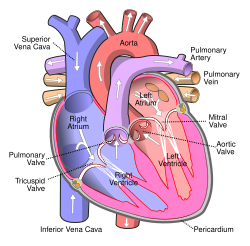
The heart is composed of two atria and two ventricles. The primary purpose of the atria is to allow uninterrupted venous blood flow to the heart during ventricular systole. This allows enough blood to get into the ventricles during atrial systole. Consequently, the atria allows a cardiac output roughly 75% greater than would be possible without them. [12] The purpose of the ventricles is to pump blood to the lungs through the right ventricle and to the rest of the body through the left ventricle. [13]

The heart has an electrical conduction system to control the contraction and relaxation of the muscles. It starts in the sinoatrial node traveling through the atria causing them to pump blood into the ventricles. It then travels to the atrioventricular node, which makes the signal slow down slightly allowing the ventricles to fill with blood before pumping it out and starting the cycle over again. [14]

Coronary artery disease is the leading cause of death worldwide, making up 16% of all deaths. [15] It is caused by the buildup of plaque in the coronary arteries supplying the heart, eventually the arteries may become so narrow that not enough blood is able to reach the myocardium, [16] a condition known as myocardial infarction or heart attack, this can cause heart failure or cardiac arrest and eventually death. [17] Risk factors for coronary artery disease include obesity, smoking, high cholesterol, high blood pressure, lack of exercise and diabetes. [18] Cancer can affect the heart, though it is exceedingly rare and has usually metastasized from another part of the body such as the lungs or breasts. This is because the heart cells quickly stop dividing and all growth occurs through size increase rather than cell division. [19]

Gallbladder

The gallbladder is a hollow pear-shaped organ located posterior to the inferior middle part of the right lobe of the liver. It is variable in shape and size. It stores bile before it is released into the small intestine via the common bile duct to help with digestion of fats. It receives bile from the liver via the cystic duct, which connects to the common hepatic duct to form the common bile duct. [20]

The gallbladder gets its blood supply from the cystic artery, which in most people, emerges from the right hepatic artery. [20]

Gallstones is a common disease in which one or more stones form in the gallbladder or biliary tract. Most people are asymptomatic but if a stone blocks the biliary tract, it causes a gallbladder attack, symptoms may include sudden pain in the upper right abdomen and or center of the abdomen. Nausea and vomiting may also occur. Typical treatment is removal of the gallbladder through a procedure called a cholecystectomy. [21] [22] Having gallstones is a risk factor for gallbladder cancer, which although quite uncommon, is rapidly fatal if not diagnosed early. [23]

Systems

The circulatory system consists of the heart and blood vessels (arteries, veins and capillaries). The heart propels the circulation of the blood, which serves as a "transportation system" to transfer oxygen, fuel, nutrients, waste products, immune cells and signaling molecules (i.e. hormones) from one part of the body to another. Paths of blood circulation within the human body can be divided into two circuits: the pulmonary circuit, which pumps blood to the lungs to receive oxygen and leave carbon dioxide, and the systemic circuit, which carries blood from the heart off to the rest of the body. The blood consists of fluid that carries cells in the circulation, including some that move from tissue to blood vessels and back, as well as the spleen and bone marrow. [24] [25] [26]

Digestive system Digestive system diagram en.svg
Digestive system

The digestive system consists of the mouth including the tongue and teeth, esophagus, stomach, (gastrointestinal tract, small and large intestines, and rectum), as well as the liver, pancreas, gallbladder, and salivary glands. It converts food into small, nutritional, non-toxic molecules for distribution and absorption into the body. These molecules take the form of proteins (which are broken down into amino acids), fats, vitamins and minerals (the last of which are mainly ionic rather than molecular). After being swallowed, food moves through the gastrointestinal tract by means of peristalsis: the systematic expansion and contraction of muscles to push food from one area to the next. [27] [28]

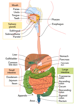
Digestion begins in the mouth, which chews food into smaller pieces for easier digestion. Then it is swallowed, and moves through the esophagus to the stomach. In the stomach, food is mixed with gastric acids to allow the extraction of nutrients. What is left is called chyme; this then moves into the small intestine, which absorbs the nutrients and water from the chyme. What remains passes on to the large intestine, where it is dried to form feces; these are then stored in the rectum until they are expelled through the anus. [28]

The endocrine system consists of the principal endocrine glands: the pituitary, thyroid, adrenals, pancreas, parathyroids, and gonads, but nearly all organs and tissues produce specific endocrine hormones as well. The endocrine hormones serve as signals from one body system to another regarding an enormous array of conditions, resulting in variety of changes of function. [29]

The immune system consists of the white blood cells, the thymus, lymph nodes and lymph channels, which are also part of the lymphatic system. The immune system provides a mechanism for the body to distinguish its own cells and tissues from outside cells and substances and to neutralize or destroy the latter by using specialized proteins such as antibodies, cytokines, and toll-like receptors, among many others. [30]

Skin Skin layers.svg
Skin

The integumentary system consists of the covering of the body (the skin), including hair and nails as well as other functionally important structures such as the sweat glands and sebaceous glands. The skin provides containment, structure, and protection for other organs, and serves as a major sensory interface with the outside world. [31] [32]

The lymphatic system extracts, transports and metabolizes lymph, the fluid found in between cells. The lymphatic system is similar to the circulatory system in terms of both its structure and its most basic function, to carry a body fluid. [33]

The musculoskeletal system consists of the human skeleton (which includes bones, ligaments, tendons, joints and cartilage) and attached muscles. It gives the body basic structure and the ability for movement. In addition to their structural role, the larger bones in the body contain bone marrow, the site of production of blood cells. Also, all bones are major storage sites for calcium and phosphate. This system can be split up into the muscular system and the skeletal system. [34]

Nervous system Nervous system diagram.png
Nervous system

The nervous system consists of the body's neurons and glial cells, which together form the nerves, ganglia and gray matter, which in turn form the brain and related structures. The brain is the organ of thought, emotion, memory, and sensory processing; it serves many aspects of communication and controls various systems and functions. The special senses consist of vision, hearing, taste, and smell. The eyes, ears, tongue, and nose gather information about the body's environment. [35]

From a structural perspective, the nervous system is typically subdivided into two component parts: the central nervous system (CNS), composed of the brain and the spinal cord; and the peripheral nervous system (PNS), composed of the nerves and ganglia outside the brain and spinal cord. The CNS is mostly responsible for organizing motion, processing sensory information, thought, memory, cognition and other such functions. [36] It remains a matter of some debate whether the CNS directly gives rise to consciousness.[ citation needed ] The peripheral nervous system (PNS) is mostly responsible for gathering information with sensory neurons and directing body movements with motor neurons. [36]

From a functional perspective, the nervous system is again typically divided into two component parts: the somatic nervous system (SNS) and the autonomic nervous system (ANS). The SNS is involved in voluntary functions like speaking and sensory processes. The ANS is involved in involuntary processes, such as digestion and regulating blood pressure. [37]

The nervous system is subject to many different diseases. In epilepsy, abnormal electrical activity in the brain can cause seizures. In multiple sclerosis, the immune system attacks the nerve linings, damaging the nerves' ability to transmit signals. Amyotrophic lateral sclerosis (ALS), also known as Lou Gehrig's disease, is a motor neuron disease which gradually reduces movement in patients. There are also many other diseases of the nervous system. [36]

Male gonad (testes, left) and female gonad (ovaries, right) Male and female gonads 1.png
Male gonad (testes, left) and female gonad (ovaries, right)

The purpose of the reproductive system is to reproduce and nurture the growth of offspring. The functions include the production of germ cells and hormones. [38] The sex organs of the male reproductive system and the female reproductive system develops and mature at puberty. These systems include the internal and external genitalia.

Internal gross anatomy of the female reproductive system Scheme female reproductive system-en.svg
Internal gross anatomy of the female reproductive system

Female puberty generally occurs between the ages of 9 and 13 and is characterized by ovulation and menstruation; the growth of secondary sex characteristics, such as growth of pubic and underarm hair, breast, uterine and vaginal growth, widening hips and increased height and weight, also occur during puberty. [39] Male puberty sees the further development of the penis and testicles. [40]

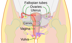
The female inner sex organs are the two ovaries, their fallopian tubes, the uterus, and the cervix. At birth there are about 70,000 immature egg cells that degenerate until at puberty there are around 40,000. No more egg cells are produced. Hormones stimulate the beginning of menstruation, and the ongoing menstrual cycles. [39] [41] The female external sex organs are the vulva (labia, clitoris, and vaginal opening). [42] [39]

The male external genitalia include the penis and scrotum that contains the testicles. The testicle is the gonad, the sex gland that produces the sperm cells. Unlike the egg cells in the female, sperm cells are produced throughout life. Other internal sex organs are the epididymides, vasa deferentia, and some accessory glands.

Diseases that affect the reproductive system include polycystic ovary syndrome, [43] a number of disorders of the testicles including testicular torsion, [44] [45] and a number of sexually transmitted infections including syphilis, HIV, chlamydia, HPV and genital warts. [46] [47] Cancer can affect most parts of the reproductive system including the penis, testicles, prostate, ovaries, cervix, vagina, fallopian, uterus and vulva. [48]

The respiratory system consists of the nose, nasopharynx, trachea, and lungs. It brings oxygen from the air and excretes carbon dioxide and water back into the air. First, air is pulled through the trachea into the lungs by the diaphragm pushing down, which creates a vacuum. Air is briefly stored inside small sacs known as alveoli (sing.: alveolus) before being expelled from the lungs when the diaphragm contracts again. Each alveolus is surrounded by capillaries carrying deoxygenated blood, which absorbs oxygen out of the air and into the bloodstream. [49] [50]

For the respiratory system to function properly, there need to be as few impediments as possible to the movement of air within the lungs. Inflammation of the lungs and excess mucus are common sources of breathing difficulties. [50] In asthma, the respiratory system is persistently inflamed, causing wheezing and/or shortness of breath. Pneumonia occurs through infection of the alveoli, and may be caused by tuberculosis. Emphysema, commonly a result of smoking, is caused by damage to connections between the alveoli. [51]

Female urinary system Urinary System (Female).png
Female urinary system

The urinary system consists of the two kidneys, two ureters, bladder, and urethra. It removes waste materials from the blood through urine, which carries a variety of waste molecules and excess ions and water out of the body.

First, the kidneys filter the blood through their respective nephrons, removing waste products like urea, creatinine and maintaining the proper balance of electrolytes and turning the waste products into urine by combining them with water from the blood. [52] The kidneys filter about 150 quarts (170 liters) of blood daily, but most of it is returned to the blood stream with only 1-2 quarts (1-2 liters) ending up as urine. [53] The urine is brought by the ureters from the kidneys down to the bladder.

The smooth muscle lining the ureter walls continuously tighten and relax through a process called peristalsis to force urine away from the kidneys and down into the bladder. Small amounts of urine are released into the bladder every 10–15 seconds.

The bladder is a hollow balloon shaped organ located in the pelvis. It stores urine until the brain signals it to relax the urinary sphincter and release the urine into the urethra starting urination. [54] A normal bladder can hold up to 16 ounces (half a liter) for 3–5 hours comfortably.

Numerous diseases affect the urinary system including kidney stones, which are formed when materials in the urine concentrate enough to form a solid mass, urinary tract infections, which are infections of the urinary tract and can cause pain when urinating, frequent urination and even death if left untreated. Renal failure occurs when the kidneys fail to adequately filter waste from the blood and can lead to death if not treated with dialysis or kidney transplantation. [55] Cancer can affect the bladder, kidneys, urethra and ureters, with the latter two being far more rare. [56]

Anatomy

Cavities of human body Body cavities.jpg
Cavities of human body

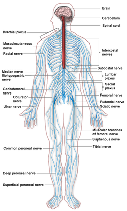
Human anatomy is the study of the shape and form of the human body. The human body has four limbs (two arms and two legs), a head and a neck, which connect to the torso. The body's shape is determined by a strong skeleton made of bone and cartilage, surrounded by fat (adipose tissue), muscle, connective tissue, organs, and other structures. The spine at the back of the skeleton contains the flexible vertebral column, which surrounds the spinal cord, which is a collection of nerve fibres connecting the brain to the rest of the body. Nerves connect the spinal cord and brain to the rest of the body. All major bones, muscles, and nerves in the body are named, with the exception of anatomical variations such as sesamoid bones and accessory muscles.

Blood vessels carry blood throughout the body, which moves because of the beating of the heart. Venules and veins collect blood low in oxygen from tissues throughout the body. These collect in progressively larger veins until they reach the body's two largest veins, the superior and inferior vena cava, which drain blood into the right side of the heart. From here, the blood is pumped into the lungs where it receives oxygen and drains back into the left side of the heart. From here, it is pumped into the body's largest artery, the aorta, and then progressively smaller arteries and arterioles until it reaches tissue. Here, blood passes from small arteries into capillaries, then small veins and the process begins again. Blood carries oxygen, waste products, and hormones from one place in the body to another. Blood is filtered at the kidneys and liver.

The body consists of a number of body cavities, separated areas which house different organ systems. The brain and central nervous system reside in an area protected from the rest of the body by the blood brain barrier. The lungs sit in the pleural cavity. The intestines, liver, and spleen sit in the abdominal cavity.

Height, weight, shape and other body proportions vary individually and with age and sex. Body shape is influenced by the distribution of bones, muscle and fat tissue. [57]

Physiology

Human physiology is the study of how the human body functions. This includes the mechanical, physical, bioelectrical, and biochemical functions of humans in good health, from organs to the cells of which they are composed. The human body consists of many interacting systems of organs. These interact to maintain homeostasis, keeping the body in a stable state with safe levels of substances such as sugar and oxygen in the blood. [58]

Each system contributes to homeostasis, of itself, other systems, and the entire body. Some combined systems are referred to by joint names. For example, the nervous system and the endocrine system operate together as the neuroendocrine system. The nervous system receives information from the body, and transmits this to the brain via nerve impulses and neurotransmitters. At the same time, the endocrine system releases hormones, such as to help regulate blood pressure and volume. Together, these systems regulate the internal environment of the body, maintaining blood flow, posture, energy supply, temperature, and acid balance (pH). [58]

Development

Baby being carried Little baby from Puno.jpg
Baby being carried

Development of the human body is the process of growth to maturity. The process begins with fertilisation, where an egg released from the ovary of a female is penetrated by sperm. The egg then lodges in the uterus, where an embryo and later fetus develop until birth. Growth and development occur after birth, and include both physical and psychological development, influenced by genetic, hormonal, environmental and other factors. Development and growth continue throughout life, through childhood, adolescence, and through adulthood to old age, and are referred to as the process of aging.

Society and culture

Professional study

Anatomical study by Leonardo da Vinci Anatomical Male Figure Showing Heart, Lungs, and Main Arteries.jpg
Anatomical study by Leonardo da Vinci

Health professionals learn about the human body from illustrations, models, and demonstrations. Medical and dental students in addition gain practical experience, for example by dissection of cadavers. Human anatomy, physiology, and biochemistry are basic medical sciences, generally taught to medical students in their first year at medical school. [59] [60] [61]

Depiction

Figure drawing by Lovis Corinth (before 1925) Corinth stehender Madchenakt.jpg
Figure drawing by Lovis Corinth (before 1925)

In Western societies, the contexts for depictions of the human body include information, art and pornography. Information includes both science and education, such as anatomical drawings. Any ambiguous image not easily fitting into one of these categories may be misinterpreted, leading to disputes. [62] The most contentious disputes are between fine art and erotic images, which define the legal distinction of which images are permitted or prohibited.

History of anatomy

Two facing pages of text with woodcuts of naked male and female figures, in the Epitome by Andreas Vesalius, 1543 Externarvm hvmani corporis sedivm partivmve, 1543..JPG
Two facing pages of text with woodcuts of naked male and female figures, in the Epitome by Andreas Vesalius, 1543

In Ancient Greece, the Hippocratic Corpus described the anatomy of the skeleton and muscles. [63] The 2nd century physician Galen of Pergamum compiled classical knowledge of anatomy into a text that was used throughout the Middle Ages. [64] In the Renaissance, Andreas Vesalius (1514–1564) pioneered the modern study of human anatomy by dissection, writing the influential book De humani corporis fabrica . [65] [66] Anatomy advanced further with the invention of the microscope and the study of the cellular structure of tissues and organs. [67] Modern anatomy uses techniques such as magnetic resonance imaging, computed tomography, fluoroscopy and ultrasound imaging to study the body in unprecedented detail. [68]

History of physiology

The study of human physiology began with Hippocrates in Ancient Greece, around 420 BCE, and with Aristotle (384–322 BCE) who applied critical thinking and emphasis on the relationship between structure and function. Galen (c.126 – c.199) was the first to use experiments to probe the body's functions. [69] The term physiology was introduced by the French physician Jean Fernel (1497–1558). In the 17th century, William Harvey (1578–1657) described the circulatory system, pioneering the combination of close observation with careful experiment. [70] In the 19th century, physiological knowledge began to accumulate at a rapid rate with the cell theory of Matthias Schleiden and Theodor Schwann in 1838, that organisms are made up of cells. Claude Bernard (1813–1878) created the concept of the milieu interieur (internal environment), which Walter Cannon (1871–1945) later said was regulated to a steady state in homeostasis. In the 20th century, the physiologists Knut Schmidt-Nielsen and George Bartholomew extended their studies to comparative physiology and ecophysiology. [71] Most recently, evolutionary physiology has become a distinct subdiscipline. [72]

See also

Human body lists

Related Research Articles

<span class="mw-page-title-main">Blood</span> Organic fluid which transports nutrients throughout the organism

Blood is a body fluid in the circulatory system of humans and other vertebrates that delivers necessary substances such as nutrients and oxygen to the cells, and transports metabolic waste products away from those same cells.

<span class="mw-page-title-main">Endocrine system</span> Hormone-producing glands of a body

The endocrine system is a messenger system in an organism comprising feedback loops of hormones that are released by internal glands directly into the circulatory system and that target and regulate distant organs. In vertebrates, the hypothalamus is the neural control center for all endocrine systems.

In biology, homeostasis is the state of steady internal physical and chemical conditions maintained by living systems. This is the condition of optimal functioning for the organism and includes many variables, such as body temperature and fluid balance, being kept within certain pre-set limits. Other variables include the pH of extracellular fluid, the concentrations of sodium, potassium, and calcium ions, as well as the blood sugar level, and these need to be regulated despite changes in the environment, diet, or level of activity. Each of these variables is controlled by one or more regulators or homeostatic mechanisms, which together maintain life.

<span class="mw-page-title-main">Kidney</span> Organ that filters blood and produces urine in humans

In humans, the kidneys are two reddish-brown bean-shaped blood-filtering organs that are a multilobar, multipapillary form of mammalian kidneys, usually without signs of external lobulation. They are located on the left and right in the retroperitoneal space, and in adult humans are about 12 centimetres in length. They receive blood from the paired renal arteries; blood exits into the paired renal veins. Each kidney is attached to a ureter, a tube that carries excreted urine to the bladder.

<span class="mw-page-title-main">Artery</span> Blood vessels that carry blood away from the heart

An artery is a blood vessel in humans and most other animals that takes oxygenated blood away from the heart in the systemic circulation to one or more parts of the body. Exceptions that carry deoxygenated blood are the pulmonary arteries in the pulmonary circulation that carry blood to the lungs for oxygenation, and the umbilical arteries in the fetal circulation that carry deoxygenated blood to the placenta.

<span class="mw-page-title-main">Heart</span> Organ found inside most animals

The heart is a muscular organ found in most animals. This organ pumps blood through the blood vessels of the circulatory system. The pumped blood carries oxygen and nutrients to the body, while carrying metabolic waste such as carbon dioxide to the lungs. In humans, the heart is approximately the size of a closed fist and is located between the lungs, in the middle compartment of the chest, called the mediastinum.

<span class="mw-page-title-main">Blood vessel</span> Tubular structure of the circulatory system which transports blood

Blood vessels are the components of the circulatory system that transport blood throughout the human body. These vessels transport blood cells, nutrients, and oxygen to the tissues of the body. They also take waste and carbon dioxide away from the tissues. Blood vessels are needed to sustain life, because all of the body's tissues rely on their functionality.

<span class="mw-page-title-main">Urinary system</span> Anatomical system consisting of the kidneys, ureters, urinary bladder, and the urethra

The human urinary system, also known as the urinary tract or renal system, consists of the kidneys, ureters, bladder, and the urethra. The purpose of the urinary system is to eliminate waste from the body, regulate blood volume and blood pressure, control levels of electrolytes and metabolites, and regulate blood pH. The urinary tract is the body's drainage system for the eventual removal of urine. The kidneys have an extensive blood supply via the renal arteries which leave the kidneys via the renal vein. Each kidney consists of functional units called nephrons. Following filtration of blood and further processing, wastes exit the kidney via the ureters, tubes made of smooth muscle fibres that propel urine towards the urinary bladder, where it is stored and subsequently expelled from the body by urination. The female and male urinary system are very similar, differing only in the length of the urethra.

<span class="mw-page-title-main">Circulatory system</span> Organ system for circulating blood in animals

The circulatory system is a system of organs that includes the heart, blood vessels, and blood which is circulated throughout the entire body of a human or other vertebrate. It includes the cardiovascular system, or vascular system, that consists of the heart and blood vessels. The circulatory system has two divisions, a systemic circulation or circuit, and a pulmonary circulation or circuit. Some sources use the terms cardiovascular system and vascular system interchangeably with circulatory system.

The excretory system is a passive biological system that removes excess, unnecessary materials from the body fluids of an organism, so as to help maintain internal chemical homeostasis and prevent damage to the body. The dual function of excretory systems is the elimination of the waste products of metabolism and to drain the body of used up and broken down components in a liquid and gaseous state. In humans and other amniotes, most of these substances leave the body as urine and to some degree exhalation, mammals also expel them through sweating.

<span class="mw-page-title-main">Organ (biology)</span> Collection of tissues with similar functions

In a multicellular organism, an organ is a collection of tissues joined in a structural unit to serve a common function. In the hierarchy of life, an organ lies between tissue and an organ system. Tissues are formed from same type cells to act together in a function. Tissues of different types combine to form an organ which has a specific function. The intestinal wall for example is formed by epithelial tissue and smooth muscle tissue. Two or more organs working together in the execution of a specific body function form an organ system, also called a biological system or body system.

<span class="mw-page-title-main">Sympathetic nervous system</span> Part of the autonomic nervous system which stimulates fight-or-flight responses

The sympathetic nervous system (SNS) is one of the three divisions of the autonomic nervous system, the others being the parasympathetic nervous system and the enteric nervous system. The enteric nervous system is sometimes considered part of the autonomic nervous system, and sometimes considered an independent system.

<span class="mw-page-title-main">Endocrine gland</span> Glands of the endocrine system that secrete hormones to blood

Endocrine glands are ductless glands of the endocrine system that secrete their products, hormones, directly into the blood. The major glands of the endocrine system include the pineal gland, pituitary gland, pancreas, ovaries, testicles, thyroid gland, parathyroid gland, hypothalamus and adrenal glands. The hypothalamus and pituitary glands are neuroendocrine organs.

<span class="mw-page-title-main">Interstitium</span> Anatomical term

The interstitium is a contiguous fluid-filled space existing between a structural barrier, such as a cell membrane or the skin, and internal structures, such as organs, including muscles and the circulatory system. The fluid in this space is called interstitial fluid, comprises water and solutes, and drains into the lymph system. The interstitial compartment is composed of connective and supporting tissues within the body – called the extracellular matrix – that are situated outside the blood and lymphatic vessels and the parenchyma of organs. The role of the interstitium in solute concentration, protein transport and hydrostatic pressure impacts human pathology and physiological responses such as edema, inflammation and shock.

A biological system is a complex network which connects several biologically relevant entities. Biological organization spans several scales and are determined based different structures depending on what the system is. Examples of biological systems at the macro scale are populations of organisms. On the organ and tissue scale in mammals and other animals, examples include the circulatory system, the respiratory system, and the nervous system. On the micro to the nanoscopic scale, examples of biological systems are cells, organelles, macromolecular complexes and regulatory pathways. A biological system is not to be confused with a living system, such as a living organism.

<span class="mw-page-title-main">Abdominopelvic cavity</span>

The abdominopelvic cavity is a body cavity that consists of the abdominal cavity and the pelvic cavity. The upper portion is the abdominal cavity, and it contains the stomach, liver, pancreas, spleen, gallbladder, kidneys, small intestine, and most of the large intestine. The lower portion is the pelvic cavity, and it contains the urinary bladder, the rest of the large intestine, and the internal reproductive organs.

<span class="mw-page-title-main">Muscle</span> Basic biological tissue present in animals

Muscle is a soft tissue, one of the four basic types of animal tissue. Muscle tissue gives skeletal muscles the ability to contract. Muscle is formed during embryonic development, in a process known as myogenesis. Muscle tissue contains special contractile proteins called actin and myosin which interact to cause movement. Among many other muscle proteins present are two regulatory proteins, troponin and tropomyosin.

In physiology, acute local blood flow regulation refers to an intrinsic regulation, or control, of the vascular tone of arteries at a local level, meaning within a certain tissue type, organ, or organ system. This intrinsic type of control means that the blood vessels can automatically adjust their own vascular tone, by dilating (widening) or constricting (narrowing), in response to some change in the environment. This change occurs in order to match up the tissue's oxygen demand with the actual oxygen supply available in the blood as closely as possible. For example, if a muscle is being utilized actively, it will require more oxygen than it was at rest, so the blood vessels supplying that muscle will vasodilate, or widen in size, to increase the amount of blood, and therefore oxygen, being delivered to that muscle.

<span class="mw-page-title-main">Glossary of medicine</span> List of definitions of terms and concepts commonly used in the study of medicine

This glossary of medical terms is a list of definitions about medicine, its sub-disciplines, and related fields.

References

  1. "Fluid Physiology". Anaesthesiamcq. Archived from the original on 3 May 2005. Retrieved 2 September 2016.
  2. Ganong's 2016, p. 5.
  3. Sender, Ron; Fuchs, Shai; Milo, Ron (2016). "Revised estimates for the number of human and bacteria cells in the body". PLOS Biology. 14 (8): e1002533. bioRxiv   10.1101/036103 . doi: 10.1371/journal.pbio.1002533 . PMC   4991899 . PMID   27541692.
  4. Hatton, Ian A.; Galbraith, Eric D.; Merleau, Nono S. C.; Miettinen, Teemu P.; Smith, Benjamin McDonald; Shander, Jeffery A. (26 September 2023). "The human cell count and size distribution". Proceedings of the National Academy of Sciences. 120 (39): e2303077120. Bibcode:2023PNAS..12003077H. doi:10.1073/pnas.2303077120. ISSN   0027-8424. PMC   10523466 . PMID   37722043.
  5. Sfriso, R; Egert, M; Gempeler, M; Voegeli, R; Campiche, R (April 2020). "Revealing the secret life of skin - with the microbiome you never walk alone". International Journal of Cosmetic Science. 42 (2): 116–126. doi:10.1111/ics.12594. PMC   7155096 . PMID   31743445.
  6. Ganong's 2016, p. 16.
  7. "Gene Expression | Learn Science at Scitable". www.nature.com. Archived from the original on 31 October 2010. Retrieved 29 July 2017.
  8. "tissue – definition of tissue in English". Oxford Dictionaries| English. Archived from the original on 5 October 2016. Retrieved 17 September 2016.
  9. 1 2 Gray's Anatomy 2008, p. 27.
  10. "organ | Definition, meaning & more". www.collinsdictionary.com. Collins Dictionary . Retrieved 17 September 2016.
  11. Jaworska-Wilczynska, Maria; Trzaskoma, Pawel; Szczepankiewicz, Andrzej A.; Hryniewiecki, Tomasz (2016). "Pericardium: structure and function in health and disease". Folia Histochemica et Cytobiologica. 54 (3): 121–125. doi: 10.5603/FHC.a2016.0014 . ISSN   1897-5631. PMID   27654013.
  12. Anderson, Robert M. (1999). "Chapter 1: Normal Physiology". The Gross Physiology of the Cardiovascular System (2nd ed.). p. 11.
  13. "Ventricle | heart". Encyclopedia Britannica. Retrieved 7 August 2021.
  14. "How the Heart Works". NHLBI, NIH. "Your Heart's Electrical System". Archived from the original on 13 August 2021. Retrieved 7 August 2021.
  15. "The top 10 causes of death". WHO. 9 December 2020. Retrieved 7 August 2021.
  16. "Coronary Artery Disease". Centers for Disease Control and Prevention. 19 July 2021. Retrieved 7 August 2021.
  17. "Heart Attack Symptoms, Risk Factors, and Recovery". Centers for Disease Control and Prevention. 11 January 2021. Retrieved 7 August 2021.
  18. "Know Your Risk for Heart Disease". Centers for Disease Control and Prevention. 9 December 2019. Retrieved 7 August 2021.
  19. "Matters of the Heart: Why Are Cardiac Tumors So Rare?". National Cancer Institute. 10 February 2009. Retrieved 7 August 2021.
  20. 1 2 Nagral, Sanjay (2005). "Anatomy relevant to cholecystectomy". Journal of Minimal Access Surgery. 1 (2): 53–8. doi: 10.4103/0972-9941.16527 . PMC   3004105 . PMID   21206646.
  21. "Gallstones – Symptoms and causes". Mayo Clinic. Retrieved 7 August 2021.
  22. "Gallstones – Diagnosis and treatment". www.mayoclinic.org. Retrieved 7 August 2021.
  23. "Gallbladder cancer – Symptoms and causes". Mayo Clinic. Retrieved 7 August 2021.
  24. "Cardiovascular System". U.S. National Cancer Institute. Archived from the original on 2 February 2007. Retrieved 16 September 2008.
  25. Human Biology and Health. Upper Saddle River, NJ: Pearson Prentice Hall. 1993. ISBN   0-13-981176-1.
  26. "The Cardiovascular System". State University of New York Downstate Medical Center. 8 March 2008. Archived from the original on 11 June 2016. Retrieved 16 September 2008.
  27. "Your Digestive System and How It Works". National Institute of Health . Retrieved 4 September 2016.
  28. 1 2 "Your Digestive System & How it Works". National Institute of Diabetes and Digestive and Kidney Diseases . Retrieved 29 June 2019.
  29. "Hormonal (endocrine) system". Victoria State Government . Retrieved 4 September 2016.
  30. Zimmermann, Kim Ann. "Immune System: Diseases, Disorders & Function". LiveScience . Retrieved 4 September 2016.
  31. Integumentary+System at the U.S. National Library of Medicine Medical Subject Headings (MeSH)
  32. Marieb, Elaine; Hoehn, Katja (2007). Human Anatomy & Physiology (7th ed.). Pearson Benjamin Cummings. p.  142. ISBN   978-0805359107.
  33. Zimmermann, Kim Anne. "Lymphatic System: Facts, Functions & Diseases". LiveScience. Retrieved 4 September 2016.
  34. Moore, Keith L.; Dalley, Arthur F.; Agur, Anne M. R. (2010). Moore's Clinically Oriented Anatomy. Phildadelphia: Lippincott Williams & Wilkins. pp. 2–3. ISBN   978-1-60547-652-0.
  35. Lagassé, Paul (2001). "Nervous System". Columbia Encyclopedia (6th ed.). New York Detroit: Columbia University Press Sold and distributed by Gale Group. ISBN   978-0-7876-5015-5.
  36. 1 2 3 Horton, James; Bradford, Alina; Zimmermann, Kim Ann (25 March 2022). "Nervous System: Facts, Function & Diseases". livescience.com. Retrieved 8 February 2023.
  37. "Visual Guide to Your Nervous System". WebMD. Retrieved 8 February 2023.
  38. "Introduction to the Reproductive System | SEER Training". training.seer.cancer.gov. Retrieved 5 March 2024.
  39. 1 2 3 "Technical Issues In Reproductive Health". www.columbia.edu. Retrieved 7 August 2021.
  40. "Accessory Glands | SEER Training". www.training.seer.cancer.gov. Retrieved 7 August 2021.
  41. "Ovaries | SEER Training". www.training.seer.cancer.gov. Retrieved 7 August 2021.
  42. "External Genitalia | SEER Training". www.training.seer.cancer.gov. Retrieved 7 August 2021.
  43. Ndefo, Uche Anadu; Eaton, Angie; Green, Monica Robinson (June 2013). "Polycystic Ovary Syndrome". Pharmacy and Therapeutics. 38 (6): 336–355. ISSN   1052-1372. PMC   3737989 . PMID   23946629.
  44. Hyun, Grace S. (2018). "Testicular Torsion". Reviews in Urology. 20 (2): 104–106. doi:10.3909/riu0800 (inactive 31 January 2024). ISSN   1523-6161. PMC   6168322 . PMID   30288149.{{cite journal}}: CS1 maint: DOI inactive as of January 2024 (link)
  45. Ringdahl, Erika; Teague, Lynn (15 November 2006). "Testicular torsion". American Family Physician. 74 (10): 1739–1743. ISSN   0002-838X. PMID   17137004.
  46. "Sexually Transmitted Diseases – Information from CDC". www.cdc.gov. 2 August 2021. Retrieved 7 August 2021.
  47. "CDC – STDs – HPV". www.cdc.gov. 23 June 2021. Retrieved 7 August 2021.
  48. "Reproductive Cancers | HHS Office of Population Affairs". opa.hhs.gov. Retrieved 7 August 2021.
  49. Maton, Anthea; Hopkins, Jean; Johnson, Susan; McLaughlin, Charles William; Warner, Maryanna Quon; LaHart, David; Wright, Jill D. (2010). Human Biology and Health. Prentice Hall. pp. 108–118. ISBN   978-0-13-423435-9.
  50. 1 2 "Respiratory System". WebMD. Retrieved 8 February 2023.
  51. Hoffman, Matthew. "Lung Diseases Overview". WebMD. Retrieved 8 February 2023.
  52. "The Kidneys – a Basic Guide" (PDF). National Health Service . Archived (PDF) from the original on 9 January 2021. Retrieved 7 August 2021.
  53. "Your Kidneys & How They Work | NIDDK". National Institute of Diabetes and Digestive and Kidney Diseases. Retrieved 7 August 2021.
  54. "The Urinary Tract & How It Works | NIDDK". National Institute of Diabetes and Digestive and Kidney Diseases. Retrieved 7 August 2021.
  55. Zimmermann, Kim Ann. "Urinary System: Facts, Functions & Diseases". LiveScience. Retrieved 4 September 2016.
  56. Yaxley, Julian P. (2016). "Urinary tract cancers: An overview for general practice". Journal of Family Medicine and Primary Care. 5 (3): 533–538. doi: 10.4103/2249-4863.197258 . ISSN   2249-4863. PMC   5290755 . PMID   28217578.
  57. Gray, Henry (1918). "Anatomy of the Human Body". Bartleby. Retrieved 4 September 2016.
  58. 1 2 "What is Physiology?". Understanding Life. Archived from the original on 19 August 2017. Retrieved 4 September 2016.
  59. "Introduction page, "Anatomy of the Human Body". Henry Gray". 1918. Retrieved 27 March 2007.
  60. Drake, Richard Lee; Gray, Henry; Vogl, Wayne; Mitchell, Adam W. M. (2004). Publisher's page for Gray's Anatomy (39th ed.). ISBN   0-443-07168-3 . Retrieved 27 March 2007.
  61. Drake, Richard Lee; Gray, Henry; Vogl, Wayne; Mitchell, Adam W. M. (2004). Publisher's page for Gray's Anatomy (39th (US) ed.). ISBN   0-443-07168-3. Archived from the original on 9 February 2007. Retrieved 27 March 2007.
  62. Eck, Beth A. (December 2001). "Nudity and Framing: Classifying Art, Pornography, Information, and Ambiguity". Sociological Forum. 16 (4). Springer: 603–632. doi:10.1023/A:1012862311849. JSTOR   684826. S2CID   143370129.
  63. Gillispie, Charles Coulston (1972). Dictionary of Scientific Biography. Vol. VI. New York: Charles Scribner's Sons. pp. 419–427.
  64. Nutton, Vivian (12 December 2023). "Galen of Pergamum". Encyclopædia Britannica 2006 Ultimate Reference Suite DVD.
  65. "Vesalius's De Humanis Corporis Fabrica". Archive.nlm.nih.gov. Retrieved 29 August 2010.
  66. "Andreas Vesalius (1514–1567)". Ingentaconnect. 1 May 1999. Archived from the original on 5 November 2011. Retrieved 29 August 2010.
  67. "Microscopic anatomy". Encyclopædia Britannica . Retrieved 14 October 2013.
  68. "Anatomical Imaging". McGraw Hill Higher Education. 1998. Archived from the original on 3 March 2016. Retrieved 25 June 2013.
  69. Fell, C.; Griffith Pearson, F. (November 2007). "Thoracic Surgery Clinics: Historical Perspectives of Thoracic Anatomy". Thorac Surg Clin. 17 (4): 443–448, v. doi:10.1016/j.thorsurg.2006.12.001. PMID   18271159.
  70. Zimmer, Carl (2004). "Soul Made Flesh: The Discovery of the Brain – and How It Changed the World". J Clin Invest. 114 (5): 604. doi:10.1172/JCI22882. PMC   514597 .
  71. Feder, Martin E. (1987). New directions in ecological physiology. New York: Cambridge University Press. ISBN   978-0-521-34938-3.
  72. Garland, Theodore Jr.; Carter, P. A. (1994). "Evolutionary physiology" (PDF). Annual Review of Physiology . 56 (1): 579–621. doi:10.1146/annurev.ph.56.030194.003051. PMID   8010752. Archived from the original (PDF) on 12 April 2021. Retrieved 20 November 2013.

Books