Capillary

Last updated

Capillary
A red blood cell in a capillary, pancreatic tissue - TEM.jpg
Transmission electron microscope image of a cross-section of a capillary occupied by a red blood cell
Capillary system CERT.jpg
A simplified illustration of a capillary network
Details
Pronunciation US: /ˈkæpəlɛri/ , UK: /kəˈpɪləri/
System Circulatory system
Identifiers
Latin vas capillare [1]
MeSH D002196
TA98 A12.0.00.025
TA2 3901
TH H3.09.02.0.02001
FMA 63194
Anatomical terminology

A capillary is a small blood vessel, from 5 to 10 micrometres in diameter, and is part of the microcirculation system. Capillaries are microvessels and the smallest blood vessels in the body. They are composed of only the tunica intima (the innermost layer of an artery or vein), consisting of a thin wall of simple squamous endothelial cells. [2] They are the site of the exchange of many substances from the surrounding interstitial fluid, and they convey blood from the smallest branches of the arteries (arterioles) to those of the veins (venules). Other substances which cross capillaries include water, oxygen, carbon dioxide, urea, [3] glucose, uric acid, lactic acid and creatinine. Lymph capillaries connect with larger lymph vessels to drain lymphatic fluid collected in microcirculation.

Contents

Etymology

Capillary comes from the Latin word capillaris, meaning "of or resembling hair", with use in English beginning in the mid-17th century. [4] The meaning stems from the tiny, hairlike diameter of a capillary. [4] While capillary is usually used as a noun, the word also is used as an adjective, as in "capillary action", in which a liquid flows without influence of external forces, such as gravity.

Structure

Diagram of capillary bed Blood vessels-en.svg
Diagram of capillary bed

Blood flows from the heart through arteries, which branch and narrow into arterioles, and then branch further into capillaries where nutrients and wastes are exchanged. The capillaries then join and widen to become venules, which in turn widen and converge to become veins, which then return blood back to the heart through the venae cavae. In the mesentery, metarterioles form an additional stage between arterioles and capillaries.

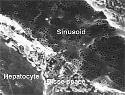
Individual capillaries are part of the capillary bed, an interweaving network of capillaries supplying tissues and organs. The more metabolically active a tissue is, the more capillaries are required to supply nutrients and carry away products of metabolism. There are two types of capillaries: true capillaries, which branch from arterioles and provide exchange between tissue and the capillary blood, and sinusoids, a type of open-pore capillary found in the liver, bone marrow, anterior pituitary gland, and brain circumventricular organs. Capillaries and sinusoids are short vessels that directly connect the arterioles and venules at opposite ends of the beds. Metarterioles are found primarily in the mesenteric microcirculation. [5]

Lymphatic capillaries are slightly larger in diameter than blood capillaries, and have closed ends (unlike the blood capillaries open at one end to the arterioles and open at the other end to the venules). This structure permits interstitial fluid to flow into them but not out. Lymph capillaries have a greater internal oncotic pressure than blood capillaries, due to the greater concentration of plasma proteins in the lymph. [6]

Types

Blood capillaries are categorized into three types: continuous, fenestrated, and sinusoidal (also known as discontinuous).

Types of capillaries: (left) continuous with no big gaps, (center) fenestrated with small pores, and (right) sinusoidal (or 'discontinuous') with intercellular gaps Different Types of Capillaries.jpg
Types of capillaries: (left) continuous with no big gaps, (center) fenestrated with small pores, and (right) sinusoidal (or 'discontinuous') with intercellular gaps

Continuous

Diagram of a continuous capillary Capillary.svg
Diagram of a continuous capillary

Continuous capillaries are continuous in the sense that the endothelial cells provide an uninterrupted lining, and they only allow smaller molecules, such as water and ions, to pass through their intercellular clefts. [7] [8] Lipid-soluble molecules can passively diffuse through the endothelial cell membranes along concentration gradients. [9] Continuous capillaries can be further divided into two subtypes:

  1. Those with numerous transport vesicles, which are found primarily in skeletal muscles, fingers, gonads, and skin. [10]
  2. Those with few vesicles, which are primarily found in the central nervous system. These capillaries are a constituent of the blood–brain barrier. [8]

Fenestrated

Fenestrated capillaries have pores known as fenestrae (Latin for "windows") in the endothelial cells that are 60–80  nanometres (nm) in diameter. They are spanned by a diaphragm of radially oriented fibrils that allows small molecules and limited amounts of protein to diffuse. [11] [12] In the renal glomerulus there are cells with no diaphragms, called podocyte foot processes or pedicels, which have slit pores with a function analogous to the diaphragm of the capillaries. Both of these types of blood vessels have continuous basal laminae and are primarily located in the endocrine glands, intestines, pancreas, and the glomeruli of the kidney.

Sinusoidal

Scanning electron micrograph of a liver sinusoid with fenestrated endothelial cells. Fenestrae are approximately 100 nm in diameter. Sinusoid.jpeg
Scanning electron micrograph of a liver sinusoid with fenestrated endothelial cells. Fenestrae are approximately 100 nm in diameter.

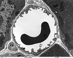
Sinusoidal capillaries or discontinuous capillaries are a special type of open-pore capillary, also known as a sinusoid, [13] that have wider fenestrations that are 30–40  micrometres (μm) in diameter, with wider openings in the endothelium. [14] Fenestrated capillaries have diaphragms that cover the pores whereas sinusoids lack a diaphragm and just have an open pore. These types of blood vessels allow red and white blood cells (7.5 μm – 25 μm diameter) and various serum proteins to pass, aided by a discontinuous basal lamina. These capillaries lack pinocytotic vesicles, and therefore use gaps present in cell junctions to permit transfer between endothelial cells, and hence across the membrane. Sinusoids are irregular spaces filled with blood and are mainly found in the liver, bone marrow, spleen, and brain circumventricular organs. [14] [15]

Annotated diagram of the exchange between capillary and body tissue through the exchange of materials between cells and fluid The exchange between capillary and body tissue diagram.svg
Annotated diagram of the exchange between capillary and body tissue through the exchange of materials between cells and fluid

Development

During early embryonic development, new capillaries are formed through vasculogenesis, the process of blood vessel formation that occurs through a novel production of endothelial cells that then form vascular tubes. [16] The term angiogenesis denotes the formation of new capillaries from pre-existing blood vessels and already-present endothelium which divides. [17] The small capillaries lengthen and interconnect to establish a network of vessels, a primitive vascular network that vascularises the entire yolk sac, connecting stalk, and chorionic villi. [18]

Function

Simplified image showing blood flow through the body, passing through capillary networks in its path 2101 Blood Flow Through the Heart.jpg
Simplified image showing blood flow through the body, passing through capillary networks in its path

The capillary wall performs an important function by allowing nutrients and waste substances to pass across it. Molecules larger than 3 nm such as albumin and other large proteins pass through transcellular transport carried inside vesicles, a process which requires them to go through the cells that form the wall. Molecules smaller than 3 nm such as water and gases cross the capillary wall through the space between cells in a process known as paracellular transport. [19] These transport mechanisms allow bidirectional exchange of substances depending on osmotic gradients. [20] Capillaries that form part of the blood–brain barrier only allow for transcellular transport as tight junctions between endothelial cells seal the paracellular space. [21]

Capillary beds may control their blood flow via autoregulation. This allows an organ to maintain constant flow despite a change in central blood pressure. This is achieved by myogenic response, and in the kidney by tubuloglomerular feedback. When blood pressure increases, arterioles are stretched and subsequently constrict (a phenomenon known as the Bayliss effect) to counteract the increased tendency for high pressure to increase blood flow. [22]

In the lungs, special mechanisms have been adapted to meet the needs of increased necessity of blood flow during exercise. When the heart rate increases and more blood must flow through the lungs, capillaries are recruited and are also distended to make room for increased blood flow. This allows blood flow to increase while resistance decreases.[ citation needed ] Extreme exercise can make capillaries vulnerable, with a breaking point similar to that of collagen. [23]

Capillary permeability can be increased by the release of certain cytokines, anaphylatoxins, or other mediators (such as leukotrienes, prostaglandins, histamine, bradykinin, etc.) highly influenced by the immune system.[ citation needed ]

Diagram of the filtration and reabsorption in capillaries 2108 Capillary Exchange.jpg
Diagram of the filtration and reabsorption in capillaries

Starling equation

The transport mechanisms can be further quantified by the Starling equation. [20] The Starling equation defines the forces across a semipermeable membrane and allows calculation of the net flux:

where:

is the net driving force,
is the proportionality constant, and
is the net fluid movement between compartments.

By convention, outward force is defined as positive, and inward force is defined as negative. The solution to the equation is known as the net filtration or net fluid movement (Jv). If positive, fluid will tend to leave the capillary (filtration). If negative, fluid will tend to enter the capillary (absorption). This equation has a number of important physiologic implications, especially when pathologic processes grossly alter one or more of the variables.[ citation needed ]

According to Starling's equation, the movement of fluid depends on six variables:

  1. Capillary hydrostatic pressure (Pc)
  2. Interstitial hydrostatic pressure (Pi)
  3. Capillary oncotic pressure (πc)
  4. Interstitial oncotic pressure (πi)
  5. Filtration coefficient (Kf)
  6. Reflection coefficient (σ)

Clinical significance

Disorders of capillary formation as a developmental defect or acquired disorder are a feature in many common and serious disorders. Within a wide range of cellular factors and cytokines, issues with normal genetic expression and bioactivity of the vascular growth and permeability factor vascular endothelial growth factor (VEGF) appear to play a major role in many of the disorders. Cellular factors include reduced number and function of bone-marrow derived endothelial progenitor cells. [24] and reduced ability of those cells to form blood vessels. [25]

Therapeutics

Major diseases where altering capillary formation could be helpful include conditions where there is excessive or abnormal capillary formation such as cancer and disorders harming eyesight; and medical conditions in which there is reduced capillary formation either for familial or genetic reasons, or as an acquired problem.

Blood sampling

Capillary blood sampling can be used to test for blood glucose (such as in blood glucose monitoring), hemoglobin, pH and lactate. [29] [30] It is generally performed by creating a small cut using a blood lancet, followed by sampling by capillary action on the cut with a test strip or small pipette. [31] It is also used to test for sexually transmitted infections that are present in the blood stream, such as HIV, syphilis, and hepatitis B and C, where a finger is lanced and a small amount of blood is sampled into a test tube. [32]

History

William Harvey did not explicitly predict the existence of capillaries, but he saw the need for some sort of connection between the arterial and venous systems. In 1653, he wrote, "...the blood doth enter into every member through the arteries, and does return by the veins, and that the veins are the vessels and ways by which the blood is returned to the heart itself; and that the blood in the members and extremities does pass from the arteries into the veins (either mediately by an anastomosis, or immediately through the porosities of the flesh, or both ways) as before it did in the heart and thorax out of the veins, into the arteries..." [33]

Marcello Malpighi was the first to observe directly and correctly describe capillaries, discovering them in a frog's lung 8 years later, in 1661. [34]

August Krogh discovered how capillaries provide nutrients to animal tissue. For his work he was awarded the 1920 Nobel Prize in Physiology or Medicine. [35]

See also

Related Research Articles

<span class="mw-page-title-main">Artery</span> Blood vessels that carry blood away from the heart

An artery is a blood vessel in humans and most other animals that takes oxygenated blood away from the heart in the systemic circulation to one or more parts of the body. Exceptions that carry deoxygenated blood are the pulmonary arteries in the pulmonary circulation that carry blood to the lungs for oxygenation, and the umbilical arteries in the fetal circulation that carry deoxygenated blood to the placenta.

<span class="mw-page-title-main">Blood vessel</span> Tubular structure of the circulatory system which transports blood

Blood vessels are the components of the circulatory system that transport blood throughout the human body. These vessels transport blood cells, nutrients, and oxygen to the tissues of the body. They also take waste and carbon dioxide away from the tissues. Blood vessels are needed to sustain life, because all of the body's tissues rely on their functionality.

<span class="mw-page-title-main">Vein</span> Blood vessels that carry blood towards the heart

Veins are blood vessels in the circulatory system of humans and most other animals that carry blood towards the heart. Most veins carry deoxygenated blood from the tissues back to the heart; exceptions are those of the pulmonary and fetal circulations which carry oxygenated blood to the heart. In the systemic circulation, arteries carry oxygenated blood away from the heart, and veins return deoxygenated blood to the heart, in the deep veins.

<span class="mw-page-title-main">Circulatory system</span> Organ system for circulating blood in animals

The blood circulatory system is a system of organs that includes the heart, blood vessels, and blood which is circulated throughout the entire body of a human or other vertebrate. It includes the cardiovascular system, or vascular system, that consists of the heart and blood vessels. The circulatory system has two divisions, a systemic circulation or circuit, and a pulmonary circulation or circuit. Some sources use the terms cardiovascular system and vascular system interchangeably with the circulatory system.

<span class="mw-page-title-main">Thrombus</span> Blood clot

A thrombus, colloquially called a blood clot, is the final product of the blood coagulation step in hemostasis. There are two components to a thrombus: aggregated platelets and red blood cells that form a plug, and a mesh of cross-linked fibrin protein. The substance making up a thrombus is sometimes called cruor. A thrombus is a healthy response to injury intended to stop and prevent further bleeding, but can be harmful in thrombosis, when a clot obstructs blood flow through healthy blood vessels in the circulatory system.

Hemodynamics or haemodynamics are the dynamics of blood flow. The circulatory system is controlled by homeostatic mechanisms of autoregulation, just as hydraulic circuits are controlled by control systems. The hemodynamic response continuously monitors and adjusts to conditions in the body and its environment. Hemodynamics explains the physical laws that govern the flow of blood in the blood vessels.

<span class="mw-page-title-main">Microcirculation</span> Circulation of the blood in the smallest blood vessels

The microcirculation is the circulation of the blood in the smallest blood vessels, the microvessels of the microvasculature present within organ tissues. The microvessels include terminal arterioles, metarterioles, capillaries, and venules. Arterioles carry oxygenated blood to the capillaries, and blood flows out of the capillaries through venules into veins.

<span class="mw-page-title-main">Arteriole</span> Small arteries in the microcirculation

An arteriole is a small-diameter blood vessel in the microcirculation that extends and branches out from an artery and leads to capillaries.

<span class="mw-page-title-main">Pulmonary artery</span> Artery in pulmonary circulation carrying deoxygenated blood from heart to lungs

A pulmonary artery is an artery in the pulmonary circulation that carries deoxygenated blood from the right side of the heart to the lungs. The largest pulmonary artery is the main pulmonary artery or pulmonary trunk from the heart, and the smallest ones are the arterioles, which lead to the capillaries that surround the pulmonary alveoli.

<span class="mw-page-title-main">Endothelium</span> Layer of cells that lining inner surface of blood vessels

The endothelium is a single layer of squamous endothelial cells that line the interior surface of blood vessels and lymphatic vessels. The endothelium forms an interface between circulating blood or lymph in the lumen and the rest of the vessel wall. Endothelial cells form the barrier between vessels and tissue and control the flow of substances and fluid into and out of a tissue.

<span class="mw-page-title-main">Venule</span> Very small blood vessel in the microcirculation

A venule is a very small vein in the microcirculation that allows blood to return from the capillary beds to drain into the venous system via increasingly larger veins. Post-capillary venules are the smallest of the veins with a diameter of between 10 and 30 micrometres (μm). When the post-capillary venules increase in diameter to 50μm they can incorporate smooth muscle and are known as muscular venules. Veins contain approximately 70% of total blood volume, while about 25% is contained in the venules. Many venules unite to form a vein.

<span class="mw-page-title-main">Glomerulus (kidney)</span> Functional unit of nephron

The glomerulus is a network of small blood vessels (capillaries) known as a tuft, located at the beginning of a nephron in the kidney. Each of the two kidneys contains about one million nephrons. The tuft is structurally supported by the mesangium, composed of intraglomerular mesangial cells. The blood is filtered across the capillary walls of this tuft through the glomerular filtration barrier, which yields its filtrate of water and soluble substances to a cup-like sac known as Bowman's capsule. The filtrate then enters the renal tubule of the nephron.

<span class="mw-page-title-main">Conjunctiva</span> Part of the eye; protective outer layer covering the sclera

In the anatomy of the eye, the conjunctiva is a thin mucous membrane that lines the inside of the eyelids and covers the sclera. It is composed of non-keratinized, stratified squamous epithelium with goblet cells, stratified columnar epithelium and stratified cuboidal epithelium. The conjunctiva is highly vascularised, with many microvessels easily accessible for imaging studies.

Mural cells are the vascular smooth muscle cells (vSMCs), and pericytes, of the microcirculation. Both types are in close contact with the endothelial cells lining the capillaries, and are important for vascular development and stability. Mural cells are involved in the formation of normal vasculature and are responsive to factors including platelet-derived growth factor B (PDGFB) and vascular endothelial growth factor (VEGF). The weakness and disorganization of tumor vasculature is partly due to the inability of tumors to recruit properly organized mural cells.

<span class="mw-page-title-main">Pericyte</span> Cells associated with capillary linings

Pericytes are multi-functional mural cells of the microcirculation that wrap around the endothelial cells that line the capillaries throughout the body. Pericytes are embedded in the basement membrane of blood capillaries, where they communicate with endothelial cells by means of both direct physical contact and paracrine signaling. The morphology, distribution, density and molecular fingerprints of pericytes vary between organs and vascular beds. Pericytes help to maintain homeostatic and hemostatic functions in the brain, one of the organs with higher pericyte coverage, and also sustain the blood–brain barrier. These cells are also a key component of the neurovascular unit, which includes endothelial cells, astrocytes, and neurons. Pericytes have been postulated to regulate capillary blood flow and the clearance and phagocytosis of cellular debris in vitro. Pericytes stabilize and monitor the maturation of endothelial cells by means of direct communication between the cell membrane as well as through paracrine signaling. A deficiency of pericytes in the central nervous system can cause increased permeability of the blood–brain barrier.

Hyperaemia is the increase of blood flow to different tissues in the body. It can have medical implications but is also a regulatory response, allowing change in blood supply to different tissues through vasodilation. Clinically, hyperaemia in tissues manifests as erythema because of the engorgement of vessels with oxygenated blood. Hyperaemia can also occur due to a fall in atmospheric pressure outside the body. The term comes from Greek ὑπέρ (hupér) 'over', and αἷμα (haîma) 'blood'.

Neovascularization is the natural formation of new blood vessels, usually in the form of functional microvascular networks, capable of perfusion by red blood cells, that form to serve as collateral circulation in response to local poor perfusion or ischemia.

<span class="mw-page-title-main">Metarteriole</span> Microvessel linking arterioles and capillaries

A metarteriole is a short microvessel in the microcirculation that links arterioles and capillaries. Instead of a continuous tunica media, they have individual smooth muscle cells placed a short distance apart, each forming a precapillary sphincter that encircles the entrance to that capillary bed. Constriction of these sphincters reduces or shuts off blood flow through their respective capillary beds. This allows the blood to be diverted to elsewhere in the body.

Microvasculature comprises the microvessels – venules and capillaries of the microcirculation, with a maximum average diameter of 0.3 millimeters. As the vessels decrease in size, they increase their surface-area-to-volume ratio. This allows surface properties to play a significant role in the function of the vessel.

A resistance artery is small diameter blood vessel in the microcirculation that contributes significantly to the creation of the resistance to flow and regulation of blood flow. Resistance arteries are usually small arteries or arterioles and include precapillary sphincters. Having thick muscular walls and narrow lumen they contribute the most to the resistance to blood flow. Degree of the contraction of vascular smooth muscle in the wall of a resistance artery is directly connected to the size of the lumen.

References

  1. Federative International Committee on Anatomical Terminology (2008). Terminologia Histologica: International Terms for Human Cytology and Histology. Baltimore: Lippincott Williams & Wilkins. p. 87. ISBN   9780781766104.
  2. "Structure and Function of Blood Vessels | Anatomy and Physiology II". courses.lumenlearning.com. Retrieved 19 November 2021.
  3. Maton, Anthea (1993). Human Biology and Health . Englewood Cliffs, New Jersey: Prentice Hall. pp. 87, 114, 120. ISBN   978-0-13-981176-0.
  4. 1 2 "Capillary". Online Etymology Dictionary. 2021. Retrieved 14 July 2021.
  5. Sakai, T; Hosoyamada, Y (2013). "Are the precapillary sphincters and metarterioles universal components of the microcirculation? An historical review". The Journal of Physiological Sciences. 63 (5): 319–31. doi:10.1007/s12576-013-0274-7. PMC   3751330 . PMID   23824465.
  6. Guyton, Arthur C.; Hall, John Edward (2006). "The Microcirculation and the Lymphatic System". Textbook of Medical Physiology (11th ed.). Philadelphia: Elsevier Saunders. pp. 187–188. ISBN   9780808923176.
  7. Stamatovic, S. M.; Johnson, A. M.; Keep, R. F.; Andjelkovic, A. V. (2016). "Junctional proteins of the blood-brain barrier: New insights into function and dysfunction". Tissue Barriers. 4 (1): e1154641. doi:10.1080/21688370.2016.1154641. PMC   4836471 . PMID   27141427.
  8. 1 2 Wilhelm, I.; Suciu, M.; Hermenean, A.; Krizbai, I. A. (2016). "Heterogeneity of the blood-brain barrier". Tissue Barriers. 4 (1): e1143544. doi:10.1080/21688370.2016.1143544. PMC   4836475 . PMID   27141424.
  9. Sarin, H. (2010). "Overcoming the challenges in the effective delivery of chemotherapies to CNS solid tumors". Therapeutic Delivery. 1 (2): 289–305. doi:10.4155/tde.10.22. PMC   3234205 . PMID   22163071.
  10. Michel, C. C. (2012). "Electron tomography of vesicles". Microcirculation. 19 (6): 473–6. doi: 10.1111/j.1549-8719.2012.00191.x . PMID   22574942. S2CID   205759387.
  11. Histology image:22401lba from Vaughan, Deborah (2002). A Learning System in Histology: CD-ROM and Guide. Oxford University Press. ISBN   978-0195151732.
  12. Pavelka, Margit; Roth, Jürgen (2005). "Fenestrated Capillary". Functional Ultrastructure: An Atlas of Tissue Biology and Pathology. Vienna: Springer. p. 232. doi:10.1007/3-211-26392-6_120. ISBN   978-3-211-26392-1.
  13. "Histology Laboratory Manual". www.columbia.edu.
  14. 1 2 Saladin, Kenneth S. (2011). Human Anatomy. McGraw-Hill. pp. 568–569. ISBN   9780071222075.
  15. Gross, P. M (1992). Circumventricular organ capillaries. Progress in Brain Research. Vol. 91. pp. 219–33. doi:10.1016/S0079-6123(08)62338-9. ISBN   9780444814197. PMID   1410407.
  16. John S. Penn (11 March 2008). Retinal and Choroidal Angiogenesis. Springer. pp. 119–. ISBN   978-1-4020-6779-2 . Retrieved 26 June 2010.
  17. Gilbert, Scott F. (2000). "Endoderm". Developmental Biology (6th ed.). Sunderland, Mass.: Sinauer Associates. ISBN   0-87893-243-7 . Retrieved 1 February 2021.
  18. Schoenwolf, Gary C. (2015). Larsen's human embryology (Fifth ed.). Philadelphia, PA. p. 306. ISBN   9781455706846.{{cite book}}: CS1 maint: location missing publisher (link)
  19. Sukriti, S; Tauseef, M; Yazbeck, P; Mehta, D (2014). "Mechanisms regulating endothelial permeability". Pulmonary Circulation. 4 (4): 535–551. doi:10.1086/677356. PMC   4278616 . PMID   25610592.
  20. 1 2 Nagy, JA; Benjamin, L; Zeng, H; Dvorak, AM; Dvorak, HF (2008). "Vascular permeability, vascular hyperpermeability and angiogenesis". Angiogenesis. 11 (2): 109–119. doi:10.1007/s10456-008-9099-z. PMC   2480489 . PMID   18293091.
  21. Bauer, HC; Krizbai, IA; Bauer, H; Traweger, A (2014). ""You Shall Not Pass"-tight junctions of the blood brain barrier". Frontiers in Neuroscience. 8: 392. doi: 10.3389/fnins.2014.00392 . PMC   4253952 . PMID   25520612.
  22. Boulpaep, Emile L. (2017). "The Microcirculation". In Boron, Walter F.; Boulpaep, Emile L. (eds.). Medical Physiology (3rd ed.). Philadelphia, PA: Elsevier. p. 481. ISBN   978-1-4557-4377-3.
  23. West, J. B. (2006). "Vulnerability of pulmonary capillaries during severe exercise". British Journal of Sports Medicine . 40 (10): 821. doi:10.1136/bjsm.2006.028886. ISSN   1473-0480. PMC   2465077 . PMID   17021008.
  24. Gittenberger-De Groot, Adriana C.; Winter, Elizabeth M.; Poelmann, Robert E. (2010). "Epicardium derived cells (EPDCs) in development, cardiac disease and repair of ischemia". Journal of Cellular and Molecular Medicine. 14 (5): 1056–60. doi:10.1111/j.1582-4934.2010.01077.x. PMC   3822740 . PMID   20646126.
  25. 1 2 Lambiase, P. D.; Edwards, RJ; Anthopoulos, P; Rahman, S; Meng, YG; Bucknall, CA; Redwood, SR; Pearson, JD; Marber, MS (2004). "Circulating Humoral Factors and Endothelial Progenitor Cells in Patients with Differing Coronary Collateral Support" (PDF). Circulation. 109 (24): 2986–92. doi: 10.1161/01.CIR.0000130639.97284.EC . PMID   15184289. S2CID   12041051.
  26. Noon, J P; Walker, B R; Webb, D J; Shore, A C; Holton, D W; Edwards, H V; Watt, G C (1997). "Impaired microvascular dilatation and capillary rarefaction in young adults with a predisposition to high blood pressure". Journal of Clinical Investigation. 99 (8): 1873–9. doi:10.1172/JCI119354. PMC   508011 . PMID   9109431.
  27. Bird, Alan C. (2010). "Therapeutic targets in age-related macular disease". Journal of Clinical Investigation. 120 (9): 3033–41. doi:10.1172/JCI42437. PMC   2929720 . PMID   20811159.
  28. Cao, Yihai (2009). "Tumor angiogenesis and molecular targets for therapy". Frontiers in Bioscience. 14 (14): 3962–73. doi: 10.2741/3504 . PMID   19273326.
  29. Krleza, Jasna Lenicek; Dorotic, Adrijana; Grzunov, Ana; Maradin, Miljenka (15 October 2015). "Capillary blood sampling: national recommendations on behalf of the Croatian Society of Medical Biochemistry and Laboratory Medicine". Biochemia Medica. 25 (3): 335–358. doi:10.11613/BM.2015.034. ISSN   1330-0962. PMC   4622200 . PMID   26524965.
  30. Moro, Christian; Bass, Jessica; Scott, Anna Mae; Canetti, Elisa F.D. (19 January 2017). "Enhancing capillary blood collection: The influence of nicotinic acid and nonivamide". Journal of Clinical Laboratory Analysis. 31 (6): e22142. doi: 10.1002/jcla.22142 . ISSN   0887-8013. PMC   6817299 . PMID   28102549.
  31. "Managing diabetes:Check your blood glucose levels". National Institute of Diabetes and Digestive and Kidney Diseases,US National Institutes of Health. 2021. Retrieved 9 September 2021.
  32. "Fettle - How to take a blood sample". Archived from the original on 16 March 2023. Retrieved 16 March 2023.
  33. Harvey, William (1653). On the motion of the Heart and Blood in Animals. pp. 59–60. Archived from the original on 1 December 2011.
  34. Cliff, Walter John (1976). Blood Vessels. Cambridge University Press. p. 14. ISBN   9780835773287.
  35. "August Krogh". July 2023.