| Tunica externa | |
|---|---|
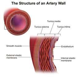 | |
 Section of a medium-sized artery. | |
| Details | |
| Part of | Wall of blood vessels |
| Identifiers | |
| Latin | tunica externa, tunica adventitia |
| TA98 | A12.0.00.017 |
| TA2 | 3920 |
| TH | H3.09.02.0.01009 |
| Anatomical terminology | |
The tunica externa (Neo-Latin "outer coat"), also known as the tunica adventitia (Neo-Latin "additional coat"), [1] [2] is the outermost tunica (layer) of a blood vessel, surrounding the tunica media. It is mainly composed of collagen and, in arteries, is supported by external elastic lamina. The collagen serves to anchor the blood vessel to nearby organs, giving it stability.
Contents
The three layers of the blood vessels are: an inner tunica intima, a middle tunica media, and an outer tunica externa.

