Related Research Articles

The aorta is the main and largest artery in the human body, originating from the left ventricle of the heart, branching upwards immediately after, and extending down to the abdomen, where it splits at the aortic bifurcation into two smaller arteries. The aorta distributes oxygenated blood to all parts of the body through the systemic circulation.

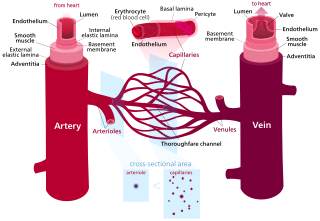
An artery is a blood vessel in humans and most other animals that takes oxygenated blood away from the heart in the systemic circulation to one or more parts of the body. Exceptions that carry deoxygenated blood are the pulmonary arteries in the pulmonary circulation that carry blood to the lungs for oxygenation, and the umbilical arteries in the fetal circulation that carry deoxygenated blood to the placenta. It consists of a multi-layered artery wall wrapped into a tube-shaped channel.
Blood vessels are the structures of the circulatory system that transport blood throughout the human body. These vessels transport blood cells, nutrients, and oxygen to the tissues of the body. They also take waste and carbon dioxide away from the tissues. Blood vessels are needed to sustain life, because all of the body's tissues rely on their functionality.

Veins are blood vessels in the circulatory system of humans and most other animals that carry blood towards the heart. Most veins carry deoxygenated blood from the tissues back to the heart; exceptions are those of the pulmonary and fetal circulations which carry oxygenated blood to the heart. In the systemic circulation, arteries carry oxygenated blood away from the heart, and veins return deoxygenated blood to the heart, in the deep veins.

The superior vena cava (SVC) is the superior of the two venae cavae, the great venous trunks that return deoxygenated blood from the systemic circulation to the right atrium of the heart. It is a large-diameter (24 mm) short length vein that receives venous return from the upper half of the body, above the diaphragm. Venous return from the lower half, below the diaphragm, flows through the inferior vena cava. The SVC is located in the anterior right superior mediastinum. It is the typical site of central venous access via a central venous catheter or a peripherally inserted central catheter. Mentions of "the cava" without further specification usually refer to the SVC.

Systole is the part of the cardiac cycle during which some chambers of the heart contract after refilling with blood.

Smooth (soft) muscle is one of the three major types of vertebrate muscle tissue, the other being skeletal and cardiac muscle. Nonetheless, it is found in invertebrates as well and is controlled by the autonomic nervous system. It is non-striated, so-called because it has no sarcomeres and therefore no striations. It can be divided into two subgroups, single-unit and multi-unit smooth muscle. Within single-unit muscle, the whole bundle or sheet of smooth muscle cells contracts as a syncytium.

An arteriole is a small-diameter blood vessel in the microcirculation that extends and branches out from an artery and leads to capillaries.

Vasa vasorum are small blood vessels that comprise a vascular network supplying the walls of large blood vessels, such as elastic arteries and large veins.
Compliance is the ability of a hollow organ (vessel) to distend and increase volume with increasing transmural pressure or the tendency of a hollow organ to resist recoil toward its original dimensions on application of a distending or compressing force. It is the reciprocal of "elastance", hence elastance is a measure of the tendency of a hollow organ to recoil toward its original dimensions upon removal of a distending or compressing force.

The tunica intima, or intima for short, is the innermost tunica (layer) of an artery or vein. It is made up of one layer of endothelial cells, and is supported by an internal elastic lamina. The endothelial cells are in direct contact with the blood flow.
The tunica media, or media for short, is the middle tunica (layer) of an artery or vein. It lies between the tunica intima on the inside and the tunica externa on the outside.

The cardiac cycle is the performance of the human heart from the beginning of one heartbeat to the beginning of the next. It consists of two periods: one during which the heart muscle relaxes and refills with blood, called diastole, following a period of robust contraction and pumping of blood, called systole. After emptying, the heart relaxes and expands to receive another influx of blood returning from the lungs and other systems of the body, before again contracting to pump blood to the lungs and those systems.

The aortic arch, arch of the aorta, or transverse aortic arch is the part of the aorta between the ascending and descending aorta. The arch travels backward, so that it ultimately runs to the left of the trachea.
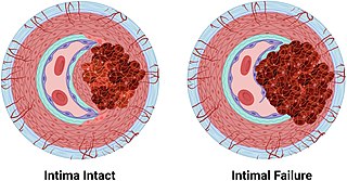
An arterial dissection is a tear within the wall of an artery, which allows blood to separate the wall layers. There are several types. Usually, a tear is in an arterial wall, but a vein wall tear has been documented.
The myogenic mechanism is how arteries and arterioles react to an increase or decrease of blood pressure to keep the blood flow constant within the blood vessel. Myogenic response refers to a contraction initiated by the myocyte itself instead of an outside occurrence or stimulus such as nerve innervation. Most often observed in smaller resistance arteries, this 'basal' myogenic tone may be useful in the regulation of organ blood flow and peripheral resistance, as it positions a vessel in a preconstricted state that allows other factors to induce additional constriction or dilation to increase or decrease blood flow.
The tunica externa, also known as the tunica adventitia, is the outermost tunica (layer) of a blood vessel, surrounding the tunica media. It is mainly composed of collagen and, in arteries, is supported by external elastic lamina. The collagen serves to anchor the blood vessel to nearby organs, giving it stability.

Windkessel effect is a term used in medicine to account for the shape of the arterial blood pressure waveform in terms of the interaction between the stroke volume and the compliance of the aorta and large elastic arteries and the resistance of the smaller arteries and arterioles. Windkessel when loosely translated from German to English means 'air chamber', but is generally taken to imply an elastic reservoir. The walls of large elastic arteries contain elastic fibers, formed of elastin. These arteries distend when the blood pressure rises during systole and recoil when the blood pressure falls during diastole. Since the rate of blood entering these elastic arteries exceeds that leaving them via the peripheral resistance, there is a net storage of blood in the aorta and large arteries during systole, which discharges during diastole. The compliance of the aorta and large elastic arteries is therefore analogous to a capacitor ; to put it another way, these arteries collectively act as a hydraulic accumulator.
A muscular artery is a medium-sized artery that draws blood from an elastic artery and branches into "resistance vessels" including small arteries and arterioles. Their walls contain larger number of smooth muscles, allowing them to contract and expand depending on peripheral blood demand.

The internal elastic lamina or internal elastic lamella is a layer of elastic tissue that forms the outermost part of the tunica intima of blood vessels. It separates tunica intima from tunica media.
References
- ↑ Shadwick RE (December 1999). "Mechanical design in arteries". J. Exp. Biol. 202 (Pt 23): 3305–13. PMID 10562513.
- ↑ Belz G G (1995). "Elastic properties and Windkessel function of the human aorta". Cardiovasc Drug Ther. 9 (1): 73–83. doi:10.1007/BF00877747.
- 1 2 3 Mescher, A.L., ed. (2024). Junqueira's Basic Histology: Text and Atlas (17th ed.). McGraw Hill.