
The aorta is the main and largest artery in the human body, originating from the left ventricle of the heart, branching upwards immediately after, and extending down to the abdomen, where it splits at the aortic bifurcation into two smaller arteries. The aorta distributes oxygenated blood to all parts of the body through the systemic circulation.

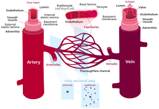
An artery is a blood vessel in humans and most other animals that takes oxygenated blood away from the heart in the systemic circulation to one or more parts of the body. Exceptions that carry deoxygenated blood are the pulmonary arteries in the pulmonary circulation that carry blood to the lungs for oxygenation, and the umbilical arteries in the fetal circulation that carry deoxygenated blood to the placenta. It consists of a multi-layered artery wall wrapped into a tube-shaped channel.
Blood vessels are the tubular structures of a circulatory system that transport blood throughout a vertebrate's body. Blood vessels transport blood cells, nutrients, and oxygen to most of the tissues of a body. They also take waste and carbon dioxide away from the tissues. Some tissues such as cartilage, epithelium, and the lens and cornea of the eye are not supplied with blood vessels and are termed avascular.

Veins are blood vessels in the circulatory system of humans and most other animals that carry blood towards the heart. Most veins carry deoxygenated blood from the tissues back to the heart; exceptions are those of the pulmonary and fetal circulations which carry oxygenated blood to the heart. In the systemic circulation, arteries carry oxygenated blood away from the heart, and veins return deoxygenated blood to the heart, in the deep veins.
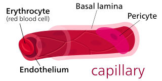
A capillary is a small blood vessel, from 5 to 10 micrometres in diameter, and is part of the microcirculation system. Capillaries are microvessels and the smallest blood vessels in the body. They are composed of only the tunica intima, consisting of a thin wall of simple squamous endothelial cells. They are the site of the exchange of many substances from the surrounding interstitial fluid, and they convey blood from the smallest branches of the arteries (arterioles) to those of the veins (venules). Other substances which cross capillaries include water, oxygen, carbon dioxide, urea, glucose, uric acid, lactic acid and creatinine. Lymph capillaries connect with larger lymph vessels to drain lymphatic fluid collected in microcirculation.

An arteriole is a small-diameter blood vessel in the microcirculation that extends and branches out from an artery and leads to capillaries.

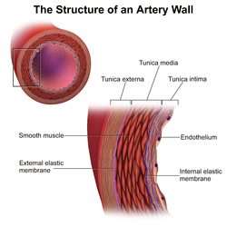
The endothelium is a single layer of squamous endothelial cells that line the interior surface of blood vessels and lymphatic vessels. The endothelium forms an interface between circulating blood or lymph in the lumen and the rest of the vessel wall.
A venule is a very small vein in the microcirculation that allows blood to return from the capillary beds to drain into the venous system via increasingly larger veins. Post-capillary venules are the smallest of the veins with a diameter of between 10 and 30 micrometres (μm). When the post-capillary venules increase in diameter to 50μm they can incorporate smooth muscle and are known as muscular venules. Veins contain approximately 70% of total blood volume, while about 25% is contained in the venules. Many venules unite to form a vein.
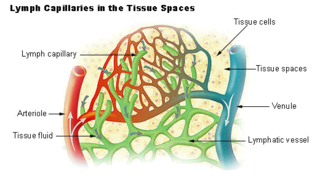
The lymphatic vessels are thin-walled vessels (tubes), structured like blood vessels, that carry lymph. As part of the lymphatic system, lymph vessels are complementary to the cardiovascular system. Lymph vessels are lined by endothelial cells, and have a thin layer of smooth muscle, and adventitia that binds the lymph vessels to the surrounding tissue. Lymph vessels are devoted to the propulsion of the lymph from the lymph capillaries, which are mainly concerned with the absorption of interstitial fluid from the tissues. Lymph capillaries are slightly bigger than their counterpart capillaries of the vascular system. Lymph vessels that carry lymph to a lymph node are called afferent lymph vessels, and those that carry it from a lymph node are called efferent lymph vessels, from where the lymph may travel to another lymph node, may be returned to a vein, or may travel to a larger lymph duct. Lymph ducts drain the lymph into one of the subclavian veins and thus return it to general circulation.

Vasa vasorum are networks of small blood vessels that supply the walls of large blood vessels, such as elastic arteries and large veins.

The basement membrane, also known as base membrane, is a thin, pliable sheet-like type of extracellular matrix that provides cell and tissue support and acts as a platform for complex signalling. The basement membrane sits between epithelial tissues including mesothelium and endothelium, and the underlying connective tissue.

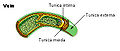
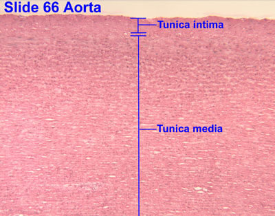
The tunica media, or media for short, is the middle tunica (layer) of an artery or vein. It lies between the internal elastic lamina of the tunica intima on the inside and the tunica externa on the outside.

The trabecular veins are the largest veins inside the spleen. They drain the blood collected in the sinuses of the pulp.
The tunica externa, also known as the tunica adventitia, is the outermost tunica (layer) of a blood vessel, surrounding the tunica media. It is mainly composed of collagen and, in arteries, is supported by external elastic lamina. The collagen serves to anchor the blood vessel to nearby organs, giving it stability.
An elastic artery is an artery with many collagen and elastin filaments in the tunica media, which gives it the ability to stretch in response to each pulse. This elasticity also gives rise to the Windkessel effect, which helps to maintain a relatively constant pressure in the arteries despite the pulsating nature of the blood flow. Elastic arteries include the largest arteries in the body, those closest to the heart. They give rise to medium-sized vessels known as distributing arteries.
A muscular artery is a medium-sized artery that draws blood from an elastic artery and branches into "resistance vessels" including small arteries and arterioles. Their walls contain larger number of smooth muscles, allowing them to contract and expand depending on peripheral blood demand.

The internal elastic lamina or internal elastic lamella is a layer of elastic tissue that forms the outermost part of the tunica intima of blood vessels. It separates tunica intima from tunica media.

Vascular remodelling is a process which occurs when an immature heart begins contracting, pushing fluid through the early vasculature. The process typically begins at day 22, and continues to the tenth week of human embryogenesis. This first passage of fluid initiates a signal cascade and cell movement based on physical cues including shear stress and circumferential stress, which is necessary for the remodelling of the vascular network, arterial-venous identity, angiogenesis, and the regulation of genes through mechanotransduction. This embryonic process is necessary for the future stability of the mature vascular network.
In biology, a tunica is a layer, coat, sheath, or similar covering. The word came to English from the Neo-Latin of science and medicine. Its literal sense is about the same as that of the word tunic, with which it is cognate. In biology, one of its senses used to be the taxonomic name of a genus of plants, but the nomenclature has been revised and those plants are now included in the genus Petrorhagia.

Anatomical terminology is used to describe microanatomical structures. This helps describe precisely the structure, layout and position of an object, and minimises ambiguity. An internationally accepted lexicon is Terminologia Histologica.

