Pulmonary vein

Last updated
Pulmonary vein
Diagram of the human heart (cropped).svg
Anterior (frontal) view of the opened heart. White arrows indicate normal blood flow.
Alveoli diagram.png
Diagram of the alveoli with both cross-section and external view.
Details
Precursor truncus arteriosus
System Circulatory system
Drains from lungs
Drains to left atrium
Artery pulmonary artery
Identifiers
Latin venae pulmonales
MeSH D011667
TA98 A12.3.02.001
TA2 4107
FMA 66643
Anatomical terminology
Openings of pulmonary veins.png

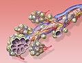
The pulmonary veins are the veins that transfer oxygenated blood from the lungs to the heart. The largest pulmonary veins are the four main pulmonary veins, two from each lung that drain into the left atrium of the heart. The pulmonary veins are part of the pulmonary circulation.

Contents

Structure

There are four main pulmonary veins, two from each lung – an inferior and a superior main vein, emerging from each hilum. The main pulmonary veins receive blood from three or four feeding veins in each lung, and drain into the left atrium. The peripheral feeding veins do not follow the bronchial tree. They run between the pulmonary segments from which they drain the blood. [1]

At the root of the lung, the right superior pulmonary vein lies in front of and a little below the pulmonary artery; the inferior is situated at the lowest part of the lung hilum. Behind the pulmonary artery is the bronchus. [2] The right main pulmonary veins (contains oxygenated blood) pass behind the right atrium and superior vena cava; the left in front of the descending thoracic aorta.

Variation

Occasionally the three lobar veins on the right side remain separate, and not infrequently the two left lobar veins end by a common opening into the left atrium. Therefore, the number of pulmonary veins opening into the left atrium can vary between three and five in the healthy population.

The two left lobar veins may be united as a single pulmonary vein in about 25% of enssaam

the two right veins may be united in about 3%. [2]

Function

The pulmonary veins play an essential role in respiration, by receiving blood that has been oxygenated in the alveoli and returning it to the left atrium.

Clinical significance

As part of the pulmonary circulation they carry oxygenated blood back to the heart, as opposed to the veins of the systemic circulation which carry deoxygenated blood.

On chest X-ray, the diameters of pulmonary veins increases from upper to lower lobes, from 3 mm at the first intercoastal space, to 6 mm just above the diaphragm. [3]

A rare genetic defect of the pulmonary veins can cause them to drain into the pulmonary circulation in whole or in part, this is known as a total anomalous pulmonary venous connection (or drainage), or partial anomalous pulmonary connection, respectively.

Additional images

See also

Related Research Articles

<span class="mw-page-title-main">Heart</span> Organ found inside most animals

The heart is a muscular organ in most animals. This organ pumps blood through the blood vessels of the circulatory system. The pumped blood carries oxygen and nutrients to the body, while carrying metabolic waste such as carbon dioxide to the lungs. In humans, the heart is approximately the size of a closed fist and is located between the lungs, in the middle compartment of the chest, called the mediastinum.

<span class="mw-page-title-main">Vein</span> Blood vessels that carry blood towards the heart

Veins are blood vessels in the circulatory system of humans and most other animals that carry blood towards the heart. Most veins carry deoxygenated blood from the tissues back to the heart; exceptions are those of the pulmonary and fetal circulations which carry oxygenated blood to the heart. In the systemic circulation, arteries carry oxygenated blood away from the heart, and veins return deoxygenated blood to the heart, in the deep veins.

<span class="mw-page-title-main">Circulatory system</span> Organ system for circulating blood in animals

The circulatory system is a system of organs that includes the heart, blood vessels, and blood which is circulated throughout the entire body of a human or other vertebrate. It includes the cardiovascular system, or vascular system, that consists of the heart and blood vessels. The circulatory system has two divisions, a systemic circulation or circuit, and a pulmonary circulation or circuit. Some sources use the terms cardiovascular system and vascular system interchangeably with circulatory system.

<span class="mw-page-title-main">Coronary circulation</span> Circulation of blood in the blood vessels of the heart muscle (myocardium)

Coronary circulation is the circulation of blood in the arteries and veins that supply the heart muscle (myocardium). Coronary arteries supply oxygenated blood to the heart muscle. Cardiac veins then drain away the blood after it has been deoxygenated. Because the rest of the body, and most especially the brain, needs a steady supply of oxygenated blood that is free of all but the slightest interruptions, the heart is required to function continuously. Therefore its circulation is of major importance not only to its own tissues but to the entire body and even the level of consciousness of the brain from moment to moment. Interruptions of coronary circulation quickly cause heart attacks, in which the heart muscle is damaged by oxygen starvation. Such interruptions are usually caused by coronary ischemia linked to coronary artery disease, and sometimes to embolism from other causes like obstruction in blood flow through vessels.

<span class="mw-page-title-main">Bronchus</span> Airway in the respiratory tract

A bronchus is a passage or airway in the lower respiratory tract that conducts air into the lungs. The first or primary bronchi to branch from the trachea at the carina are the right main bronchus and the left main bronchus. These are the widest bronchi, and enter the right lung, and the left lung at each hilum. The main bronchi branch into narrower secondary bronchi or lobar bronchi, and these branch into narrower tertiary bronchi or segmental bronchi. Further divisions of the segmental bronchi are known as 4th order, 5th order, and 6th order segmental bronchi, or grouped together as subsegmental bronchi. The bronchi, when too narrow to be supported by cartilage, are known as bronchioles. No gas exchange takes place in the bronchi.

<span class="mw-page-title-main">Pulmonary artery</span> Artery in pulmonary circulation carrying deoxygenated blood from heart to lungs

A pulmonary artery is an artery in the pulmonary circulation that carries deoxygenated blood from the right side of the heart to the lungs. The largest pulmonary artery is the main pulmonary artery or pulmonary trunk from the heart, and the smallest ones are the arterioles, which lead to the capillaries that surround the pulmonary alveoli.

<span class="mw-page-title-main">Pulmonary circulation</span> Part of the circulatory system which carries blood from heart to lungs and back to the heart

The pulmonary circulation is a division of the circulatory system in all vertebrates. The circuit begins with deoxygenated blood returned from the body to the right atrium of the heart where it is pumped out from the right ventricle to the lungs. In the lungs the blood is oxygenated and returned to the left atrium to complete the circuit.

<span class="mw-page-title-main">Azygos vein</span> Human blood vessel by the spine

The azygos vein is a vein running up the right side of the thoracic vertebral column draining itself towards the superior vena cava. It connects the systems of superior vena cava and inferior vena cava and can provide an alternative path for blood to the right atrium when either of the venae cavae is blocked.

<span class="mw-page-title-main">Atrium (heart)</span> Part of the human heart

The atrium is one of the two upper chambers in the heart that receives blood from the circulatory system. The blood in the atria is pumped into the heart ventricles through the atrioventricular mitral and tricuspid heart valves.

<span class="mw-page-title-main">Bronchial artery</span> Blood vessels supplying the lungs

In human anatomy, the bronchial arteries supply the lungs with oxygenated blood, and nutrition. Although there is much variation, there are usually two bronchial arteries that run to the left lung, and one to the right lung, and are a vital part of the respiratory system.

<span class="mw-page-title-main">Fetal circulation</span> Circulatory system of fetuses

In humans, the circulatory system is different before and after birth. The fetal circulation is composed of the placenta, umbilical blood vessels encapsulated by the umbilical cord, heart and systemic blood vessels. A major difference between the fetal circulation and postnatal circulation is that the lungs are not used during the fetal stage resulting in the presence of shunts to move oxygenated blood and nutrients from the placenta to the fetal tissue. At birth, the start of breathing and the severance of the umbilical cord prompt various changes that quickly transform fetal circulation into postnatal circulation.

<span class="mw-page-title-main">Thoracic aorta</span> Part of the aorta located in the thorax

The thoracic aorta is a part of the aorta located in the thorax. It is a continuation of the aortic arch. It is located within the posterior mediastinal cavity, but frequently bulges into the left pleural cavity. The descending thoracic aorta begins at the lower border of the fourth thoracic vertebra and ends in front of the lower border of the twelfth thoracic vertebra, at the aortic hiatus in the diaphragm where it becomes the abdominal aorta.

<span class="mw-page-title-main">Coronary sinus</span> Set of veins which drain blood from the myocardium (heart muscle)

The coronary sinus is the largest vein of the heart. It drains over half of the deoxygenated blood from the heart muscle into the right atrium. It begins on the backside of the heart, in between the left atrium, and left ventricle; it begins at the junction of the great cardiac vein, and oblique vein of the left atrium. It receives multiple tributaries. It passes across the backside of the heart along a groove between left atrium and left ventricle, then drains into the right atrium at the orifice of the coronary sinus.

<span class="mw-page-title-main">Scimitar syndrome</span> Medical condition

Scimitar syndrome, or congenital pulmonary venolobar syndrome, is a rare congenital heart defect characterized by anomalous venous return from the right lung. This anomalous pulmonary venous return can be either partial (PAPVR) or total (TAPVR). The syndrome associated with PAPVR is more commonly known as Scimitar syndrome after the curvilinear pattern created on a chest radiograph by the pulmonary veins that drain to the inferior vena cava. This radiographic density often has the shape of a scimitar, a type of curved sword. The syndrome was first described by Catherine Neill in 1960.

The bronchial veins are small vessels that return blood from the larger bronchi and structures at the roots of the lungs. The right side drains into the azygos vein, while the left side drains into the left superior intercostal vein or the accessory hemiazygos vein. Bronchial veins are thereby part of the bronchial circulation, carrying waste products away from the cells that constitute the lungs.

<span class="mw-page-title-main">Root of the lung</span> Anatomical structure

The root of the lung is a group of structures that emerge at the hilum of each lung, just above the middle of the mediastinal surface and behind the cardiac impression of the lung. It is nearer to the back than the front. The root of the lung is connected by the structures that form it to the heart and the trachea. The rib cage is separated from the lung by a two-layered membranous coating, the pleura. The hilum is the large triangular depression where the connection between the parietal pleura and the visceral pleura is made, and this marks the meeting point between the mediastinum and the pleural cavities.

<span class="mw-page-title-main">Smallest cardiac veins</span> Small veins in the walls of all four heart chambers

The smallest cardiac veins are small, valveless veins in the walls of all four heart chambers that drain venous blood from the myocardium directly into any of the heart chambers.

<span class="mw-page-title-main">Anomalous pulmonary venous connection</span> Medical condition

Anomalous pulmonary venous connection is a congenital defect of the pulmonary veins.

<span class="mw-page-title-main">Bronchial circulation</span> Circulation of blood supplying lungs tissues

The bronchial circulation is the part of the systemic circulation that supplies nutrients and oxygen to the cells that constitute the lungs, as well as carrying waste products away from them. It is complementary to the pulmonary circulation that brings deoxygenated blood to the lungs and carries oxygenated blood away from them in order to oxygenate the rest of the body.

The heart is a muscular organ situated in the mediastinum. It consists of four chambers, four valves, two main arteries, and the conduction system. The left and right sides of the heart have different functions: the right side receives de-oxygenated blood through the superior and inferior venae cavae and pumps blood to the lungs through the pulmonary artery, and the left side receives saturated blood from the lungs.

References

PD-icon.svgThis article incorporates text in the public domain from page 642 of the 20th edition of Gray's Anatomy (1918)

  1. Drake, Richard L.; Vogl, Wayne; Tibbitts, Adam W.M. Mitchell; illustrations by Richard; Richardson, Paul (2005). Gray's anatomy for students (Pbk. ed.). Philadelphia: Elsevier/Churchill Livingstone. ISBN   978-0-443-06612-2.
  2. 1 2 Skandalakis, editor in chief John E. (2004). "Chapter 7. Pericardium, Heart, and Great Vessels in the Thorax". Skandalakis' surgical anatomy : the embryologic and anatomic basis of modern surgery. Athens, Greece: PMP. pp. section titled 'Pulmonary veins'. ISBN   9603990744.{{cite book}}: |first= has generic name (help)
  3. Porres, Diego Varona; Morenza, Óscar Persiva; Pallisa, Esther; Roque, Alberto; Andreu, Jorge; Martínez, Manel (July 2013). "Learning from the Pulmonary Veins". RadioGraphics. 33 (4): 999–1022. doi:10.1148/rg.334125043. ISSN   0271-5333. PMID   23842969.