Thoracic aorta | |
---|---|
![]() Schematic view of the aorta, showing major branches and segments with thoracic aorta shown as continuation of descending aorta | |
![]() The thoracic aorta, viewed from the left side. | |
Details | |
Source | Descending aorta |
Branches | Bronchial arteries, esophageal arteries, posterior intercostal arteries, abdominal aorta, superior phrenic artery, pericardial arteries |
Identifiers | |
Latin | aorta thoracalis |
MeSH | D001013 |
TA98 | A12.2.11.001 |
TA2 | 4186 |
FMA | 3786 |
Anatomical terminology |
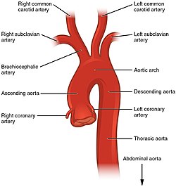
The thoracic aorta is a part of the aorta located in the thorax. It is a continuation of the aortic arch. It is located within the posterior mediastinal cavity, but frequently bulges into the left pleural cavity. The descending thoracic aorta begins at the lower border of the fourth thoracic vertebra and ends in front of the lower border of the twelfth thoracic vertebra, at the aortic hiatus in the diaphragm where it becomes the abdominal aorta.
At its commencement, it is situated on the left of the vertebral column; it approaches the median line as it descends; and, at its termination, lies directly in front of the column.
The thoracic aorta has a curved shape that faces forward, and has small branches. It has a radius of approximately 1.16 cm. [1]
The thoracic aorta is part of the descending aorta, which has different parts named according to their structure or location. The thoracic aorta is a continuation of the descending aorta and becomes the abdominal aorta when it passes through the diaphragm. The initial part of the aorta, the ascending aorta, rises out of the left ventricle, from which it is separated by the aortic valve. The two coronary arteries of the heart arise from the aortic root, just above the cusps of the aortic valve. The aorta then arches back over the right pulmonary artery. Three vessels come out of the aortic arch: the brachiocephalic artery, the left common carotid artery, and the left subclavian artery. These vessels supply blood to the head, neck, thorax and upper limbs.
Behind the descending thoracic aorta is the vertebral column and the hemiazygos vein. To the right is the azygos veins and thoracic duct, and to the left is the left pleura and lung. In front of the thoracic aorta lies the root of the left lung, the pericardium, the esophagus, and the diaphragm.
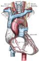
The esophagus, which is covered by a nerve plexus lies to the right of the descending thoracic aorta. Lower, the esophagus passes in front of the aorta, and ultimately is situated on the left.
The aorta is an artery that conveys oxygenated blood from the heart to other parts of the body. It is one of the largest arteries in the body. [2] The aorta gives off several paired branches as it descends. In descending order, these include the
Note: The posterior intercostal arteries are branches that originate throughout the length of the posterior aspect of the descending thoracic aorta.
The aorta is the main and largest artery in the human body, originating from the left ventricle of the heart, branching upwards immediately after, and extending down to the abdomen, where it splits at the aortic bifurcation into two smaller arteries. The aorta distributes oxygenated blood to all parts of the body through the systemic circulation.
The thoracic cavity is the chamber of the body of vertebrates that is protected by the thoracic wall. The central compartment of the thoracic cavity is the mediastinum. There are two openings of the thoracic cavity, a superior thoracic aperture known as the thoracic inlet and a lower inferior thoracic aperture known as the thoracic outlet.
The inferior vena cava is a large vein that carries the deoxygenated blood from the lower and middle body into the right atrium of the heart. It is formed by the joining of the right and the left common iliac veins, usually at the level of the fifth lumbar vertebra.
In human anatomy, the subclavian arteries are paired major arteries of the upper thorax, below the clavicle. They receive blood from the aortic arch. The left subclavian artery supplies blood to the left arm and the right subclavian artery supplies blood to the right arm, with some branches supplying the head and thorax. On the left side of the body, the subclavian comes directly off the aortic arch, while on the right side it arises from the relatively short brachiocephalic artery when it bifurcates into the subclavian and the right common carotid artery.
In human anatomy, the thoracic duct is the larger of the two lymph ducts of the lymphatic system. The thoracic duct usually begins from the upper aspect of the cisterna chyli, passing out of the abdomen through the aortic hiatus into first the posterior mediastinum and then the superior mediastinum, extending as high up as the root of the neck before descending to drain into the systemic (blood) circulation at the venous angle.
The thoracic diaphragm, or simply the diaphragm, is a sheet of internal skeletal muscle in humans and other mammals that extends across the bottom of the thoracic cavity. The diaphragm is the most important muscle of respiration, and separates the thoracic cavity, containing the heart and lungs, from the abdominal cavity: as the diaphragm contracts, the volume of the thoracic cavity increases, creating a negative pressure there, which draws air into the lungs. Its high oxygen consumption is noted by the many mitochondria and capillaries present; more than in any other skeletal muscle.
A pulmonary artery is an artery in the pulmonary circulation that carries deoxygenated blood from the right side of the heart to the lungs. The largest pulmonary artery is the main pulmonary artery or pulmonary trunk from the heart, and the smallest ones are the arterioles, which lead to the capillaries that surround the pulmonary alveoli.
The azygos vein is a vein running up the right side of the thoracic vertebral column draining itself towards the superior vena cava. It connects the systems of superior vena cava and inferior vena cava and can provide an alternative path for blood to the right atrium when either of the venae cavae is blocked.
In human anatomy, the abdominal aorta is the largest artery in the abdominal cavity. As part of the aorta, it is a direct continuation of the descending aorta.
The mediastinum is the central compartment of the thoracic cavity. Surrounded by loose connective tissue, it is an undelineated region that contains a group of structures within the thorax, namely the heart and its vessels, the esophagus, the trachea, the phrenic and cardiac nerves, the thoracic duct, the thymus and the lymph nodes of the central chest.
In human anatomy, the descending aorta is part of the aorta, the largest artery in the body. The descending aorta begins at the aortic arch and runs down through the chest and abdomen. The descending aorta anatomically consists of two portions or segments, the thoracic and the abdominal aorta, in correspondence with the two great cavities of the trunk in which it is situated. Within the abdomen, the descending aorta branches into the two common iliac arteries which serve the pelvis and eventually legs.
In anatomy, the left and right common carotid arteries (carotids) are arteries that supply the head and neck with oxygenated blood; they divide in the neck to form the external and internal carotid arteries.
The abdomen is the part of the body between the thorax (chest) and pelvis, in humans and in other vertebrates. The abdomen is the front part of the abdominal segment of the torso. The area occupied by the abdomen is called the abdominal cavity. In arthropods, it is the posterior tagma of the body; it follows the thorax or cephalothorax.
The aortic arch, arch of the aorta, or transverse aortic arch is the part of the aorta between the ascending and descending aorta. The arch travels backward, so that it ultimately runs to the left of the trachea.
The hemiazygos vein is a vein running superiorly in the lower thoracic region, just to the left side of the vertebral column.
The aortic hiatus is a midline opening in the posterior part of the diaphragm giving passage to the descending aorta as well as the thoracic duct, and variably the azygos and hemiazygos veins. It is the lowest and most posterior of the large apertures.
The intercostal arteries are a group of arteries passing within an intercostal space. There are 9 anterior and 11 posterior intercostal arteries on each side of the body. The anterior intercostal arteries are branches of the internal thoracic artery and its terminal branch - the musculophrenic artery. The posterior intercostal arteries are branches of the supreme intercostal artery and thoracic aorta.
The following outline is provided as an overview of and topical guide to human anatomy:
The heart is a muscular organ situated in the mediastinum. It consists of four chambers, four valves, two main arteries, and the conduction system. The left and right sides of the heart have different functions: the right side receives de-oxygenated blood through the superior and inferior venae cavae and pumps blood to the lungs through the pulmonary artery, and the left side receives saturated blood from the lungs.
This article incorporates text in the public domain from page 598 of the 20th edition of Gray's Anatomy (1918)