
The aorta is the main and largest artery in the human body, originating from the left ventricle of the heart and extending down to the abdomen, where it splits into two smaller arteries. The aorta distributes oxygenated blood to all parts of the body through the systemic circulation.

The heart is a muscular organ in most animals, which pumps blood through the blood vessels of the circulatory system. Blood provides the body with oxygen and nutrients, as well as assisting in the removal of metabolic wastes. In humans, the heart is located between the lungs, in the middle compartment of the chest.

Veins are blood vessels that carry blood toward the heart. Most veins carry deoxygenated blood from the tissues back to the heart; exceptions are the pulmonary and umbilical veins, both of which carry oxygenated blood to the heart. In contrast to veins, arteries carry blood away from the heart.
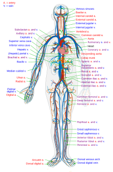
The circulatory system, also called the cardiovascular system or the vascular system, is an organ system that permits blood to circulate and transport nutrients, oxygen, carbon dioxide, hormones, and blood cells to and from the cells in the body to provide nourishment and help in fighting diseases, stabilize temperature and pH, and maintain homeostasis.

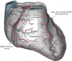
Coronary circulation is the circulation of blood in the blood vessels that supply the heart muscle (myocardium).
Coronary arteries supply oxygenated blood to the heart muscle, and cardiac veins drain away the blood once it has been deoxygenated.
Because the rest of the body, and most especially the brain, needs a steady supply of oxygenated blood that is free of all but the slightest interruptions, the heart works constantly and sometimes works quite hard. Therefore its circulation is of major importance not only to its own tissues but to the entire body and even the level of consciousness of the brain from moment to moment.
Interruptions of coronary circulation quickly cause heart attacks, in which the heart muscle is damaged by oxygen starvation. Such interruptions are usually caused by ischemic heart disease and sometimes by embolism from other causes like obstruction in blood flow through vessels.

The coronary arteries are the arteries of the coronary circulation, which transports blood into and out of the cardiac muscle. They are mainly composed of the left and right coronary arteries, both of which give off branches. Coronary arteries can also be categorized as epicardial and microvascular.

The atrioventricular node, or AV node is a part of the electrical conduction system of the heart that coordinates the top of the heart. It electrically connects the atria and ventricles. The AV node lies at the lower back section of the interatrial septum near the opening of the coronary sinus, which conducts the normal electrical impulse from the atria to the ventricles. The AV node is quite compact.

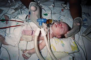
Cardiac catheterization is the insertion of a catheter into a chamber or vessel of the heart. This is done both for diagnostic and interventional purposes. A common example of cardiac catheterization is coronary catheterization that involves catheterization of the coronary arteries for coronary artery disease and myocardial infarctions. Catheterization is most often performed in special laboratories with fluoroscopy and highly maneuverable tables. These "cath labs" are often equipped with cabinets of catheters, stents, balloons, etc of various sizes to increase efficiency. Monitors show the fluoroscopy imaging, EKG, pressure waves, and more.

The interventricular septum, is the stout wall separating the ventricles, the lower chambers of the heart, from one another.

The coronary sinus is a collection of veins joined together to form a large vessel that collects blood from the heart muscle (myocardium). It delivers less-oxygenated blood to the right atrium, as do the superior and inferior vena cavae. It is present in all mammals, including humans.
The Jatene procedure, arterial switch operation or arterial switch, is an open heart surgical procedure used to correct dextro-transposition of the great arteries (d-TGA); its development was pioneered by Canadian cardiac surgeon William Mustard and it was named for Brazilian cardiac surgeon Adib Jatene, who was the first to use it successfully. It was the first method of d-TGA repair to be attempted, but the last to be put into regular use because of technological limitations at the time of its conception. Use of the arterial switch is historically preceded by two atrial switch methods: the Senning and Mustard procedures.

In the heart's conduction system, Bachmann's bundle is a branch of the anterior internodal tract that resides on the inner wall of the left atrium. It is a broad band of cardiac muscle that passes from the right atrium, between the superior vena cava and the ascending aorta. Bachmann's bundle is, during normal sinus rhythm, the preferential path for electrical activation of the left atrium. It is therefore considered to be part of the "atrial conduction system" of the heart.

The cardiac plexus is a plexus of nerves situated at the base of the heart that innervates the heart.

The atria of the heart are separated from the ventricles by the coronary sulcus. The structure contains the trunks of the nutrient vessels of the heart, and is deficient in front, where it is crossed by the root of the pulmonary trunk. On the posterior surface of the heart, the coronary sulcus contains the coronary sinus.
The left anterior descending artery is a branch of the left coronary artery. Occlusion of this artery is often called the widow-maker infarction due to a high death risk.

The sinoatrial nodal artery is an artery of the heart which supplies the sinoatrial node, the natural pacemaker center of the heart, and arises from the right coronary artery in around 60% of people. In about 40% of cases, the sinoatrial artery is a branch of the left circumflex coronary artery. In less than 1% of humans, the artery has an anomalous origin directly from the coronary sinus, descending aorta, or distal right coronary artery.
The atrioventricular nodal branch is a cardiac artery that is crucial because it feeds the atrioventricular node, necessary for the excitation and contraction of the ventricles. Most frequently it arises as a distal branch from the right coronary artery near the crux of the heart. In some people, the atrioventricular node instead receives blood from the circumflex branch of the left coronary artery, otherwise known as the left circumflex coronary artery or LCX. Very rarely, in approximately 2% of people, the vascular supply to the atrioventricular node arises from both the right coronary artery and the left circumflex branch.
Coronary steal is a phenomenon where an alteration of circulation patterns leads to a reduction in the blood directed to the coronary circulation. It is caused when there is narrowing of the coronary arteries and a coronary vasodilator is used – "stealing" blood away from those parts of the heart. This happens as a result of the narrowed coronary arteries being always maximally dilated to compensate for the decreased upstream blood supply. Thus, dilating the resistance vessels in the coronary circulation causes blood to be shunted away from the coronary vessels supplying the ischemic zones, creating more ischemia.

