Abdominal aorta | |
---|---|
Details | |
Source | Thoracic aorta |
Branches | Celiac artery, superior mesenteric artery, inferior mesenteric artery, common iliac, ovarian artery, median sacral artery |
Vein | Inferior vena cava |
Identifiers | |
Latin | aorta abdominali, pars abdominalis aortae |
MeSH | D001012 |
TA98 | A12.2.12.001 |
TA2 | 4205 |
FMA | 3789 |
Anatomical terminology |
In human anatomy, the abdominal aorta is the largest artery in the abdominal cavity. As part of the aorta, it is a direct continuation of the descending aorta (of the thorax). [1]
The abdominal aorta begins at the level of the diaphragm, crossing it via the aortic hiatus, technically behind the diaphragm, at the vertebral level of T12. [1] It travels down the posterior wall of the abdomen, anterior to the vertebral column. It thus follows the curvature of the lumbar vertebrae, that is, convex anteriorly. The peak of this convexity is at the level of the third lumbar vertebra (L3). It runs parallel to the inferior vena cava, which is located just to the right of the abdominal aorta, and becomes smaller in diameter as it gives off branches. This is thought to be due to the large size of its principal branches. At the 11th rib, the diameter is 122mm long and 55mm wide and this is because of the constant pressure. [2] The abdominal aorta is clinically divided into 2 segments:
The abdominal aorta supplies blood to much of the abdominal cavity. It begins at T12 and ends at L4 with its bifurcation into the common iliac arteries [1] and usually has the following branches:
Artery Branch | Vertebra | Type | Paired? | A/P | Description |
---|---|---|---|---|---|
inferior phrenic | T12 | Parietal | yes | post. | 
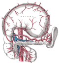
Originates above the celiac trunk, below the diaphragm. Passes upward and medially to the suprarenal gland, and crosses crus of diaphragm of corresponding side. Supplies diaphragm and gives superior suprarenal arteries. |
celiac | T12 | Visceral | no | ant. | Large anterior branch |
superior mesenteric | L1 | Visceral | no | ant. | Large anterior branch, arises just below celiac trunk |
middle suprarenal | L1 | Visceral | yes | post. | Crosses crus of diaphragm laterally on each side; supplies the suprarenal gland. |
renal | In between L1 and L2 | Visceral | yes | post. | Arises just below the superior mesenteric artery. Right renal artery passes deep to the inferior vena cava to right kidney; here it divides into branches. Left renal artery passes deep to the left renal vein. Divides in hilum of kidney. Both arteries give inferior suprarenal arteries and ureteral branches. |
gonadal | L2 | Visceral | yes | ant. | Ovarian artery in females; testicular artery in males |
lumbar | L1-L4 | Parietal | yes | post. | Four on each side that supply the abdominal wall and spinal cord. The fifth pair is the lumbar branches of the iliolumbar arteries. They pass deep to the crura on side of vertebral bodies and pass deep to the psoas major and quadratus lumborum to enter the space between the internal oblique and transversus abdominis muscles. Each artery gives off a small dorsal branch, which gives a spinal branch to the vertebral canal and then continues to supply the muscles of the back. |
inferior mesenteric | L3 | Visceral | no | ant. | Large anterior branch |
median sacral | L4 | Parietal | no | post. | Artery arising from the middle of the aorta at its lowest part. Represents the continuation of the primitive dorsal aorta; quite large in animals with tails but smaller in humans. |
common iliac | L4 | Terminal | yes | post. | Branches (bifurcations) to supply blood to the lower limbs and the pelvis, ending the abdominal aorta |
The bifurcation (union) of the inferior vena cava is at L5 and therefore below that of the bifurcation of the aorta.
The abdominal aorta lies slightly to the left of the midline of the body. It is covered, anteriorly, by the lesser omentum and stomach, behind which are the branches of the celiac artery and the celiac plexus; below these, by the lienal vein (splenic vein), are the pancreas, the left renal vein, the inferior part of the duodenum, the mesentery, and the aortic plexus.
Posteriorly, it is separated from the lumbar vertebrae and intervertebral fibrocartilages by the anterior longitudinal ligament and left lumbar veins.
On the right side it is in relation above with the azygos vein, cisterna chyli, thoracic duct, and the right crus of the diaphragm—the last separating it from the upper part of the inferior vena cava, and from the right celiac ganglion; the inferior vena cava is in contact with the aorta below.
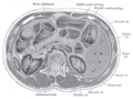
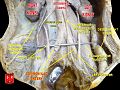
On the left side are the left crus of the diaphragm, the left celiac ganglion, the ascending part of the duodenum, and some coils of the small intestine.
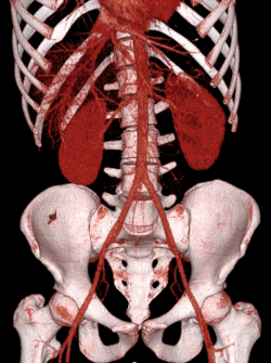
The abdominal aorta's venous counterpart, the inferior vena cava (IVC), travels parallel to it on its right side.
The collateral circulation would be carried on by the anastomoses between the internal thoracic artery and the inferior epigastric artery; by the free communication between the superior and inferior mesenterics, if the ligature were placed between these vessels; or by the anastomosis between the inferior mesenteric artery and the internal pudendal artery, when (as is more common) the point of ligature is below the origin of the inferior mesenteric artery; and possibly by the anastomoses of the lumbar arteries with the branches of the internal iliac artery.
The aorta is the main and largest artery in the human body, originating from the left ventricle of the heart, branching upwards immediately after, and extending down to the abdomen, where it splits at the aortic bifurcation into two smaller arteries. The aorta distributes oxygenated blood to all parts of the body through the systemic circulation.
Articles related to anatomy include:
The inferior vena cava is a large vein that carries the deoxygenated blood from the lower and middle body into the right atrium of the heart. It is formed by the joining of the right and the left common iliac veins, usually at the level of the fifth lumbar vertebra.
The celiac plexus, also known as the solar plexus because of its radiating nerve fibers, is a complex network of nerves located in the abdomen, near where the celiac trunk, superior mesenteric artery, and renal arteries branch from the abdominal aorta. It is behind the stomach and the omental bursa, and in front of the crura of the diaphragm, on the level of the first lumbar vertebra.
The renal arteries are paired arteries that supply the kidneys with blood. Each is directed across the crus of the diaphragm, so as to form nearly a right angle.
The periaortic lymph nodes are a group of lymph nodes that lie in front of the lumbar vertebrae near the aorta. These lymph nodes receive drainage from the gastrointestinal tract and the abdominal organs.
The renal veins in the renal circulation, are large-calibre veins that drain blood filtered by the kidneys into the inferior vena cava. There is one renal vein draining each kidney. Each renal vein is formed by the convergence of the interlobar veins of one kidney.
The celiacartery, also known as the celiac trunk or truncus coeliacus, is the first major branch of the abdominal aorta. It is about 1.25 cm in length. Branching from the aorta at thoracic vertebra 12 (T12) in humans, it is one of three anterior/ midline branches of the abdominal aorta.
In human anatomy, the superior mesenteric artery (SMA) is an artery which arises from the anterior surface of the abdominal aorta, just inferior to the origin of the celiac trunk, and supplies blood to the intestine from the lower part of the duodenum through two-thirds of the transverse colon, as well as the pancreas.
In human anatomy, the inferior mesenteric artery (IMA) is the third main branch of the abdominal aorta and arises at the level of L3, supplying the large intestine from the distal transverse colon to the upper part of the anal canal. The regions supplied by the IMA are the descending colon, the sigmoid colon, and part of the rectum.
The common iliac artery is a large artery of the abdomen paired on each side. It originates from the aortic bifurcation at the level of the 4th lumbar vertebra. It ends in front of the sacroiliac joint, one on either side, and each bifurcates into the external and internal iliac arteries.
The inferior phrenic artery is a bilaterally paired artery of the abdominal cavity which represents the main source of arterial supply to the diaphragm. Each artery usually arises either from the coeliac trunk or the abdominal aorta, however, their origin is highly variable and the different sites of origin are different for the left artery and right artery. The superior suprarenal artery is a branch of the inferior phrenic artery.
The middle suprarenal artery is a paired artery in the abdomen. It is a branch of the aorta. It supplies the adrenal gland.
The superior hypogastric plexus is a plexus of nerves situated on the vertebral bodies anterior to the bifurcation of the abdominal aorta. It bifurcates to form the left and the right hypogastric nerve. The SHP is the continuation of the abdominal aortic plexus.
The lumbar veins are four pairs of veins running along the inside of the posterior abdominal wall, and drain venous blood from parts of the abdominal wall. Each lumbar vein accompanies a single lumbar artery. The lower two pairs of lumbar veins all drain directly into the inferior vena cava, whereas the fate of the upper two pairs is more variable.
The following outline is provided as an overview of and topical guide to human anatomy:
The aortic bifurcation is the point at which the abdominal aorta bifurcates (forks) into the left and right common iliac arteries. The aortic bifurcation is usually seen at the level of L4, just above the junction of the left and right common iliac veins.