Common iliac vein

Last updated

Common iliac vein
Iliac veins.gif
Veins of the abdomen and lower limbinferior vena cava, common iliac vein, external iliac vein, internal iliac vein, femoral vein and their tributaries. The aorta and its bifurcation (unlabeled) appear in red.
Gray847.png
Abdominal portion of the sympathetic trunk, with the celiac and hypogastric plexuses. (Common iliac vein labeled at lower right.)
Details
Drains from Pelvis and lower limbs
Source External iliac veins and internal iliac veins
Drains to Inferior vena cava
Artery Common iliac arteries
Identifiers
Latin vena iliaca communis
MeSH D007084
TA98 A12.3.10.001
TA2 5021
FMA 14333
Anatomical terminology

In human anatomy, the common iliac veins are formed by the external iliac veins and internal iliac veins. The left and right common iliac veins come together in the abdomen at the level of the fifth lumbar vertebra, [1] forming the inferior vena cava. They drain blood from the pelvis and lower limbs.

Contents

Both common iliac veins are accompanied along their course by common iliac arteries.

Structure

The external iliac vein and internal iliac vein unite in front of the sacroiliac joint to form the common iliac veins. [2] Both common iliac veins ascend to form the inferior vena cava behind the right common iliac artery at the level of the fifth lumbar vertebra. [3]

The vena cava is to the right of the midline and therefore the left common iliac vein is longer than the right. [2] The left common iliac vein occasionally travels upwards to the left of the aorta to the level of the kidney, where it receives the left renal vein and crosses in front of the aorta to join the inferior vena cava. [4] The right common iliac vein is virtually vertical and lies behind and then lateral to its artery. Each common iliac vein receives iliolumbar veins, while the left also receives the median sacral vein which lies on the right of the corresponding artery. [2]

Clinical significance

Overlying arterial structures may cause compression of the upper part of the left common iliac vein. [3]

Compression of the left common iliac vein against the fifth lumbar vertebral body by the right common iliac artery as the artery crosses in front of it traditionally happens in May–Thurner syndrome. [5]

Continuous pulsation of the common iliac artery may trigger an inflammatory response within the common iliac vein. The resulting intraluminal elastin and collagen deposition can cause intimal fibrosis and the formation of venous spurs and webs. This can lead to narrowing of the vein and cause persistent unilateral leg swelling, contributing to venous thromboembolism. [5]

Related Research Articles

<span class="mw-page-title-main">Vein</span> Blood vessels that carry blood towards the heart

Veins are blood vessels in the circulatory system of humans and most other animals that carry blood towards the heart. Most veins carry deoxygenated blood from the tissues back to the heart; exceptions are those of the pulmonary and fetal circulations which carry oxygenated blood to the heart. In the systemic circulation, arteries carry oxygenated blood away from the heart, and veins return deoxygenated blood to the heart, in the deep veins.

<span class="mw-page-title-main">Brachiocephalic vein</span> Vein

The left and right brachiocephalic veins are major veins in the upper chest, formed by the union of the ipsilateral internal jugular vein and subclavian vein behind the sternoclavicular joint. The left brachiocephalic vein is more than twice the length of the right brachiocephalic vein.

<span class="mw-page-title-main">Inferior vena cava</span> One of two veinous trunks bringing deoxygenated blood back to the heart

The inferior vena cava is a large vein that carries the deoxygenated blood from the lower and middle body into the right atrium of the heart. It is formed by the joining of the right and the left common iliac veins, usually at the level of the fifth lumbar vertebra.

<span class="mw-page-title-main">Azygos vein</span> Human blood vessel by the spine

The azygos vein is a vein running up the right side of the thoracic vertebral column draining itself towards the superior vena cava. It connects the systems of superior vena cava and inferior vena cava and can provide an alternative path for blood to the right atrium when either of the venae cavae is blocked.

<span class="mw-page-title-main">Abdominal aorta</span> Largest artery in the abdomen

In human anatomy, the abdominal aorta is the largest artery in the abdominal cavity. As part of the aorta, it is a direct continuation of the descending aorta.

<span class="mw-page-title-main">Renal artery</span> Vessel supplying blood to kidney

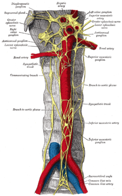
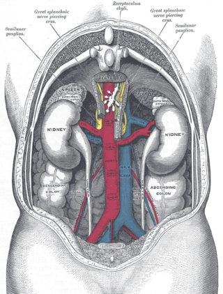
The renal arteries are paired arteries that supply the kidneys with blood. Each is directed across the crus of the diaphragm, so as to form nearly a right angle.

<span class="mw-page-title-main">Periaortic lymph nodes</span> Human lymph nodes

The periaortic lymph nodes are a group of lymph nodes that lie in front of the lumbar vertebrae near the aorta. These lymph nodes receive drainage from the gastrointestinal tract and the abdominal organs.

<span class="mw-page-title-main">Renal vein</span> Short thick veins which return blood from the kidneys to the vena cava

The renal veins in the renal circulation, are large-calibre veins that drain blood filtered by the kidneys into the inferior vena cava. There is one renal vein draining each kidney. Each renal vein is formed by the convergence of the interlobar veins of one kidney.

<span class="mw-page-title-main">Celiac artery</span> First major branch of the abdominal aorta

The celiacartery, also known as the celiac trunk or truncus coeliacus, is the first major branch of the abdominal aorta. It is about 1.25 cm in length. Branching from the aorta at thoracic vertebra 12 (T12) in humans, it is one of three anterior/ midline branches of the abdominal aorta.

<span class="mw-page-title-main">External iliac vein</span> Blood vessels connecting the thigh veins to the pelvis

The external iliac veins are large veins that connect the femoral veins to the common iliac veins. Their origin is at the inferior margin of the inguinal ligaments and they terminate when they join the internal iliac veins.

<span class="mw-page-title-main">External iliac artery</span> Arteries of the pelvis

The external iliac arteries are two major arteries which bifurcate off the common iliac arteries anterior to the sacroiliac joint of the pelvis.

<span class="mw-page-title-main">Common iliac artery</span> Artery in the abdomen

The common iliac artery is a large artery of the abdomen paired on each side. It originates from the aortic bifurcation at the level of the 4th lumbar vertebra. It ends in front of the sacroiliac joint, one on either side, and each bifurcates into the external and internal iliac arteries.

<span class="mw-page-title-main">May–Thurner syndrome</span> Medical condition

May–Thurner syndrome (MTS), also known as the iliac vein compression syndrome, is a condition in which compression of the common venous outflow tract of the left lower extremity may cause discomfort, swelling, pain or iliofemoral deep vein thrombosis.

<span class="mw-page-title-main">Gonadal vein</span>

In medicine, gonadal vein refers to the blood vessel that carries blood away from the gonad toward the heart. These are different arteries in women and men, but share the same embryological origin.

<span class="mw-page-title-main">Median sacral artery</span>

The median sacral artery is a small artery that arises posterior to the abdominal aorta and superior to its bifurcation.

<span class="mw-page-title-main">Superior hypogastric plexus</span>

The superior hypogastric plexus is a plexus of nerves situated on the vertebral bodies anterior to the bifurcation of the abdominal aorta. It bifurcates to form the left and the right hypogastric nerve. The SHP is the continuation of the abdominal aortic plexus.

<span class="mw-page-title-main">Internal iliac vein</span> Large blood vessel of the pelvis

The internal iliac vein begins near the upper part of the greater sciatic foramen, passes upward behind and slightly medial to the internal iliac artery and, at the brim of the pelvis, joins with the external iliac vein to form the common iliac vein.

<span class="mw-page-title-main">Testicular vein</span>

The testicular vein, the male gonadal vein, carries deoxygenated blood from its corresponding testis to the inferior vena cava or one of its tributaries. It is the male equivalent of the ovarian vein, and is the venous counterpart of the testicular artery.

<span class="mw-page-title-main">Lumbar veins</span> Veins that drain the posterior abdominal wall

The lumbar veins are four pairs of veins running along the inside of the posterior abdominal wall, and drain venous blood from parts of the abdominal wall. Each lumbar vein accompanies a single lumbar artery. The lower two pairs of lumbar veins all drain directly into the inferior vena cava, whereas the fate of the upper two pairs is more variable.

<span class="mw-page-title-main">Aortic bifurcation</span>

The aortic bifurcation is the point at which the abdominal aorta bifurcates (forks) into the left and right common iliac arteries. The aortic bifurcation is usually seen at the level of L4, just above the junction of the left and right common iliac veins.

References

  1. Henry Gray (1918), Anatomy of the Human Body, p. 677, archived from the original on December 22, 2009, retrieved June 15, 2008
  2. 1 2 3 Sinnatamby, Chummy S. (2011). "5". Last's Anatomy: Regional and Applied (12th ed.). Great Britain: Churchill Livingstone Elsevier. p. 277. ISBN   978-0-7020-4839-5 . Retrieved March 24, 2018.
  3. 1 2 Mozes, Geza; Gloviczki, Peter (2014). John J. Bergan & Nisha Bunke-Paquette (ed.). The Vein Book (2nd ed.). New York: Oxford University Press. p. 21. ISBN   978-0-19-539963-9.
  4. Delancey, John O.L. (2016). "73, True pelvis, pelvic floor and perineum". In Standring, Susan (ed.). Gray's Anatomy: The Anatomical Basis of Clinical Practice (41st ed.). Elsevier. pp. 1221–1236. ISBN   978-0-7020-6851-5.
  5. 1 2 DeRubertis, Brian; Patel, Rhasheet (2017). "35, May-Thurner Syndrome: Diagnosis and Management". In Cassius Iyad Ochoa Chaar (ed.). Current Management of Venous Diseases. Springer International Publishing. pp. 464–465. ISBN   978-3-319-65226-9 . Retrieved March 24, 2018.