This page is based on this
Wikipedia article Text is available under the
CC BY-SA 4.0 license; additional terms may apply.
Images, videos and audio are available under their respective licenses.

Coronary circulation is the circulation of blood in the blood vessels that supply the heart muscle (myocardium).
Coronary arteries supply oxygenated blood to the heart muscle, and cardiac veins drain away the blood once it has been deoxygenated.
Because the rest of the body, and most especially the brain, needs a steady supply of oxygenated blood that is free of all but the slightest interruptions, the heart works constantly and sometimes works quite hard. Therefore its circulation is of major importance not only to its own tissues but to the entire body and even the level of consciousness of the brain from moment to moment.
Interruptions of coronary circulation quickly cause heart attacks, in which the heart muscle is damaged by oxygen starvation. Such interruptions are usually caused by ischemic heart disease and sometimes by embolism from other causes like obstruction in blood flow through vessels.

The external iliac arteries are two major arteries which bifurcate off the common iliac arteries anterior to the sacroiliac joint of the pelvis. They proceed anterior and inferior along the medial border of the psoas major muscles. They exit the pelvic girdle posterior and inferior to the inguinal ligament about one third laterally from the insertion point of the inguinal ligament on the pubic tubercle at which point they are referred to as the femoral arteries. The external iliac artery is usually the artery used to attach the renal artery to the recipient of a kidney transplant.

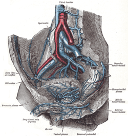
The internal iliac artery is the main artery of the pelvis.

The iliolumbar artery is the first branch of the posterior trunk of the internal iliac artery.

The lateral sacral arteries arise from the posterior division of the internal iliac artery; there are usually two, a superior and an inferior.

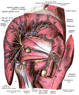
The inferior gluteal artery, the smaller of the two terminal branches of the anterior trunk of the internal iliac artery, is distributed chiefly to the buttock and back of the thigh.

The inferior gluteal veins, or venæ comitantes of the inferior gluteal artery, begin on the upper part of the back of the thigh, where they anastomose with the medial femoral circumflex and first perforating veins.

The lateral superior genicular artery is a branch of the popliteal artery that supplies a portion of the knee joint.

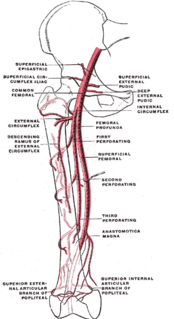
The lateral circumflex femoral artery is an artery in the upper thigh.

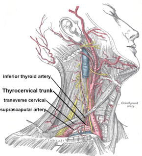
The suprascapular artery is a branch of the thyrocervical trunk on the neck.

The crest of the ilium is the superior border of the wing of ilium and the superiolateral margin of the greater pelvis.
The cruciate anastomosis is a circulatory anastomosis in the upper thigh of the inferior gluteal artery, the lateral and medial circumflex femoral arteries, and the first perforating artery of the profunda femoris artery. Also, the anastomotic branch of the posterior branch of the obturator artery. The cruciate anastomosis is clinically relevant because if there is a blockage between the femoral artery and external iliac artery, blood can reach the popliteal artery by means of the anastomosis. The route of blood is through the internal iliac, to the inferior gluteal artery, to a perforating branch of the deep femoral artery, to the lateral circumflex femoral artery, then to its descending branch into the superior lateral genicular artery and thus into the popliteal artery.
In anatomy, arterial tree is used to refer to all arteries and/or the branching pattern of the arteries. This article regards the human arterial tree. Starting from the aorta:

The deep circumflex iliac artery is an artery in the pelvis that travels along the iliac crest of the pelvic bone.

The perforating arteries, usually three in number, are so named because they perforate the tendon of the Adductor magnus to reach the back of the thigh.

The superficial iliac circumflex artery, the smallest of the cutaneous branches of the femoral artery, arises close to the superficial epigastric artery, and, piercing the fascia lata, runs lateralward, parallel with the inguinal ligament, as far as the crest of the ilium.

The following outline is provided as an overview of and topical guide to human anatomy:

The articular branches of descending genicular artery descend in the substance of the vastus medialis muscle, and in front of the tendon of the adductor magnus muscle, to the medial side of the knee, where they anastomose with the medial superior genicular and anterior recurrent tibial artery.


