Superior gluteal artery

Last updated
Superior gluteal artery
Gray1244.png
Left gluteal region, showing surface markings for arteries and sciatic nerve
Internal iliac branches.PNG
Internal iliac artery and some of its branches
(superior gluteal artery labeled at right)
Details
Source Internal iliac artery
Vein Superior gluteal veins
Supplies Gluteus maximus, gluteus medius, gluteus minimus and tensor fasciae latae
Identifiers
Latin arteria glutaea superior
TA98 A12.2.15.013
TA2 4310
FMA 18868
Anatomical terminology

The superior gluteal artery is the terminal branch of the posterior division of the internal iliac artery. It exits the pelvis through the greater sciatic foramen before splitting into a superficial branch and a deep branch.

Contents

Structure

Origin

The superior gluteal artery is the largest and final branch of the internal iliac artery. [1] [2] It branches from the posterior division of the internal iliac artery; [1] it represents the continuation of the posterior division.

Course, relations and branches

It is a short artery. It passes posterior-ward between the lumbosacral trunk and the first sacral nerve (S1). [3] Within the pelvis, it gives branches to the iliacus, piriformis, and obturator internus muscles. Just prior to exiting the pelvic cavity, it also gives off a nutrient artery which enters the ilium. [4]

It exits the pelvis through the greater sciatic foramen [2] [3] superior to the piriformis muscle, then promptly [3] divides into a superficial branch and a deep branch. [2] [3]

Superficial branch

The superficial branch passes over the piriformis muscle. [2] It enters the deep surface of the gluteus maximus muscle, and divides into numerous branches. [2] Some branches supply the muscle and anastomose with the inferior gluteal artery, while others perforate its tendinous origin, and supply the integument covering the posterior surface of the sacrum, anastomosing with the posterior branches of the lateral sacral arteries. [4] The superficial branch also supplies the skin over the origin of the gluteus maximus muscle. [3]

Deep branch

The deep branch passes deep to the gluteus medius. It almost immediately subdivides into the superior and inferior divisions. [3]

The deep branch supplies the gluteus medius, gluteus minimus, and tensor fasciae latae muscles, [3] as well as the hip joint. [4]

Superior division

The superior division continues the original course of the vessel, passing along the superior border of the gluteus minimus muscle to the anterior superior spine of the ilium (ASIS), anastomosing with the deep iliac circumflex artery and the ascending branch of the lateral femoral circumflex artery. [3]

Inferior division

The inferior division crosses the gluteus minimus obliquely to the greater trochanter, distributing branches to the gluteal muscles, and anastomoses with the lateral femoral circumflex artery. [3]

Some branches pierce the gluteus minimus and supply the hip joint. [4]

Distribution

Within the pelvis, it supplies the iliacus, piriformis, and obturator internus muscles, and the ilium. [4]

In the gluteal region, the superior gluteal artery supplies the gluteus maximus and overlying skin, gluteus medius, gluteus minimus, and tensor fascia lata. [5] [ better source needed ]

Anastomoses

The superior gluteal artery forms anastomoses with the inferior gluteal artery, and the medial circumflex femoral artery. [3]

The it participates in the formation of the trochanteric anastomoses, forming a connection between internal iliac and femoral artery. [5] [ better source needed ] It contributes to anastomoses at the anterior superior iliac spine and the hip joint. [5] [ better source needed ]

Additional images

See also

Related Research Articles

<span class="mw-page-title-main">Gluteus maximus</span> Largest and most superficial of the three gluteal muscles

The gluteus maximus is the main extensor muscle of the hip in humans. It is the largest and outermost of the three gluteal muscles and makes up a large part of the shape and appearance of each side of the hips. It is the single largest muscle in the human body. Its thick fleshy mass, in a quadrilateral shape, forms the prominence of the buttocks. The other gluteal muscles are the medius and minimus, and sometimes informally these are collectively referred to as the glutes.

<span class="mw-page-title-main">Gluteus medius</span> One of the three gluteal muscles

The gluteus medius, one of the three gluteal muscles, is a broad, thick, radiating muscle. It is situated on the outer surface of the pelvis.

<span class="mw-page-title-main">Gluteus minimus</span> Smallest of the three gluteal muscles

The gluteus minimus, or glutæus minimus, the smallest of the three gluteal muscles, is situated immediately beneath the gluteus medius.

<span class="mw-page-title-main">Greater trochanter</span> Quadrilateral part of the thigh bone

The greater trochanter of the femur is a large, irregular, quadrilateral eminence and a part of the skeletal system.

<span class="mw-page-title-main">Piriformis muscle</span> One of six small hip muscles in the lateral rotator group

The piriformis muscle is a flat, pyramidally-shaped muscle in the gluteal region of the lower limbs. It is one of the six muscles in the lateral rotator group.

<span class="mw-page-title-main">Hip</span> Anatomical region between the torso and the legs, holding the buttocks and genital region

In vertebrate anatomy, the hip, or coxa(pl.: coxae) in medical terminology, refers to either an anatomical region or a joint on the outer (lateral) side of the pelvis.

<span class="mw-page-title-main">Gluteal sulcus</span> Fold separating the thigh from the buttock

The gluteal sulcus is an area of the body of humans and anthropoid apes, described by a horizontal crease formed by the inferior aspect of the buttocks and the posterior upper thigh. The gluteal sulcus is formed by the posterior horizontal skin crease of the hip joint and overlying fat and is not formed by the lower border of the gluteus maximus muscle, which crosses the fold obliquely. It is one of the major defining features of the buttocks. Children with developmental dysplasia of the hips are born with uneven gluteal folds and can be diagnosed with a physical examination and sonogram.

<span class="mw-page-title-main">Internal iliac artery</span> Main artery of the pelvis

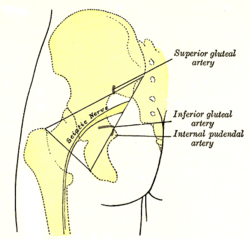
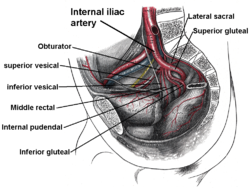
The internal iliac artery is the main artery of the pelvis.

<span class="mw-page-title-main">Gluteal muscles</span> Group of three muscles which make up the buttocks

The gluteal muscles, often called glutes, are a group of three muscles which make up the gluteal region commonly known as the buttocks: the gluteus maximus, gluteus medius and gluteus minimus. The three muscles originate from the ilium and sacrum and insert on the femur. The functions of the muscles include extension, abduction, external rotation, and internal rotation of the hip joint.

<span class="mw-page-title-main">Muscles of the hip</span> Causes movement in the hip

In human anatomy, the muscles of the hip joint are those muscles that cause movement in the hip. Most modern anatomists define 17 of these muscles, although some additional muscles may sometimes be considered. These are often divided into four groups according to their orientation around the hip joint: the gluteal group; the lateral rotator group; the adductor group; and the iliopsoas group.

<span class="mw-page-title-main">Posterior cutaneous nerve of thigh</span> Sensory nerve that supplies the back of the thigh, leg, buttock, and perineum

The posterior cutaneous nerve of the thigh is a sensory nerve of the thigh. It is a branch of the sacral plexus. It supplies the skin of the posterior surface of the thigh, leg, buttock, and also the perineum.

<span class="mw-page-title-main">Inferior gluteal nerve</span>

The inferior gluteal nerve is the main motor neuron that innervates the gluteus maximus muscle. It is responsible for the movement of the gluteus maximus in activities requiring the hip to extend the thigh, such as climbing stairs. Injury to this nerve is rare but often occurs as a complication of posterior approach to the hip during hip replacement. When damaged, one would develop gluteus maximus lurch, which is a gait abnormality which causes the individual to 'lurch' backwards to compensate lack in hip extension.

<span class="mw-page-title-main">Superior gluteal nerve</span> Nerve on the pelvis and leg

The superior gluteal nerve is a mixed nerve of the sacral plexus that originates in the pelvis. It provides motor innervation to the gluteus medius, gluteus minimus, tensor fasciae latae, and piriformis muscles; it also has a cutaneous branch.

<span class="mw-page-title-main">Inferior gluteal artery</span>

The inferior gluteal artery is a terminal branch of the anterior trunk of the internal iliac artery. It exits the pelvis through the greater sciatic foramen. It is distributed chiefly to the buttock and the back of the thigh.

<span class="mw-page-title-main">Lateral circumflex femoral artery</span>

The lateral circumflex femoral artery is an artery in the upper thigh. It is usually a branch of the profunda femoris artery, and produces three branches. It is mostly distributed to the muscles of the lateral thigh, supplying arterial blood to muscles of the knee extensor group.

<span class="mw-page-title-main">Deep circumflex iliac artery</span> Artery in the pelvis

The deep circumflex iliac artery is an artery in the pelvis that travels along the iliac crest of the pelvic bone.

<span class="mw-page-title-main">Superficial circumflex iliac artery</span>

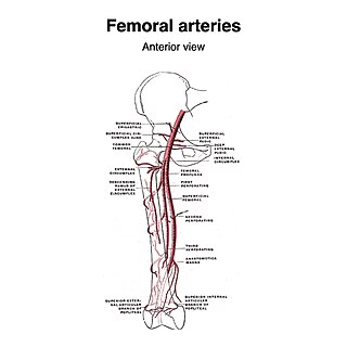
The superficial iliac circumflex artery, the smallest of the cutaneous branches of the femoral artery, arises close to the superficial epigastric artery, and, piercing the fascia lata, runs lateralward, parallel with the inguinal ligament, as far as the crest of the ilium.

<span class="mw-page-title-main">Iliac branch of iliolumbar artery</span>

The iliac branch of the iliolumbar artery descends to supply the iliacus muscle; some offsets, running between the muscle and the bone, anastomose with the iliac branches of the obturator artery; one of these enters an oblique canal to supply the bone, while others run along the crest of the ilium, distributing branches to the gluteal and abdominal muscles, and anastomosing in their course with the superior gluteal artery, iliac circumflex artery, and the lateral circumflex femoral artery. This anastamosis occurs around the anterior superior iliac spine.

<span class="mw-page-title-main">Hip bone</span> Bone of the pelvis

The hip bone is a large flat bone, constricted in the center and expanded above and below. In some vertebrates it is composed of three parts: the ilium, ischium, and the pubis.

<span class="mw-page-title-main">Gluteal lines</span> Lines of the human ilium

The gluteal lines are three curved lines outlined from three bony ridges on the exterior surface of the ilium in the gluteal region. They are the anterior gluteal line; the inferior gluteal line, and the posterior gluteal line.

References

PD-icon.svgThis article incorporates text in the public domain from page 622 of the 20th edition of Gray's Anatomy (1918)

  1. 1 2 Elias Soueid, Nassif; Mountcastle, Timothy S.; Levine, Joshua L.; Allen, Robert J.; Chiu, Ernest S.; Vasile, Julie (2009-01-01), Wei, Fu-Chan; Mardini, Samir (eds.), "CHAPTER 36 - Superior and inferior gluteal artery perforator flaps", Flaps and Reconstructive Surgery, Edinburgh: W.B. Saunders, pp. 525–539, ISBN   978-0-7216-0519-7 , retrieved 2021-01-13
  2. 1 2 3 4 5 Hamdi, Moustapha; Gagnon, Alain R. (2009-01-01), Wei, Fu-Chan; Mardini, Samir (eds.), "CHAPTER 28 - Gluteus flap", Flaps and Reconstructive Surgery, Edinburgh: W.B. Saunders, pp. 377–395, ISBN   978-0-7216-0519-7 , retrieved 2021-01-14
  3. 1 2 3 4 5 6 7 8 9 10 Moore, Keith L.; Dalley, Arthur F.; Agur, Anne M. R. (2018). Clinically Oriented Anatomy (8th ed.). Wolters Kluwer. p. 734. ISBN   978-1-4963-4721-3.
  4. 1 2 3 4 5 "Gray, Henry. 1918. Anatomy of the Human Body. Page 622". www.bartleby.com. Retrieved 2018-05-03.
  5. 1 2 3 Florescu, Grace. "Superior gluteal artery | Radiology Reference Article | Radiopaedia.org". Radiopaedia. Retrieved 2022-06-01.