Internal iliac artery

Last updated
Internal iliac
Iliac artery bifurcation.PNG
Front of abdomen, showing surface markings for arteries and inguinal canal.
Internaliliac.png
Details
Source Common iliac artery
Branches iliolumbar artery, lateral sacral artery, superior gluteal artery, inferior gluteal artery, middle rectal artery, uterine artery, obturator artery, inferior vesical artery, superior vesical artery, obliterated umbilical artery, internal pudendal artery, Vaginal artery
Vein Internal iliac vein
Identifiers
Latin arteria iliaca interna
MeSH D007083
TA98 A12.2.15.001
TA2 4302
FMA 18808
Anatomical terminology

The internal iliac artery (formerly known as the hypogastric artery) is the main artery of the pelvis.

Contents

Structure

The internal iliac artery supplies the walls and viscera of the pelvis, the buttock, the reproductive organs, and the medial compartment of the thigh. The vesicular branches of the internal iliac arteries supply the bladder. [1]

It is a short, thick vessel, smaller than the external iliac artery, and about 3 to 4 cm in length.

Course

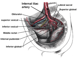
The internal iliac artery arises at the bifurcation of the common iliac artery, opposite the lumbosacral articulation, and, passing downward to the upper margin of the greater sciatic foramen, divides into two large trunks, an anterior and a posterior.

It is posterior to the ureter, [2] anterior to the internal iliac vein, [2] anterior to the lumbosacral trunk, and anterior to the piriformis muscle. Near its origin, it is medial to the external iliac vein, which lies between it and the psoas major muscle. It is above the obturator nerve.

Branches

The arrangement of branches of the internal iliac artery is extremely variable. [3] Typically, the artery divides into an anterior division and a posterior division, with the posterior division giving rise to the superior gluteal, iliolumbar, and lateral sacral arteries. The rest usually arise from the anterior division. Because it is variable, an artery may not be a direct branch, but instead might arise off a direct branch.

In recent years the devolopement of techniques like Prostate artery embolisation and angiografy led to an increased understanding of the prostate vascularisation. Regarding the arterial supply M. de Assis et al has suggested an anatomic classification for the origin of the inferior vesical artery [4]

The following are the branches of internal iliac artery:

DivisionTypeBranchSub-branchesTo/through
AnteriorVesical Superior vesical artery [2] (usually from the umbilical artery [5] )Sometimes middle vesicularupper urinary bladder and ureters [2]
Umbilical artery [2] Artery to vas deferens (male) and Superior vesical artery (usually, but sometimes it branches directly from anterior trunk) medial umbilical ligament [2]
Inferior vesical artery (male) [2] -lower urinary bladder and ureters [2]
Visceral Middle rectal artery [2] -lower rectum [2]
Vaginal artery (female); [2] the artery usually takes the place of the inferior vesical artery present in the male- vagina [2]
Uterine artery (female) [2] vaginal branch uterus and cervix [2]
Parietal Obturator artery [2] (occasionally from the inferior epigastric artery)- obturator canal [2]
Internal pudendal artery [2] many branches - see article for details greater sciatic foramen and lesser sciatic foramen to perineum [2]
Inferior gluteal artery [2] - greater sciatic foramen (inferior to piriformis muscle) and gluteus maximus muscle [2]
PosteriorParietal Iliolumbar artery [2] lumbar and iliac branches psoas major muscle, quadratus lumborum muscle, iliacus muscle [ citation needed ]
Lateral sacral artery [2] superior and inferior branches anterior sacral foramina [ citation needed ]
Superior gluteal artery [2] - gluteus maximus muscle [2]

Anastamoses

In individuals assigned female at birth, the ovarian artery (a branch of the abdominal aorta) and uterine arteries form an anastomoses. [6]

Right (distal from spectator) internal iliac artery and branches, except for iliolumbar artery, umbilical artery, uterine artery/deferential artery and vaginal artery/inferior vesical artery. Internal iliac branches.PNG
Right (distal from spectator) internal iliac artery and branches, except for iliolumbar artery, umbilical artery, uterine artery/deferential artery and vaginal artery/inferior vesical artery.

Fetal structure

In the fetus, the internal iliac artery is twice as large as the external iliac, and is the direct continuation of the common iliac. It ascends along the side of the bladder, and runs upward on the back of the anterior wall of the abdomen to the umbilicus, converging toward its fellow of the opposite side.

Having passed through the umbilical opening, the two arteries, now termed umbilical, enter the umbilical cord, where they coil around the umbilical vein, and ultimately ramify in the placenta.

At birth, when the placental circulation ceases, the pelvic portion only of the umbilical artery remains patent gives rise to the superior vesical artery (or arteries) of the adult; the remainder of the vessel is converted into a solid fibrous cord, the medial umbilical ligament (otherwise known as the obliterated hypogastric artery) which extends from the pelvis to the umbilicus.

Variation

In two-thirds of a large number of cases, the length of the internal iliac varied between 2.25 and 3.4 cm.; in the remaining third it was more frequently longer than shorter, the maximum length being about 7 cm. the minimum about 1 cm.[ citation needed ]

The lengths of the common iliac and internal iliac arteries bear an inverse proportion to each other, the internal iliac artery being long when the common iliac is short, and vice versa.

The place of division of the internal iliac artery varies between the upper margin of the sacrum and the upper border of the greater sciatic foramen.

The right and left hypogastric arteries in a series of cases often differed in length, but neither seemed constantly to exceed the other.[ citation needed ]

Common branching variations

Collateral circulation

The circulation after ligature of the internal iliac artery is carried on by the anastomoses of: [8]

Additional images

See also

Related Research Articles

<span class="mw-page-title-main">External iliac artery</span> Arteries of the pelvis

The external iliac arteries are two major arteries which bifurcate off the common iliac arteries anterior to the sacroiliac joint of the pelvis.

<span class="mw-page-title-main">Common iliac artery</span> Artery in the abdomen

The common iliac artery is a large artery of the abdomen paired on each side. It originates from the aortic bifurcation at the level of the 4th lumbar vertebra. It ends in front of the sacroiliac joint, one on either side, and each bifurcates into the external and internal iliac arteries.

<span class="mw-page-title-main">Sacral plexus</span> Nerve plexus

In human anatomy, the sacral plexus is a nerve plexus which provides motor and sensory nerves for the posterior thigh, most of the lower leg and foot, and part of the pelvis. It is part of the lumbosacral plexus and emerges from the lumbar vertebrae and sacral vertebrae (L4-S4). A sacral plexopathy is a disorder affecting the nerves of the sacral plexus, usually caused by trauma, nerve compression, vascular disease, or infection. Symptoms may include pain, loss of motor control, and sensory deficits.

<span class="mw-page-title-main">Femoral nerve</span> Long nerve down the thigh and inner leg

The femoral nerve is a nerve in the thigh that supplies skin on the upper thigh and inner leg, and the muscles that extend the knee. It is the largest branch of the lumbar plexus.

<span class="mw-page-title-main">Lumbar plexus</span> Web of nerves in the lower spine

The lumbar plexus is a web of nerves in the lumbar region of the body which forms part of the larger lumbosacral plexus. It is formed by the divisions of the first four lumbar nerves (L1-L4) and from contributions of the subcostal nerve (T12), which is the last thoracic nerve. Additionally, the ventral rami of the fourth lumbar nerve pass communicating branches, the lumbosacral trunk, to the sacral plexus. The nerves of the lumbar plexus pass in front of the hip joint and mainly support the anterior part of the thigh.

<span class="mw-page-title-main">Obturator nerve</span>

The obturator nerve in human anatomy arises from the ventral divisions of the second, third, and fourth lumbar nerves in the lumbar plexus; the branch from the third is the largest, while that from the second is often very small.

<span class="mw-page-title-main">Iliolumbar artery</span>

The iliolumbar artery is the first branch of the posterior trunk of the internal iliac artery.

<span class="mw-page-title-main">Superior gluteal artery</span>

The superior gluteal artery is the terminal branch of the posterior division of the internal iliac artery. It exits the pelvis through the greater sciatic foramen before splitting into a superficial branch and a deep branch.

<span class="mw-page-title-main">Inferior gluteal artery</span>

The inferior gluteal artery is a terminal branch of the anterior trunk of the internal iliac artery. It exits the pelvis through the greater sciatic foramen. It is distributed chiefly to the buttock and the back of the thigh.

<span class="mw-page-title-main">Obturator artery</span>

The obturator artery is a branch of the internal iliac artery that passes antero-inferiorly on the lateral wall of the pelvis, to the upper part of the obturator foramen, and, escaping from the pelvic cavity through the obturator canal, it divides into an anterior branch and a posterior branch.

<span class="mw-page-title-main">Median sacral artery</span>

The median sacral artery is a small artery that arises posterior to the abdominal aorta and superior to its bifurcation.

<span class="mw-page-title-main">Superior hypogastric plexus</span>

The superior hypogastric plexus is a plexus of nerves situated on the vertebral bodies anterior to the bifurcation of the abdominal aorta. It bifurcates to form the left and the right hypogastric nerve. The SHP is the continuation of the abdominal aortic plexus.

<span class="mw-page-title-main">Internal iliac vein</span> Large blood vessel of the pelvis

The internal iliac vein begins near the upper part of the greater sciatic foramen, passes upward behind and slightly medial to the internal iliac artery and, at the brim of the pelvis, joins with the external iliac vein to form the common iliac vein.

In anatomy, arterial tree is used to refer to all arteries and/or the branching pattern of the arteries. This article regards the human arterial tree. Starting from the aorta:

<span class="mw-page-title-main">Deep circumflex iliac artery</span> Artery in the pelvis

The deep circumflex iliac artery is an artery in the pelvis that travels along the iliac crest of the pelvic bone.

<span class="mw-page-title-main">Hypogastric nerve</span> Human nerves

The hypogastric nerves are the continuation of the superior hypogastric plexus that descend into the pelvis anterior the sacrum and become the inferior hypogastric plexuses on either side of pelvic organs. The hypogastric nerves serve as a pathway for autonomic fibers to communicate between the lower abdomen and pelvis.

<span class="mw-page-title-main">Outline of human anatomy</span> Overview of and topical guide to human anatomy

The following outline is provided as an overview of and topical guide to human anatomy:

<span class="mw-page-title-main">Hip bone</span> Bone of the pelvis

The hip bone is a large flat bone, constricted in the center and expanded above and below. In some vertebrates it is composed of three parts: the ilium, ischium, and the pubis.

References

PD-icon.svgThis article incorporates text in the public domain from page 614 of the 20th edition of Gray's Anatomy (1918)

  1. Kaplan Qbook - USMLE Step 1 - 5th edition - page 52
  2. 1 2 3 4 5 6 7 8 9 10 11 12 13 14 15 16 17 18 19 20 21 22 23 24 Paterson-Brown, Sara (2010-01-01), Bennett, Phillip; Williamson, Catherine (eds.), "Chapter Five - Applied anatomy", Basic Science in Obstetrics and Gynaecology (Fourth Edition), Churchill Livingstone, pp. 57–95, ISBN   978-0-443-10281-3 , retrieved 2021-01-13
  3. Tunstall R (2016-05-06). "Internal iliac arteries". In Tubbs RS, Shoja MM, Loukas M (eds.). Bergman's Comprehensive Encyclopedia of Human Anatomic Variation. Wiley. p. 1456. doi:10.1002/9781118430309. ISBN   978-1-118-43035-4.
  4. de Assis, André Moreira; Moreira, Airton Mota; de Paula Rodrigues, Vanessa Cristina; Harward, Sardis Honoria; Antunes, Alberto Azoubel; Srougi, Miguel; Carnevale, Francisco Cesar (August 2015). "Pelvic Arterial Anatomy Relevant to Prostatic Artery Embolisation and Proposal for Angiographic Classification" . CardioVascular and Interventional Radiology. 38 (4): 855–861. doi:10.1007/s00270-015-1114-3. ISSN   0174-1551. PMID   25962991. S2CID   9680972.
  5. Drake, Richard L.; Wayne Vogl; Adam W. M. Mitchell (2020). Gray's anatomy for students (4th ed.). Philadelphia. p. 490. ISBN   978-0-323-39304-1. OCLC   1085137919.{{cite book}}: CS1 maint: location missing publisher (link)
  6. Uterine artery and ovarian artery anatomy , retrieved 2022-11-30
  7. Essential Clinical Anatomy. K.L. Moore & A.M. Agur. Lippincott, 2 ed. 2002. Page 224
  8. Arisudhan Anantharachagan, Sarris, I. and Ugwumadu, A. (2011). Revision Notes for the MRCOG Part 1. Oxford Oxford University Press -07-01. pages 90-91