
The papillary muscles are muscles located in the ventricles of the heart. They attach to the cusps of the atrioventricular valves via the chordae tendineae and contract to prevent inversion or prolapse of these valves on systole.

A pulmonary artery is an artery in the pulmonary circulation that carries deoxygenated blood from the right side of the heart to the lungs. The largest pulmonary artery is the main pulmonary artery or pulmonary trunk from the heart, and the smallest ones are the arterioles, which lead to the capillaries that surround the pulmonary alveoli.

In human anatomy, the superior mesenteric artery (SMA) is an artery which arises from the anterior surface of the abdominal aorta, just inferior to the origin of the celiac trunk, and supplies blood to the intestine from the lower part of the duodenum through two-thirds of the transverse colon, as well as the pancreas.

In human anatomy, the inferior mesenteric artery (IMA) is the third main branch of the abdominal aorta and arises at the level of L3, supplying the large intestine from the distal transverse colon to the upper part of the anal canal. The regions supplied by the IMA are the descending colon, the sigmoid colon, and part of the rectum.

In human anatomy, the marginal artery of the colon, also known as the marginal artery of Drummond, the artery of Drummond, and simply as the marginal artery, is an artery that connects the inferior mesenteric artery with the superior mesenteric artery. It is sometimes absent, as an anatomical variant.

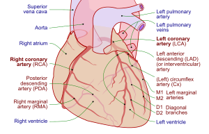
The left coronary artery is a coronary artery that arises from the aorta above the left cusp of the aortic valve, and supplies blood to the left side of the heart muscle. The left coronary artery typically runs for 10–25 mm, then bifurcates into the left anterior descending artery, and the left circumflex artery.

In the blood supply of the heart, the right coronary artery (RCA) is an artery originating above the right cusp of the aortic valve, at the right aortic sinus in the heart. It travels down the right coronary sulcus, towards the crux of the heart. It gives off many branches, including the sinoatrial nodal artery, right marginal artery, posterior interventricular artery, conus artery, and atrioventricular nodal branch. It contributes the right side of the heart, and parts of the interventricular septum.

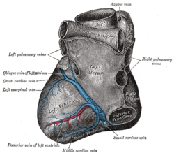
The coronary sinus is the largest vein of the heart. It drains over half of the deoxygenated blood from the heart muscle into the right atrium. It begins on the backside of the heart, in between the left atrium, and left ventricle; it begins at the junction of the great cardiac vein, and oblique vein of the left atrium. It receives multiple tributaries. It passes across the backside of the heart along a groove between left atrium and left ventricle, then drains into the right atrium at the orifice of the coronary sinus.

The great cardiac vein is a vein of the heart. It begins at the apex of the heart and ascends along the anterior interventricular sulcus before joining the oblique vein of the left atrium to form the coronary sinus upon the posterior surface of the heart.

The right gastroepiploic artery is one of the two terminal branches of the gastroduodenal artery. It runs from right to left along the greater curvature of the stomach, between the layers of the greater omentum, anastomosing with the left gastroepiploic artery, a branch of the splenic artery.
The left gastroepiploic artery, the largest branch of the splenic artery, runs from left to right about a finger's breadth or more from the greater curvature of the stomach, between the layers of the greater omentum, and anastomoses with the right gastroepiploic.

The middle colic artery is an artery of the abdomen; a branch of the superior mesenteric artery distributed to parts of the ascending and transverse colon. It usually divides into two terminal branches - a left one and a right one - which go on to form anastomoses with the left colic artery, and right colic artery (respectively), thus participating in the formation of the marginal artery of the colon.
The left colic artery is a branch of the inferior mesenteric artery distributed to the descending colon, and left part of the transverse colon. It ends by dividing into an ascending branch and a descending branch; the terminal branches of the two branches go on to form anastomoses with the middle colic artery, and a sigmoid artery (respectively).

The right gastric artery usually arises from the proper hepatic artery. It descends to the pyloric end of the stomach before passing from right to left along its lesser curvature, supplying it with branches, and finally anastomosing with the left gastric artery.

In the coronary circulation, the posterior descending artery (PDA), also called the posterior interventricular artery, is an artery running in the posterior interventricular sulcus to the apex of the heart where it meets with the left anterior descending artery also known as the anterior interventricular artery. The PDA supplies the posterior third of the interventricular septum. The remaining anterior two-thirds is supplied by the left anterior descending artery, which is a branch of left coronary artery.
The circumflex branch of left coronary artery is a branch of the left coronary artery. It winds around the left side of the heart along the atrioventricular groove. It supplies the posterolateral portion of the left ventricle.

The anterior interventricular sulcus is one of two grooves separating the ventricles of the heart. They can also be known as paraconal interventricular groove or subsinosal interventricular groove respectively. It is situated on the sternocostal surface of the heart, close to the left margin of the heart. It extends between the coronary sulcus, and the apex of the heart; upon reaching the diaphragmatic surface of the heart, it ends at the notch of cardiac apex. It contains the anterior interventricular branch of the left coronary artery, and great cardiac vein.

The right marginal branch of right coronary artery is the largest marginal branch of the right coronary artery. It follows the acute margin of the heart. It supplies blood to both surfaces of the right ventricle.
The left anterior descending artery is a branch of the left coronary artery. It supplies the anterior portion of the left ventricle. It provides about half of the arterial supply to the left ventricle and is thus considered the most important vessel supplying the left ventricle. Blockage of this artery is often called the widow-maker infarction due to a high risk of death.
The left border of heart is formed from the rounded lateral wall of the left ventricle. It is called the 'obtuse' margin because of the obtuse angle created between the anterior part of the heart and the left side, which is formed from the rounded lateral wall of the left ventricle. Within this margin can be found the obtuse marginal artery, which is the a branch of the left circumflex artery.
