
Coronary circulation is the circulation of blood in the arteries and veins that supply the heart muscle (myocardium). Coronary arteries supply oxygenated blood to the heart muscle. Cardiac veins then drain away the blood after it has been deoxygenated. Because the rest of the body, and most especially the brain, needs a steady supply of oxygenated blood that is free of all but the slightest interruptions, the heart is required to function continuously. Therefore its circulation is of major importance not only to its own tissues but to the entire body and even the level of consciousness of the brain from moment to moment. Interruptions of coronary circulation quickly cause heart attacks, in which the heart muscle is damaged by oxygen starvation. Such interruptions are usually caused by coronary ischemia linked to coronary artery disease, and sometimes to embolism from other causes like obstruction in blood flow through vessels.

The papillary muscles are muscles located in the ventricles of the heart. They attach to the cusps of the atrioventricular valves via the chordae tendineae and contract to prevent inversion or prolapse of these valves on systole.

A pulmonary artery is an artery in the pulmonary circulation that carries deoxygenated blood from the right side of the heart to the lungs. The largest pulmonary artery is the main pulmonary artery or pulmonary trunk from the heart, and the smallest ones are the arterioles, which lead to the capillaries that surround the pulmonary alveoli.

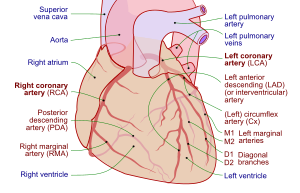
The coronary arteries are the arterial blood vessels of coronary circulation, which transport oxygenated blood to the heart muscle. The heart requires a continuous supply of oxygen to function and survive, much like any other tissue or organ of the body.
In the blood supply of the heart, the right coronary artery (RCA) is an artery originating above the right cusp of the aortic valve, at the right aortic sinus in the heart. It travels down the right coronary sulcus, towards the crux of the heart. It gives off many branches, including the sinoatrial nodal artery, right marginal artery, posterior interventricular artery, conus artery, and atrioventricular nodal branch. It contributes the right side of the heart, and parts of the interventricular septum.

The interventricular septum is the stout wall separating the ventricles, the lower chambers of the heart, from one another.

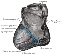
The coronary sinus is the largest vein of the heart. It drains over half of the deoxygenated blood from the heart muscle into the right atrium. It begins on the backside of the heart, in between the left atrium, and left ventricle; it begins at the junction of the great cardiac vein, and oblique vein of the left atrium. It receives multiple tributaries. It passes across the backside of the heart along a groove between left atrium and left ventricle, then drains into the right atrium at the orifice of the coronary sinus.

The great cardiac vein is a vein of the heart. It begins at the apex of the heart and ascends along the anterior interventricular sulcus before joining the oblique vein of the left atrium to form the coronary sinus upon the posterior surface of the heart.

The transverse cervical artery is an artery in the neck and a branch of the thyrocervical trunk, running at a higher level than the suprascapular artery.

The coronary sulcus is a groove on the surface of the heart at the base of right auricle that separates the atria from the ventricles. The structure contains the trunks of the nutrient vessels of the heart, and is deficient in front, where it is crossed by the root of the pulmonary trunk. On the posterior surface of the heart, the coronary sulcus contains the coronary sinus. The right coronary artery, circumflex branch of left coronary artery, and small cardiac vein all travel along parts of the coronary sulcus.

The circumflex branch of left coronary artery is a branch of the left coronary artery. It winds around the left side of the heart along the atrioventricular groove. It supplies the posterolateral portion of the left ventricle.
The right marginal branch of right coronary artery is the largest marginal branch of the right coronary artery. It follows the acute margin of the heart. It supplies blood to both surfaces of the right ventricle.
Coronary artery anomalies are variations of the coronary circulation, affecting <1% of the general population. Symptoms include chest pain, shortness of breath and syncope, although cardiac arrest may be the first clinical presentation. Several varieties are identified, with a different potential to cause sudden cardiac death.
A circulatory anastomosis is a connection between two blood vessels, such as between arteries, between veins or between an artery and a vein. Anastomoses between arteries and between veins result in a multitude of arteries and veins, respectively, serving the same volume of tissue. Such anastomoses occur normally in the body in the circulatory system, serving as back-up routes in a collateral circulation that allow blood to flow if one link is blocked or otherwise compromised, but may also occur pathologically.
The left anterior descending artery is a branch of the left coronary artery. It supplies the anterior portion of the left ventricle. It provides about half of the arterial supply to the left ventricle and is thus considered the most important vessel supplying the left ventricle. Blockage of this artery is often called the widow-maker infarction due to a high risk of death.

The sinoatrial nodal artery, sinuatrial nodal artery or sinoatrial artery is an artery of the heart which supplies the sinoatrial node, the natural pacemaker center of the heart. It is usually a branch of the right coronary artery. It passes between the right atrium, and the opening of the superior vena cava.
The atrioventricular nodal branch is a coronary artery that supplies arterial blood to the atrioventricular node, which is responsible for initiating muscular contraction of the ventricles. The AV nodal branch is most often a branch of the right coronary artery.
The crux cordis or crux of the heart is the area on the lower back side of the heart where the coronary sulcus and the posterior interventricular sulcus meet. It is important surgically because the atrioventricular nodal artery, a small but vital vessel, passes in proximity to the crux of the heart. It is the anastomotic point of right and left coronary artery.
The heart is a muscular organ situated in the mediastinum. It consists of four chambers, four valves, two main arteries, and the conduction system. The left and right sides of the heart have different functions: the right side receives de-oxygenated blood through the superior and inferior venae cavae and pumps blood to the lungs through the pulmonary artery, and the left side receives saturated blood from the lungs.

