This article needs additional citations for verification .(May 2015) |
Interventricular septum | |
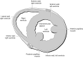
---|---|
Details | |
Part of | Heart |
Artery | anterior interventricular branch of left coronary artery and Posterior interventricular artery |
Identifiers | |
Latin | septum interventriculare cordis |
MeSH | D054088 |
TA98 | A12.1.00.013 |
TA2 | 3970 |
FMA | 7133 |
Anatomical terminology |
The interventricular septum (IVS, or ventricular septum, or during development septum inferius) is the stout wall separating the ventricles, the lower chambers of the heart, from one another.
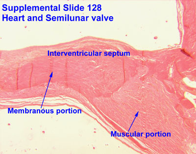
The interventricular septum is directed obliquely backward to the right and curved with the convexity toward the right ventricle; its margins correspond with the anterior and posterior interventricular sulci. The lower part of the septum, which is the major part, is thick and muscular, and its much smaller upper part is thin and membraneous. [1]
During each cardiac cycle the interventricular septum contracts by shortening longitudinally and becoming thicker.
The interventricular septum is the stout wall separating the ventricles, the lower chambers of the heart, from one another.
The ventricular septum is directed obliquely backward to the right and curved with the convexity toward the right ventricle; its margins correspond with the anterior and posterior interventricular sulci. The greater portion of it is thick and muscular and constitutes the muscular interventricular septum. Its upper and posterior part, which separates the aortic vestibule from the lower part of the right atrium and upper part of the right ventricle, is thin and fibrous, and is termed the membranous ventricular septum.
The posterior interventricular artery, a branch of right coronary artery, supplies the posterior 1/3 of the interventricular septum. The remaining anterior 2/3 is supplied by the anterior interventricular artery, which is a septal branch of the left anterior descending artery, which is a branch of left coronary artery. [2]
The muscular part of the interventricular septum derives from the bulboventricular flange which is developed due to differential growth of primitive ventricle and bulbous cordis. Membranous part has a neural crest origin which connects the upper free margin of the bulboventricular flange and anterior and posterior endocardial cushions of atrio ventricular canal. It also gets attached to lower border of spiral septum or the aorticopulmonary septum.
In the final stages of the heart development, the interatrial septum aligns in the same plane as the interventricular septum. The gap between the interatrial septum and interventricular septum forms the membranous part of interventricular septum. [3]
A ventricular septal defect (VSD), a hole in the interventricular septum is one of the four congenital defects of the condition of tetralogy of Fallot. A VSD can cause a left-to-right shunt of blood flow in the heart and is one of the most common of the congenital heart defects. This type of shunt is an acyanotic disorder that can result in ventricular hypertrophy. [4]
The alignment of interventricular septum and interatrial septum is disturbed in various congenital heart diseases. [5]
Coronary circulation is the circulation of blood in the arteries and veins that supply the heart muscle (myocardium). Coronary arteries supply oxygenated blood to the heart muscle. Cardiac veins then drain away the blood after it has been deoxygenated. Because the rest of the body, and most especially the brain, needs a steady supply of oxygenated blood that is free of all but the slightest interruptions, the heart is required to function continuously. Therefore its circulation is of major importance not only to its own tissues but to the entire body and even the level of consciousness of the brain from moment to moment. Interruptions of coronary circulation quickly cause heart attacks, in which the heart muscle is damaged by oxygen starvation. Such interruptions are usually caused by coronary ischemia linked to coronary artery disease, and sometimes to embolism from other causes like obstruction in blood flow through vessels.
Tetralogy of Fallot (TOF), formerly known as Steno-Fallot tetralogy, is a congenital heart defect characterized by four specific cardiac defects. Classically, the four defects are:
A ventricle is one of two large chambers located toward the bottom of the heart that collect and expel blood towards the peripheral beds within the body and lungs. The blood pumped by a ventricle is supplied by an atrium, an adjacent chamber in the upper heart that is smaller than a ventricle. Interventricular means between the ventricles, while intraventricular means within one ventricle.
dextro-Transposition of the great arteries is a potentially life-threatening birth defect in the large arteries of the heart. The primary arteries are transposed.
Atrial septal defect (ASD) is a congenital heart defect in which blood flows between the atria of the heart. Some flow is a normal condition both pre-birth and immediately post-birth via the foramen ovale; however, when this does not naturally close after birth it is referred to as a patent (open) foramen ovale (PFO). It is common in patients with a congenital atrial septal aneurysm (ASA).
A congenital heart defect (CHD), also known as a congenital heart anomaly, congenital cardiovascular malformation, and congenital heart disease, is a defect in the structure of the heart or great vessels that is present at birth. A congenital heart defect is classed as a cardiovascular disease. Signs and symptoms depend on the specific type of defect. Symptoms can vary from none to life-threatening. When present, symptoms are variable and may include rapid breathing, bluish skin (cyanosis), poor weight gain, and feeling tired. CHD does not cause chest pain. Most congenital heart defects are not associated with other diseases. A complication of CHD is heart failure.
An acyanotic heart defect, is a class of congenital heart defects. In these, blood is shunted (flows) from the left side of the heart to the right side of the heart, most often due to a structural defect (hole) in the interventricular septum. People often retain normal levels of oxyhemoglobin saturation in systemic circulation.
A ventricular septal defect (VSD) is a defect in the ventricular septum, the wall dividing the left and right ventricles of the heart. The extent of the opening may vary from pin size to complete absence of the ventricular septum, creating one common ventricle. The ventricular septum consists of an inferior muscular and superior membranous portion and is extensively innervated with conducting cardiomyocytes.
Pulmonary atresia is a congenital malformation of the pulmonary valve in which the valve orifice fails to develop. The valve is completely closed thereby obstructing the outflow of blood from the heart to the lungs. The pulmonary valve is located on the right side of the heart between the right ventricle and pulmonary artery. In a normal functioning heart, the opening to the pulmonary valve has three flaps that open and close.
Atrioventricular septal defect (AVSD) or atrioventricular canal defect (AVCD), also known as "common atrioventricular canal" or "endocardial cushion defect" (ECD), is characterized by a deficiency of the atrioventricular septum of the heart that creates connections between all four of its chambers. It is a very specific combination of 3 defects:
The interatrial septum is the wall of tissue that separates the right and left atria of the heart.
Levo-Transposition of the great arteries is an acyanotic congenital heart defect in which the primary arteries are transposed, with the aorta anterior and to the left of the pulmonary artery; the morphological left and right ventricles with their corresponding atrioventricular valves are also transposed.
A right-to-left shunt is a cardiac shunt which allows blood to flow from the right heart to the left heart. This terminology is used both for the abnormal state in humans and for normal physiological shunts in reptiles.
Arterial switch operation (ASO) or arterial switch, is an open heart surgical procedure used to correct dextro-transposition of the great arteries (d-TGA).
The aorticopulmonary septum is developmentally formed from neural crest, specifically the cardiac neural crest, and actively separates the aorta and pulmonary arteries and fuses with the interventricular septum within the heart during heart development.
The heart is the first functional organ in a vertebrate embryo. There are 5 stages to heart development.
In cardiology, a cardiac shunt is a pattern of blood flow in the heart that deviates from the normal circuit of the circulatory system. It may be described as right-left, left-right or bidirectional, or as systemic-to-pulmonary or pulmonary-to-systemic. The direction may be controlled by left and/or right heart pressure, a biological or artificial heart valve or both. The presence of a shunt may also affect left and/or right heart pressure either beneficially or detrimentally.
The Senning procedure is an atrial switch heart operation performed to treat transposition of the great arteries. It is named after its inventor, the Swedish cardiac surgeon Åke Senning (1915–2000), also known for implanting the first permanent cardiac pacemaker in 1958.
Heart development, also known as cardiogenesis, refers to the prenatal development of the heart. This begins with the formation of two endocardial tubes which merge to form the tubular heart, also called the primitive heart tube. The heart is the first functional organ in vertebrate embryos.
The heart is a muscular organ situated in the mediastinum. It consists of four chambers, four valves, two main arteries, and the conduction system. The left and right sides of the heart have different functions: the right side receives de-oxygenated blood through the superior and inferior venae cavae and pumps blood to the lungs through the pulmonary artery, and the left side receives saturated blood from the lungs.