Fossa ovalis (heart)

Last updated
Fossa ovalis
Gray493.png
Heart viewed from the front, with right atrium and right ventricle opened. Fossa ovalis is labeled in the right atrium.
Details
Precursor Foramen ovale
Identifiers
Latin fossa ovalis cordis
TA98 A12.1.01.005
TA2 3965
FMA 9246
Anatomical terminology

The fossa ovalis is a depression in the right atrium of the heart, at the level of the interatrial septum, the wall between right and left atrium. The fossa ovalis is the remnant of a thin fibrous sheet that covered the foramen ovale during fetal development.

Contents

Function

During fetal development, the foramen ovale allows blood to pass from the right atrium to the left atrium, bypassing the nonfunctional fetal lungs while the fetus obtains its oxygen from the placenta. A flap of tissue called the septum primum acts as a valve over the foramen ovale during that time. After birth, the introduction of air into the lungs causes the pressure in the pulmonary circulatory system to drop. This change in pressure pushes the septum primum against the atrial septum, closing the foramen. [1] The septum primum and atrial septum eventually fuse together to form a complete seal, leaving a depression called the fossa ovalis. By age two, about 75% of people have a completely sealed fossa ovalis. An unfused fossa ovalis is called a patent foramen ovale. Depending on the circumstances, a patent foramen ovale may be completely asymptomatic, or may require surgery. [1] The limbus of fossa ovalis (annulus ovalis) is the prominent oval margin of the fossa ovalis in the right atrium. It is most distinct above and at the sides of the fossa ovalis; below, it is deficient. A small slit-like valvular opening is occasionally found, at the upper margin of the fossa, leading upward beneath the limbus, into the left atrium; it is the remains of the fetal aperture the foramen ovale between the two atria.

Closure

Almost immediately after the infant is born, the foramen ovale and ductus arteriosus close. The major changes that are made by the body occur at the first breath (in the case of heart and lung functions) and up to weeks after birth (such as the liver's enzyme synthesis). The foramen ovale becomes the fossa ovalis as the foramen closes while edge of the septum secundum in right atrium becomes anulus ovalis, so the depression beneath it becomes the fossa ovalis. [2] [ unreliable medical source? ] This enables respiration and circulation independent from the mother's placenta.

With the child's first breath, the lung sends oxygenated blood to the left atrium. As a result, pressure in the left atrium is higher than that of the right, and the increased pressure holds the interatrial flap (which covers the foramen ovale) shut, therefore closing the foramen ovale as well. [2] In normal development, the closed foramen ovale fuses with the interatrial wall. During the first breath, vasoconstriction causes the ductus arteriosus to close, and during adult years, tissue occludes what once was the ductus arterious, creating the ligamentum arteriosum. [3]

Clinical significance

Aneurysm

Aneurysms can occur in adulthood if the foramen ovale is not closed correctly. An aneurysm happens when an artery becomes enlarged in a localized area due to weakening of the arterial wall. [4]

When this type of aneurysm occurs in the area of the fossa ovalis, an enlarged pouch is formed. This pouch can protrude into the right atrium or the left atrium. The cause of this aneurysm is the result of abnormal, increased pressure within the heart. Even if the foramen ovale does seal shut, an aneurysm may occur, usually on the side of the right atrium. If the aneurysm stretches too far, it can narrow the opening of the inferior vena cava. [5] This type of aneurysm can be a result of plaque build-up in the arteries from coronary heart disease, as well as diseases of the aortic valve or mitral valve. Surgery may be useful in helping to cope with the aneurysm.

Patent foramen ovale

If the atrial septum does not close properly, it leads to a patent foramen ovale (PFO). This type of defect generally works like a flap valve, opening during certain conditions of increased pressure in the chest, such as during strain while having a bowel movement, cough, or sneeze. With enough pressure, blood may travel from the right atrium to the left. If there is a clot in the right side of the heart, it can cross the PFO, enter the left atrium, and travel out of the heart and to the brain, causing a stroke. If the clot travels into a coronary artery it can cause a heart attack. [6]

See also

Related Research Articles

dextro-Transposition of the great arteries Medical condition

dextro-Transposition of the great arteries is a potentially life-threatening birth defect in the large arteries of the heart. The primary arteries are transposed.

<span class="mw-page-title-main">Atrial septal defect</span> Human heart defect present at birth

Atrial septal defect (ASD) is a congenital heart defect in which blood flows between the atria of the heart. Some flow is a normal condition both pre-birth and immediately post-birth via the foramen ovale; however, when this does not naturally close after birth it is referred to as a patent (open) foramen ovale (PFO). It is common in patients with a congenital atrial septal aneurysm (ASA).

<span class="mw-page-title-main">Congenital heart defect</span> Defect in the structure of the heart that is present at birth

A congenital heart defect (CHD), also known as a congenital heart anomaly, congenital cardiovascular malformation, and congenital heart disease, is a defect in the structure of the heart or great vessels that is present at birth. A congenital heart defect is classed as a cardiovascular disease. Signs and symptoms depend on the specific type of defect. Symptoms can vary from none to life-threatening. When present, symptoms are variable and may include rapid breathing, bluish skin (cyanosis), poor weight gain, and feeling tired. CHD does not cause chest pain. Most congenital heart defects are not associated with other diseases. A complication of CHD is heart failure.

<span class="mw-page-title-main">Atrium (heart)</span> Part of the human heart

The atrium is one of the two upper chambers in the heart that receives blood from the circulatory system. The blood in the atria is pumped into the heart ventricles through the atrioventricular mitral and tricuspid heart valves.

<span class="mw-page-title-main">Tricuspid atresia</span> Medical condition

Tricuspid atresia is a form of congenital heart disease whereby there is a complete absence of the tricuspid valve. Therefore, there is an absence of right atrioventricular connection. This leads to a hypoplastic (undersized) or absent right ventricle. This defect is contracted during prenatal development, when the heart does not finish developing. It causes the systemic circulation to be filled with relatively deoxygenated blood. The causes of tricuspid atresia are unknown.

<span class="mw-page-title-main">Foramen ovale (heart)</span> Passageway between the atria of the human heart

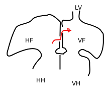
In the fetal heart, the foramen ovale, also foramen Botalli, or the ostium secundum of Born, allows blood to enter the left atrium from the right atrium. It is one of two fetal cardiac shunts, the other being the ductus arteriosus. Another similar adaptation in the fetus is the ductus venosus. In most individuals, the foramen ovale closes at birth. It later forms the fossa ovalis.

<span class="mw-page-title-main">Interatrial septum</span> Wall of tissue separating atria of human heart

The interatrial septum is the wall of tissue that separates the right and left atria of the heart.

<span class="mw-page-title-main">Fetal circulation</span> Circulatory system of fetuses

In humans, the circulatory system is different before and after birth. The fetal circulation is composed of the placenta, umbilical blood vessels encapsulated by the umbilical cord, heart and systemic blood vessels. A major difference between the fetal circulation and postnatal circulation is that the lungs are not used during the fetal stage resulting in the presence of shunts to move oxygenated blood and nutrients from the placenta to the fetal tissue. At birth, the start of breathing and the severance of the umbilical cord prompt various changes that quickly transform fetal circulation into postnatal circulation.

Levo-Transposition of the great arteries is an acyanotic congenital heart defect in which the primary arteries are transposed, with the aorta anterior and to the left of the pulmonary artery; the morphological left and right ventricles with their corresponding atrioventricular valves are also transposed.

Pulmonic stenosis, is a dynamic or fixed obstruction of flow from the right ventricle of the heart to the pulmonary artery. It is usually first diagnosed in childhood.

<span class="mw-page-title-main">Primitive atrium</span>

The primitive atrium is a stage in the embryonic development of the human heart. It grows rapidly and partially encircles the bulbus cordis; the groove against which the bulbus cordis lies is the first indication of a division into right and left atria.

<span class="mw-page-title-main">Septum primum</span>

During heart development of a human embryo, the single primitive atrium becomes divided into right and left by a septum, the septum primum. The septum primum grows downward into the single atrium.

<span class="mw-page-title-main">Septum secundum</span>

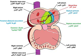
The septum secundum is a muscular flap that is important in heart development. It is semilunar in shape, and grows downward from the upper wall of the atrium immediately to the right of the septum primum and ostium secundum. It is important in the closure of the foramen ovale after birth.

<span class="mw-page-title-main">Foramen secundum</span>

The foramen secundum, or ostium secundum is a foramen in the septum primum, a precursor to the interatrial septum of the human heart.

<span class="mw-page-title-main">Valve of inferior vena cava</span>

The valve of the inferior vena cava is a venous valve that lies at the junction of the inferior vena cava and right atrium.

The heart is the first functional organ in a vertebrate embryo. There are 5 stages to heart development.

A cardiac shunt is a pattern of blood flow in the heart that deviates from the normal circuit of the circulatory system. It may be described as right-left, left-right or bidirectional, or as systemic-to-pulmonary or pulmonary-to-systemic. The direction may be controlled by left and/or right heart pressure, a biological or artificial heart valve or both. The presence of a shunt may also affect left and/or right heart pressure either beneficially or detrimentally.

<span class="mw-page-title-main">Lutembacher's syndrome</span> Medical condition

Lutembacher's syndrome is a very rare form of congenital heart disease that affects one of the chambers of the heart as well as a valve. It is commonly known as both congenital atrial septal defect (ASD) and acquired mitral stenosis (MS). Congenital atrial septal defect refers to a hole being in the septum or wall that separates the two atria; this condition is usually seen in fetuses and infants. Mitral stenosis refers to mitral valve leaflets sticking to each other making the opening for blood to pass from the atrium to the ventricles very small. With the valve being so small, blood has difficulty passing from the left atrium into the left ventricle. Septal defects that may occur with Lutembacher's syndrome include: Ostium primum atrial septal defect or ostium secundum which is more prevalent.

<span class="mw-page-title-main">Atrial septostomy</span> Surgical procedure on the heart

Atrial septostomy is a surgical procedure in which a small hole is created between the upper two chambers of the heart, the atria. This procedure is primarily used to palliate dextro-Transposition of the great arteries or d-TGA, a life-threatening cyanotic congenital heart defect seen in infants. It is performed prior to an arterial switch operation. Atrial septostomy has also seen limited use as a surgical treatment for pulmonary hypertension. The first atrial septostomy was developed by Vivien Thomas in a canine model and performed in humans by Alfred Blalock. The Rashkind balloon procedure, a common atrial septostomy technique, was developed in 1966 by American cardiologist William Rashkind at the Children's Hospital of Philadelphia.

<span class="mw-page-title-main">Heart development</span> Prenatal development of the heart

Heart development, also known as cardiogenesis, refers to the prenatal development of the heart. This begins with the formation of two endocardial tubes which merge to form the tubular heart, also called the primitive heart tube. The heart is the first functional organ in vertebrate embryos.

References

  1. 1 2 Hara, H; Virmani, R; Ladich, E; Mackey-Bojack, S; Titus, J; Reisman, M; Gray, W; Nakamura, M; Mooney, M; Poulose, A; Schwartz, RS (Nov 1, 2005). "Patent foramen ovale: current pathology, pathophysiology, and clinical status". Journal of the American College of Cardiology. 46 (9): 1768–1776. doi: 10.1016/j.jacc.2005.08.038 . PMID   16256883.
  2. 1 2 Johnson, Donna. "What is the Fossa Ovalis?". Conjecture Corporation. Retrieved 16 March 2012.
  3. Dryden, Richard. "Respiratory system" . Retrieved 16 April 2012.
  4. Shirani, J.; Zafari, A.M.; Roberts, W.C. (August 1995). "Morphologic features of fossa ovalis membrane aneurysm in the adult and its clinical significance". Journal of the American College of Cardiology. 26 (2): 466–471. doi: 10.1016/0735-1097(95)80024-b . PMID   7608452.
  5. Topaz, O.; Edwards, J.E.; Bojack-Mackey, S; Titus, J.L. (Jul–Aug 2003). "Aneurysm of fossa ovalis in adults: a pathologic study". Cardiovascular Pathology. 12 (4): 219–225. doi:10.1016/s1054-8807(03)00041-3. PMID   12826292.
  6. "Patent Foramen Ovale". The Cleveland Clinic. Retrieved 15 April 2012.