The inguinal ligament is a band running from the pubic tubercle to the anterior superior iliac spine. It forms the base of the inguinal canal through which an indirect inguinal hernia may develop.

The lesser omentum is the double layer of peritoneum that extends from the liver to the lesser curvature of the stomach and the first part of the duodenum.

The sacrotuberous ligament is situated at the lower and back part of the pelvis. It is flat, and triangular in form; narrower in the middle than at the ends.

The round ligament of the uterus originates at the uterine horns, in the parametrium. The round ligament exits the pelvis via the deep inguinal ring, passes through the inguinal canal and continues on to the labia majora where its fibers spread and mix with the tissue of the mons pubis.

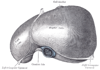
The falciform ligament is a ligament that attaches the liver to the anterior (ventral) body wall, and separates the liver into the right lobe and left lobe. The falciform ligament, from Latin, meaning 'sickle-shaped', is a broad and thin fold of peritoneum, its base being directed downward and backward and its apex upward and backward. The falciform ligament droops down from the hilum of the liver.

The greater omentum is a large apron-like fold of visceral peritoneum that hangs down from the stomach. It extends from the greater curvature of the stomach, passing in front of the small intestines and doubles back to ascend to the transverse colon before reaching to the posterior abdominal wall. The greater omentum is larger than the lesser omentum, which hangs down from the liver to the lesser curvature. The common anatomical term "epiploic" derives from "epiploon", from the Greek epipleein, meaning to float or sail on, since the greater omentum appears to float on the surface of the intestines. It is the first structure observed when the abdominal cavity is opened anteriorly.

The broad ligament of the uterus is the wide fold of peritoneum that connects the sides of the uterus to the walls and floor of the pelvis.

In the sphenoid bone, the anterior boundary of the sella turcica is completed by two small eminences, one on either side, called the anterior clinoid processes, while the posterior boundary is formed by a square-shaped plate of bone, the dorsum sellæ, ending at its superior angles in two tubercles, the posterior clinoid processes, the size and form of which vary considerably in different individuals. The posterior clinoid processes deepen the sella turcica, and give attachment to the tentorium cerebelli.

The suspensory ligament of the ovary, also infundibulopelvic ligament, is a fold of peritoneum that extends out from the ovary to the wall of the pelvis.
The parametrium is the fibrous and fatty connective tissue that surrounds the uterus. This tissue separates the supravaginal portion of the cervix from the bladder. The parametrium lies in front of the cervix and extends laterally between the layers of the broad ligaments. It conects the uterus to other tissues in the pelvis. It is different from the perimetrium, which is the outermost layer of the uterus.

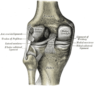
The fibular collateral ligament is a ligament located on the lateral (outer) side of the knee, and thus belongs to the extrinsic knee ligaments and posterolateral corner of the knee.

The round ligament of the liver is a degenerative string of tissue that exists in the free edge of the falciform ligament of the liver. The round ligament divides the left part of the liver into medial and lateral sections.

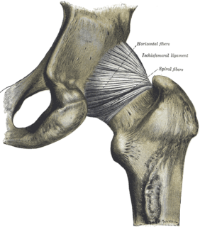
The pubofemoral ligament is a ligament on the inferior side of the hip joint.

The ischiocapsular ligament consists of a triangular band of strong fibers on the posterior side of the hip joint. Its fibers span from the ischium at a point below and behind the acetabulum to blend with the circular fibers at the posterior end of the joint capsule and attach at the intertrochanteric line of the femur.

The interspinous ligaments are thin and membranous ligaments, that connect adjoining spinous processes of the vertebra in the spine. They extend from the root to the apex of each spinous process. They meet the ligamenta flava in front and blend with the supraspinous ligament behind.

The ovarian ligament is a fibrous ligament that connects the ovary to the lateral surface of the uterus.

The anterior talofibular ligament is a ligament in the ankle. It passes from the anterior margin of the fibular malleolus, anteriorly and medially, to the talus bone, in front of its lateral articular facet. It is one of the lateral ligaments of the ankle and prevents the foot from sliding forward in relation to the shin. It is the most commonly injured ligament in a sprained ankle—from an inversion injury—and will allow a positive anterior drawer test of the ankle if completely torn.

The uterosacral ligaments belong to the major ligaments of uterus.

The cardinal ligament is a major ligament of the uterus. It is located at the base of the broad ligament of the uterus. There are a pair of cardinal ligaments in the female human body.

The pelvis is either the lower part of the trunk of the human body between the abdomen and the thighs or the skeleton embedded in it.
The public domain consists of all the creative works to which no exclusive intellectual property rights apply. Those rights may have expired, been forfeited, expressly waived, or may be inapplicable.

Gray's Anatomy is an English language textbook of human anatomy originally written by Henry Gray and illustrated by Henry Vandyke Carter. Earlier editions were called Anatomy: Descriptive and Surgical and Gray's Anatomy: Descriptive and Applied, but the book's name is commonly shortened to, and later editions are titled, Gray's Anatomy. The book is widely regarded as an extremely influential work on the subject, and has continued to be revised and republished from its initial publication in 1858 to the present day. The latest edition of the book, the 41st, was published in September 2015.


