Tricuspid valve

Last updated
Tricuspid valve
Diagram of the human heart (cropped).svg
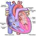
Anterior (frontal) view of the opened heart. White arrows indicate normal blood flow. (Tricuspid valve labeled at bottom left.)
CG Heart.gif
Heart in motion: the anterior walls of the ventricles are removed. The action of the tricuspid valve, located in the right ventricle, is seen on the left portion of this illustration. The three leaflets with their attached chordae tendineae and papillary muscles can be seen.
Details
Identifiers
Latin valvula tricuspidalis,
valva atrioventricularis dextra
MeSH D014261
TA98 A12.1.02.003
TA2 3982
FMA 7234
Anatomical terminology

The tricuspid valve, or right atrioventricular valve, is on the right dorsal side of the mammalian heart, at the superior portion of the right ventricle. The function of the valve is to allow blood to flow from the right atrium to the right ventricle during diastole, and to close to prevent backflow (regurgitation) from the right ventricle into the right atrium during right ventricular contraction (systole).

Contents

Structure

The tricuspid valve usually has three cusps or leaflets, named the anterior, posterior, and septal cusps. [1] Each leaflet is connected via chordae tendineae to the anterior, posterior, and septal papillary muscles of the right ventricle, respectively. Tricuspid valves may also occur with two or four leaflets; the number may change over a lifetime. [2]

Function

The tricuspid valve functions as a one-way valve that closes during ventricular systole to prevent regurgitation of blood from the right ventricle back into the right atrium. It opens during ventricular diastole, allowing blood to flow from the right atrium into the right ventricle. The back flow of blood is also known as regression or tricuspid regurgitation. Tricuspid regurgitation can result in increased ventricular preload because the blood refluxed back into the atrium is added to the volume of blood that must be pumped back into the ventricle during the next cycle of ventricular diastole. Increased right ventricular preload over a prolonged period of time may lead to right ventricular enlargement (dilatation), [3] which can progress to right heart failure if left uncorrected. [4]

Clinical significance

Video explanation of tricuspid valve disease

Infected valves can result in endocarditis in intravenous drug users. [5] [6] Patients who inject narcotics or other drugs intravenously may introduce infection, which can travel to the right side of the heart, most often caused by the bacteria S. aureus . [7] In patients without a history of intravenous exposure, endocarditis is more frequently left-sided. [7]

The tricuspid valve can be affected by rheumatic fever, which can cause tricuspid stenosis or tricuspid regurgitation. [8] Some individuals are born with congenital abnormalities of the tricuspid valve. Congenital apical displacement of the tricuspid valve is called Ebstein's anomaly and typically causes significant tricuspid regurgitation.

Certain carcinoid syndromes can affect the tricuspid valve by producing fibrosis due to serotonin production by those tumors.

The first endovascular tricuspid valve implant was performed by surgeons at the Cleveland Clinic. [9]

Tricuspid regurgitation

Tricuspid regurgitation is common and is estimated to occur in 65–85% of the population. [10] In the Framingham Heart Study presence of any severity of tricuspid regurgitation, ranging from trace to above moderate was in 82% of men and in 85.7% of women. [11] Mild tricuspid regurgitation tends to be common, benign, and in structurally normal tricuspid valve apparatus can be considered a normal variant. [10] Moderate or severe tricuspid regurgitation is usually associated with tricuspid valve leaflet abnormalities and/or possibly annular dilation and is usually pathologic which can lead to irreversible damage of cardiac muscle and worse outcomes due to chronic prolonged right ventricular volume overload. [10]

Additional images

See also

Related Research Articles

<span class="mw-page-title-main">Heart valve</span> A flap of tissue that prevent backflow of blood around the heart

A heart valve is a biological one-way valve that allows blood to flow in one direction through the chambers of the heart. Four valves are usually present in a mammalian heart and together they determine the pathway of blood flow through the heart. A heart valve opens or closes according to differential blood pressure on each side.

<span class="mw-page-title-main">Heart sounds</span> Noise generated by the beating heart

Heart sounds are the noises generated by the beating heart and the resultant flow of blood through it. Specifically, the sounds reflect the turbulence created when the heart valves snap shut. In cardiac auscultation, an examiner may use a stethoscope to listen for these unique and distinct sounds that provide important auditory data regarding the condition of the heart.

<span class="mw-page-title-main">Aortic valve</span> Valve in the human heart between the left ventricle and the aorta

The aortic valve is a valve in the heart of humans and most other animals, located between the left ventricle and the aorta. It is one of the four valves of the heart and one of the two semilunar valves, the other being the pulmonary valve. The aortic valve normally has three cusps or leaflets, although in 1–2% of the population it is found to congenitally have two leaflets. The aortic valve is the last structure in the heart the blood travels through before stopping the flow through the systemic circulation.

<span class="mw-page-title-main">Mitral valve</span> Valve in the heart connecting the left atrium and left ventricle

The mitral valve, also known as the bicuspid valve or left atrioventricular valve, is one of the four heart valves. It has two cusps or flaps and lies between the left atrium and the left ventricle of the heart. The heart valves are all one-way valves allowing blood flow in just one direction. The mitral valve and the tricuspid valve are known as the atrioventricular valves because they lie between the atria and the ventricles.

<span class="mw-page-title-main">Systole</span> Part of the cardiac cycle when a heart chamber contracts

Systole is the part of the cardiac cycle during which some chambers of the heart contract after refilling with blood.

<span class="mw-page-title-main">Ventricle (heart)</span> Chamber of the heart

A ventricle is one of two large chambers located toward the bottom of the heart that collect and expel blood towards the peripheral beds within the body and lungs. The blood pumped by a ventricle is supplied by an atrium, an adjacent chamber in the upper heart that is smaller than a ventricle. Interventricular means between the ventricles, while intraventricular means within one ventricle.

<span class="mw-page-title-main">Papillary muscle</span> Heart ventricle muscles

The papillary muscles are muscles located in the ventricles of the heart. They attach to the cusps of the atrioventricular valves via the chordae tendineae and contract to prevent inversion or prolapse of these valves on systole.

<span class="mw-page-title-main">Diastole</span> Part of the cardiac cycle

Diastole is the relaxed phase of the cardiac cycle when the chambers of the heart are refilling with blood. The contrasting phase is systole when the heart chambers are contracting. Atrial diastole is the relaxing of the atria, and ventricular diastole the relaxing of the ventricles.

<span class="mw-page-title-main">Mitral stenosis</span> Heart disease with narrowing of valve

Mitral stenosis is a valvular heart disease characterized by the narrowing of the opening of the mitral valve of the heart. It is almost always caused by rheumatic valvular heart disease. Normally, the mitral valve is about 5 cm2 during diastole. Any decrease in area below 2 cm2 causes mitral stenosis. Early diagnosis of mitral stenosis in pregnancy is very important as the heart cannot tolerate increased cardiac output demand as in the case of exercise and pregnancy. Atrial fibrillation is a common complication of resulting left atrial enlargement, which can lead to systemic thromboembolic complications such as stroke.

<span class="mw-page-title-main">Aortic regurgitation</span> Medical condition

Aortic regurgitation (AR), also known as aortic insufficiency (AI), is the leaking of the aortic valve of the heart that causes blood to flow in the reverse direction during ventricular diastole, from the aorta into the left ventricle. As a consequence, the cardiac muscle is forced to work harder than normal.

<span class="mw-page-title-main">Ventricular septal defect</span> Medical condition

A ventricular septal defect (VSD) is a defect in the ventricular septum, the wall dividing the left and right ventricles of the heart. The extent of the opening may vary from pin size to complete absence of the ventricular septum, creating one common ventricle. The ventricular septum consists of an inferior muscular and superior membranous portion and is extensively innervated with conducting cardiomyocytes.

<span class="mw-page-title-main">Mitral regurgitation</span> Form of valvular heart disease

Mitral regurgitation (MR), also known as mitral insufficiency or mitral incompetence, is a form of valvular heart disease in which the mitral valve is insufficient and does not close properly when the heart pumps out blood. It is the abnormal leaking of blood backwards – regurgitation from the left ventricle, through the mitral valve, into the left atrium, when the left ventricle contracts. Mitral regurgitation is the most common form of valvular heart disease.

<span class="mw-page-title-main">Ebstein's anomaly</span> Congenital heart defect

Ebstein's anomaly is a congenital heart defect in which the septal and posterior leaflets of the tricuspid valve are displaced downwards towards the apex of the right ventricle of the heart. EA has great anatomical heterogeneity that generates a wide spectrum of clinical features at presentation and is complicated by the fact that the lesion is often accompanied by other congenital cardiac lesions. It is classified as a critical congenital heart defect accounting for less than 1% of all congenital heart defects presenting in around 1 per 200,000 live births. Ebstein's anomaly usually presents with a systolic murmur and frequently with a gallop rhythm.

<span class="mw-page-title-main">Chordae tendineae</span> Inelastic cords of fibrous connective tissue connecting papillary muscles to heart valves

The chordae tendineae or tendinous cords, colloquially known as the heart strings, are inelastic cords of fibrous connective tissue that connect the papillary muscles to the tricuspid valve and the mitral valve in the heart.

A transthoracic echocardiogram (TTE) is the most common type of echocardiogram, which is a still or moving image of the internal parts of the heart using ultrasound. In this case, the probe is placed on the chest or abdomen of the subject to get various views of the heart. It is used as a non-invasive assessment of the overall health of the heart, including a patient's heart valves and degree of heart muscle contraction. The images are displayed on a monitor for real-time viewing and then recorded.

<span class="mw-page-title-main">Valvular heart disease</span> Disease in the valves of the heart

Valvular heart disease is any cardiovascular disease process involving one or more of the four valves of the heart. These conditions occur largely as a consequence of aging, but may also be the result of congenital (inborn) abnormalities or specific disease or physiologic processes including rheumatic heart disease and pregnancy.

<span class="mw-page-title-main">Tricuspid regurgitation</span> Type of valvular heart disease

Tricuspid regurgitation (TR), also called tricuspid insufficiency, is a type of valvular heart disease in which the tricuspid valve of the heart, located between the right atrium and right ventricle, does not close completely when the right ventricle contracts (systole). TR allows the blood to flow backwards from the right ventricle to the right atrium, which increases the volume and pressure of the blood both in the right atrium and the right ventricle, which may increase central venous volume and pressure if the backward flow is sufficiently severe.

The following outline is provided as an overview of and topical guide to cardiology, the branch of medicine dealing with disorders of the human heart. The field includes medical diagnosis and treatment of congenital heart defects, coronary artery disease, heart failure, valvular heart disease and electrophysiology. Physicians who specialize in cardiology are called cardiologists.

<span class="mw-page-title-main">Third heart sound</span> Medical condition

The third heart sound or S3 is a rare extra heart sound that occurs soon after the normal two "lub-dub" heart sounds (S1 and S2). S3 is associated with heart failure.

Cardiac physiology or heart function is the study of healthy, unimpaired function of the heart: involving blood flow; myocardium structure; the electrical conduction system of the heart; the cardiac cycle and cardiac output and how these interact and depend on one another.

References

  1. "Anatomy of the Tricuspid Valve". e-echocardiography.com. Retrieved 2018-03-30.
  2. Richard Van Pragh: Cardiac anatomy in A. C. Chang et al.: Pediatric Cardiac Intensive Care, Philadelphia 1998.
  3. Reynertson, Sandra I.; Kundur, Ramesh; Mullen, G. Martin; Costanzo, Maria Rosa; McKiernan, Thomas L.; Louie, Eric K. (1999-08-03). "Asymmetry of Right Ventricular Enlargement in Response to Tricuspid Regurgitation". Circulation. 100 (5): 465–467. doi: 10.1161/01.CIR.100.5.465 . ISSN   0009-7322. PMID   10430758.
  4. "Enlarged heart - Symptoms and causes". Mayo Clinic. Retrieved 2018-03-30.
  5. Demin AA, Drobysheva VP, Vel'ter OIu (2000). "[Infectious endocarditis in intravenous drug abusers]". Klinicheskaia Meditsina (in Russian). 78 (8): 47–51. PMID   11019526.
  6. Butany J, Dev V, Leong SW, Soor GS, Thangaroopan M, Borger MA (2006). "Infective endocarditis of the tricuspid valve". Journal of Cardiac Surgery. 21 (6): 603–4. doi: 10.1111/j.1540-8191.2006.00313.x . PMID   17073968. S2CID   32603989.
  7. 1 2 Mitchell RS, Kumar V, Robbins SL, Abbas AK, Fausto N (2007). Robbins Basic Pathology (8th ed.). Saunders/Elsevier. pp. 406–8. ISBN   978-1-4160-2973-1.
  8. Tricuspid valve disease Mount Sinai Hospital, New York
  9. University Circle Inc. Archived 2008-06-17 at the Wayback Machine
  10. 1 2 3 Arsalan, Mani; Walther, Thomas; Smith, Robert L.; Grayburn, Paul A. (2015-09-10). "Tricuspid regurgitation diagnosis and treatment". European Heart Journal. 38 (9): 634–638. doi: 10.1093/eurheartj/ehv487 . ISSN   0195-668X. PMID   26358570.
  11. Prihadi', 'Edgard A. "Tricuspid valve regurgitation: no longer the "forgotten valve"". www.escardio.org. Retrieved 2021-11-27.