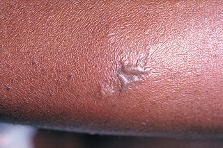
Group A streptococcal infections are a number of infections with Streptococcus pyogenes, a group A streptococcus (GAS). S. pyogenes is a species of beta-hemolytic Gram-positive bacteria that is responsible for a wide range of infections that are mostly common and fairly mild. If the bacteria enters the bloodstream, the infection can become severe and life-threatening, and is called an invasive GAS (iGAS).
Bloodstream infections (BSIs) are infections of blood caused by blood-borne pathogens. The detection of microbes in the blood is always abnormal. A bloodstream infection is different from sepsis, which is characterized by severe inflammatory or immune responses of the host organism to pathogens.

Rheumatic fever (RF) is an inflammatory disease that can involve the heart, joints, skin, and brain. The disease typically develops two to four weeks after a streptococcal throat infection. Signs and symptoms include fever, multiple painful joints, involuntary muscle movements, and occasionally a characteristic non-itchy rash known as erythema marginatum. The heart is involved in about half of the cases. Damage to the heart valves, known as rheumatic heart disease (RHD), usually occurs after repeated attacks but can sometimes occur after one. The damaged valves may result in heart failure, atrial fibrillation and infection of the valves.

Mitral valve prolapse (MVP) is a valvular heart disease characterized by the displacement of an abnormally thickened mitral valve leaflet into the left atrium during systole. It is the primary form of myxomatous degeneration of the valve. There are various types of MVP, broadly classified as classic and nonclassic. In severe cases of classic MVP, complications include mitral regurgitation, infective endocarditis, congestive heart failure, and, in rare circumstances, cardiac arrest.

Infective endocarditis is an infection of the inner surface of the heart (endocardium), usually the valves. Signs and symptoms may include fever, small areas of bleeding into the skin, heart murmur, feeling tired, and low red blood cell count. Complications may include backward blood flow in the heart, heart failure – the heart struggling to pump a sufficient amount of blood to meet the body's needs, abnormal electrical conduction in the heart, stroke, and kidney failure.

Procalcitonin (PCT) is a peptide precursor of the hormone calcitonin, the latter being involved with calcium homeostasis. It arises once preprocalcitonin is cleaved by endopeptidase. It was first identified by Leonard J. Deftos and Bernard A. Roos in the 1970s. It is composed of 116 amino acids and is produced by parafollicular cells of the thyroid and by the neuroendocrine cells of the lung and the intestine.

A boil, also called a furuncle, is a deep folliculitis, which is an infection of the hair follicle. It is most commonly caused by infection by the bacterium Staphylococcus aureus, resulting in a painful swollen area on the skin caused by an accumulation of pus and dead tissue. Boils are therefore basically pus-filled nodules. Individual boils clustered together are called carbuncles. Most human infections are caused by coagulase-positive S. aureus strains, notable for the bacteria's ability to produce coagulase, an enzyme that can clot blood. Almost any organ system can be infected by S. aureus.
Libman–Sacks endocarditis is a form of non-bacterial endocarditis that is seen in association with systemic lupus erythematosus, antiphospholipid syndrome, and malignancies. It is one of the most common cardiac manifestations of lupus.
Nonbacterial thrombotic endocarditis (NBTE) is a form of endocarditis in which small sterile vegetations are deposited on the valve leaflets. Formerly known as marantic endocarditis, which comes from the Greek marantikos, meaning "wasting away". The term "marantic endocarditis" is still sometimes used to emphasize the association with a wasting state such as cancer.

Valvular heart disease is any cardiovascular disease process involving one or more of the four valves of the heart. These conditions occur largely as a consequence of aging, but may also be the result of congenital (inborn) abnormalities or specific disease or physiologic processes including rheumatic heart disease and pregnancy.

Subacute bacterial endocarditis, abbreviated SBE, is a type of endocarditis. Subacute bacterial endocarditis can be considered a form of type III hypersensitivity.
Loeffler endocarditis is a form of heart disease characterized by a stiffened, poorly-functioning heart caused by infiltration of the heart by white blood cells known as eosinophils. Restrictive cardiomyopathy is a disease of the heart muscle which results in impaired diastolic filling of the heart ventricles, i.e. the large heart chambers which pump blood into the pulmonary or systemic circulation. Diastole is the part of the cardiac contraction-relaxation cycle in which the heart fills with venous blood after the emptying done during its previous systole.

Eikenella corrodens is a Gram-negative facultative anaerobic bacillus that can cause severe invasive disease in humans. It was first identified by M. Eiken in 1958, who called it Bacteroides corrodens. E. corrodens is a rare pericarditis associated pathogen. It is a fastidious, slow growing, human commensal bacillus, capable of acting as an opportunistic pathogen and causing abscesses in several anatomical sites, including the liver, lung, spleen, and submandibular region. E. corrodens could independently cause serious infection in both immunocompetent and immunocompromised hosts.

Gonorrhoea or gonorrhea, colloquially known as the clap, is a sexually transmitted infection (STI) caused by the bacterium Neisseria gonorrhoeae. Infection may involve the genitals, mouth, or rectum. Infected males may experience pain or burning with urination, discharge from the penis, or testicular pain. Infected females may experience burning with urination, vaginal discharge, vaginal bleeding between periods, or pelvic pain. Complications in females include pelvic inflammatory disease and in males include inflammation of the epididymis. Many of those infected, however, have no symptoms. If untreated, gonorrhea can spread to joints or heart valves.

Kingella kingae is a species of Gram-negative facultative anaerobic β-hemolytic coccobacilli. First isolated in 1960 by Elizabeth O. King, it was not recognized as a significant cause of infection in young children until the 1990s, when culture techniques had improved enough for it to be recognized. It is best known as a cause of septic arthritis, osteomyelitis, spondylodiscitis, bacteraemia, and endocarditis, and less frequently lower respiratory tract infections and meningitis.
Cardiobacterium hominis /ˌkɑːrdiəʊbækˈtɪəriəm ˈhɒmɪnɪs/ is a microaerophilic, pleomorphic, fastidious, Gram-negative bacterium part of the Cardiobacteriaceae family and the HACEK group. It is most commonly found in the human microbiota, specifically the oropharyngeal region including the mouth and upper part of the respiratory tract. It is one of the causes of endocarditis, a life-threatening inflammation close to the heart's inner lining and valves. While infections caused by Cardiobacterium hominis are uncommon, various clinical manifestations are linked to the bacterium, including meningitis, septicemia, and bone infections.

Austrian syndrome, also known as Osler's triad, is a medical condition that was named after Robert Austrian in 1957. The presentation of the condition consists of pneumonia, endocarditis, and meningitis, all caused by Streptococcus pneumoniae. It is associated with alcoholism due to hyposplenism and can be seen in males between the ages of 40 and 60 years old. Robert Austrian was not the first one to describe the condition, but Richard Heschl or William Osler were not able to link the signs to the bacteria because microbiology was not yet developed.
Dental antibiotic prophylaxis is the administration of antibiotics to a dental patient for prevention of harmful consequences of bacteremia, that may be caused by invasion of the oral flora into an injured gingival or peri-apical vessel during dental treatment.
Eosinophilic myocarditis is inflammation in the heart muscle that is caused by the infiltration and destructive activity of a type of white blood cell, the eosinophil. Typically, the disorder is associated with hypereosinophilia, i.e. an eosinophil blood cell count greater than 1,500 per microliter. It is distinguished from non-eosinophilic myocarditis, which is heart inflammation caused by other types of white blood cells, i.e. lymphocytes and monocytes, as well as the respective descendants of these cells, NK cells and macrophages. This distinction is important because the eosinophil-based disorder is due to a particular set of underlying diseases and its preferred treatments differ from those for non-eosinophilic myocarditis.
Granulicatella adiacens is a fastidious Gram-positive cocci and is part of the nutritionally variant streptococci (NVS). Like other constituents of the NVS, it can cause bacteremia and infective endocarditis (IE), with significant morbidity and mortality. NVS has less often been implicated in a variety of other infections, including those of the orbit, nasolacrimal duct and breast implants. It is a commensal of the human mouth, genital, and intestinal tracts, although it is rarely implicated in infections, in part due to it being a fastidious organism and rarely being identified in the laboratory environment. However, its identification has become more frequent with use of commercial mediums and automated identification systems. Because it has been difficult to identify, it has been considered one of the causes of culture negative IE. Identifying G. adiacens can allow more appropriate selection of antibiotics, especially when susceptibility testing is not available.


