Second-degree atrioventricular block

Last updated
Second-degree atrioventricular block
Other namesSecond-degree heart block
Second degree heart block.png
ECGs demonstrating forms of second-degree AV block
Specialty Cardiology
Symptoms Dizziness, Fainting, Shortness of breath
TypesType 1 (Wenckebach), Type 2
CausesFibrosis in AV node, medication, vagal tone, electrolyte disturbances
Diagnostic method Electrocardiogram
TreatmentAvoidance of AV-nodal-blocking medication, pacemaker

Second-degree atrioventricular block (AV block) is a disease of the electrical conduction system of the heart. It is a conduction block between the atria and ventricles. The presence of second-degree AV block is diagnosed when one or more (but not all) of the atrial impulses fail to conduct to the ventricles due to impaired conduction. It is classified as a block of the AV node, falling between first-degree (slowed conduction) and third degree blocks (complete block). [1]

Contents

Signs and symptoms

Most people with Wenckebach (Type I Mobitz) do not show symptoms. However, those that do usually display one or more of the following: [2]

Types

There are two non-distinct types of second-degree AV block, called Type 1 and Type 2. In both types, a P wave is blocked from initiating a QRS complex; but, in Type 1, there are increasing delays in each cycle before the omission, whereas, in Type 2, there is no such pattern. [3]

Type 1 second-degree heart block is considered more benign than type 2 second-degree heart block. [4] The type 1 does not have structural changes found on histology.

Both types are named after Woldemar Mobitz. [5] [6] Type I is also named after Karel Frederik Wenckebach, [7] and type II is also named after John Hay. [8] [9]

Type 1 (Mobitz I/Wenckebach)

Type 1 Second-degree AV block, also known as Mobitz I or Wenckebach periodicity, is almost always a disease of the AV node. Wenckebach published a paper in 1906 on progressively lengthening PR intervals [10] that was later classified as Type I in Mobitz's 1924 paper. [11] Thus, both "Mobitz type I" and "Wenckebach block" refer to the same pattern and pathophysiology.

In Wenckebach's 1906 paper, his original observations were of increasing delay in contraction of the atria and ventricles that shortened after a brief pause. He later observed this in the electrocardiogram (ECG) after Einthoven's 1901 invention. In modern practice, Mobitz I heart block is characterized by progressive prolongation of the PR interval on consecutive beats followed by a blocked P wave (i.e., a dropped QRS complex). After the dropped QRS complex, the PR interval resets and the cycle repeats. This grouped beating was described as "Luciani periods" after Luigi Luciani's work in 1873. [12] The result is a lengthening of the R-R intervals as each subsequent P-wave reaches an increasingly refractory AV node until the impulse fails to conduct, which ultimately results in a blocked QRS complex. [13]

One of the baseline assumptions when determining if an individual has Mobitz I heart block is that the atrial rhythm has to be regular. If the atrial rhythm is not regular, there could be alternative explanations as to why certain P waves do not conduct to the ventricles.[ citation needed ]

Mobitz I heart block is almost always a benign condition for which no specific treatment is needed for the rhythm itself. It can be seen in myocardial ischemia, propranolol use, digitalis use, rheumatic fever, and chronically in ischemic heart disease and other structural diseases (amyloidosis, mitral valve prolapse, aortic valve disease, and atrial septal defect). In symptomatic cases, intravenous atropine or isoproterenol may transiently improve conduction. [14]

Sinus rhythm with acute inferior infarction complicated by Type I A-V block manifest in the form of 5:4 Wenckebach periods; R-P/P-R reciprocity. Type I A-V block 5-to-4 Wenckebach periods.png
Sinus rhythm with acute inferior infarction complicated by Type I A-V block manifest in the form of 5:4 Wenckebach periods; R-P/P-R reciprocity.
Sinus rhythm (rate = 100/min) with 3:2 and 2:1 Type II A-V block; RBBB Sinus rhythm with 3-to-2 and 2-to-1 Type II A-V block.png
Sinus rhythm (rate = 100/min) with 3:2 and 2:1 Type II A-V block; RBBB

Type 2 (Mobitz II/Hay)

Type 2 Second-degree AV block, also known as Mobitz II, is almost always a disease of the distal conduction system (His-Purkinje System).

Mobitz II heart block is characterized on a surface ECG by intermittently non-conducted P waves not preceded by PR prolongation and not followed by PR shortening. There is usually a fixed number of non-conducted P waves for every successfully conducted QRS complex, and this ratio is often specified in describing Mobitz II blocks. For example, Mobitz II block in which there are two P waves for every one QRS complex may be referred to as 2:1 Mobitz II block. [15] :181

The medical significance of this type of AV block is that it may progress rapidly to complete heart block, in which no escape rhythm may emerge. In this case, the person may experience a Stokes-Adams attack, cardiac arrest, or sudden cardiac death. The definitive treatment for this form of AV Block is an implanted pacemaker.

The impairment is usually below the AV node. [16] Although the terms infranodal block or infrahisian block are often applied to this disorder, they refer to the anatomic location of the block, whereas Mobitz II refers to an electrocardiographic pattern.

P:QRS ratios

Because type I Mobitz block occurs in regular cycles, there is always a fixed ratio between the number of P waves and the number of QRS complexes per cycle. This ratio is often specified when describing the block. For example, a Mobitz type I block which has 4 P waves and 3 QRS complexes per cycle may be referred to as 4:3 Mobitz Type I block. [15] :179

Type II Mobitz block also usually occurs with a fixed P:QRS ratio, with a set number of P waves for every successfully elicited QRS. [15] :179 This ratio is also frequently specified in referring to 3:1, 4:1, 5:1, or higher Mobitz type II block. Higher numbers of P waves for every QRS indicate more severe block [15] :181 and ratios of 3:1 and above are also referred to as "High Grade" AV Block [17] e60. Of course, because type II Mobitz block is unstable by nature, it is common for the P:QRS ratio in Mobitz type II block to change over time.[ citation needed ]

The P:QRS ratio is always of the form X:(X  1) in type I Mobitz block.

2:1 AV block

In the case of 2:1 block (2 P waves for every QRS complex) it is impossible to differentiate type I from type II Mobitz block-based solely on the P:QRS ratio or on a pattern of lengthening PR intervals. [15] :182 In this case, a lengthened PR interval with a normal QRS width is most likely indicative of a type I-like pathology, and a normal PR interval with a widened QRS is most likely indicative of a type II-like pathology. [15] :182

See also

Related Research Articles

<span class="mw-page-title-main">Bradycardia</span> Heart rate below the normal range

Bradycardia is a medical term used to describe a resting heart rate under 60 beats per minute (BPM). While bradycardia can result from a variety of pathologic processes, it is commonly a physiologic response to cardiovascular conditioning, or due to asymptomatic type 1 atrioventricular block. Resting heart rates less than 50 BPM are often normal during sleep in young and healthy adults, and in athletes. In large population studies of adults without underlying heart disease, resting heart rates of 45-50 BPM appear to be the lower limits of normal, dependent on age and sex. Bradycardia is most likely to be discovered in the elderly, as both age and underlying cardiac disease progression contribute to its development.

<span class="mw-page-title-main">Electrocardiography</span> Examination of the hearts electrical activity

Electrocardiography is the process of producing an electrocardiogram, a recording of the heart's electrical activity through repeated cardiac cycles. It is an electrogram of the heart which is a graph of voltage versus time of the electrical activity of the heart using electrodes placed on the skin. These electrodes detect the small electrical changes that are a consequence of cardiac muscle depolarization followed by repolarization during each cardiac cycle (heartbeat). Changes in the normal ECG pattern occur in numerous cardiac abnormalities, including cardiac rhythm disturbances, inadequate coronary artery blood flow, and electrolyte disturbances.

<span class="mw-page-title-main">Wolff–Parkinson–White syndrome</span> Medical condition

Wolff–Parkinson–White syndrome (WPWS) is a disorder due to a specific type of problem with the electrical system of the heart involving an accessory pathway able to conduct electrical current between the atria and the ventricles, thus bypassing the atrioventricular node. About 60% of people with the electrical problem developed symptoms, which may include an abnormally fast heartbeat, palpitations, shortness of breath, lightheadedness, or syncope. Rarely, cardiac arrest may occur. The most common type of irregular heartbeat that occurs is known as paroxysmal supraventricular tachycardia.

<span class="mw-page-title-main">Third-degree atrioventricular block</span> Medical condition

Third-degree atrioventricular block is a medical condition in which the electrical impulse generated in the sinoatrial node in the atrium of the heart can not propagate to the ventricles.

<span class="mw-page-title-main">Cardiac conduction system</span> Aspect of heart function

The cardiac conduction system transmits the signals generated by the sinoatrial node – the heart's pacemaker, to cause the heart muscle to contract, and pump blood through the body's circulatory system. The pacemaking signal travels through the right atrium to the atrioventricular node, along the bundle of His, and through the bundle branches to Purkinje fibers in the walls of the ventricles. The Purkinje fibers transmit the signals more rapidly to stimulate contraction of the ventricles.

<span class="mw-page-title-main">First-degree atrioventricular block</span> Medical condition

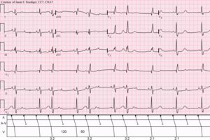
First-degree atrioventricular block is a disease of the electrical conduction system of the heart in which electrical impulses conduct from the cardiac atria to the ventricles through the atrioventricular node more slowly than normal. First degree AV block does not generally cause any symptoms, but may progress to more severe forms of heart block such as second- and third-degree atrioventricular block. It is diagnosed using an electrocardiogram, and is defined as a PR interval greater than 200 milliseconds. First degree AV block affects 0.65-1.1% of the population with 0.13 new cases per 1000 persons each year.

Heart block (HB) is a disorder in the heart's rhythm due to a fault in the natural pacemaker. This is caused by an obstruction – a block – in the electrical conduction system of the heart. Sometimes a disorder can be inherited. Despite the severe-sounding name, heart block may cause no symptoms at all in some cases, or occasional missed heartbeats in other cases, or may require the implantation of an artificial pacemaker, depending upon exactly where in the heart conduction is being impaired and how significantly it is affected.

<span class="mw-page-title-main">Supraventricular tachycardia</span> Abnormally fast heart rhythm

Supraventricular tachycardia (SVT) is an umbrella term for fast heart rhythms arising from the upper part of the heart. This is in contrast to the other group of fast heart rhythms – ventricular tachycardia, which start within the lower chambers of the heart. There are four main types of SVT: atrial fibrillation, atrial flutter, paroxysmal supraventricular tachycardia (PSVT), and Wolff–Parkinson–White syndrome. The symptoms of SVT include palpitations, feeling of faintness, sweating, shortness of breath, and/or chest pain.

<span class="mw-page-title-main">AV nodal reentrant tachycardia</span> Medical condition

AV-nodal reentrant tachycardia (AVNRT) is a type of abnormal fast heart rhythm. It is a type of supraventricular tachycardia (SVT), meaning that it originates from a location within the heart above the bundle of His. AV nodal reentrant tachycardia is the most common regular supraventricular tachycardia. It is more common in women than men. The main symptom is palpitations. Treatment may be with specific physical maneuvers, medications, or, rarely, synchronized cardioversion. Frequent attacks may require radiofrequency ablation, in which the abnormally conducting tissue in the heart is destroyed.

<span class="mw-page-title-main">Bundle branch block</span> Medical condition

A bundle branch block is a defect in one of the bundle branches in the electrical conduction system of the heart.

<span class="mw-page-title-main">Atrioventricular block</span> Medical condition

Atrioventricular block is a type of heart block that occurs when the electrical signal traveling from the atria, or the upper chambers of the heart, to ventricles, or the lower chambers of the heart, is impaired. Normally, the sinoatrial node produces an electrical signal to control the heart rate. The signal travels from the SA node to the ventricles through the atrioventricular node. In an AV block, this electrical signal is either delayed or completely blocked. When the signal is completely blocked, the ventricles produce their own electrical signal to control the heart rate. The heart rate produced by the ventricles is much slower than that produced by the SA node.

Premature atrial contraction (PAC), also known as atrial premature complexes (APC) or atrial premature beats (APB), are a common cardiac dysrhythmia characterized by premature heartbeats originating in the atria. While the sinoatrial node typically regulates the heartbeat during normal sinus rhythm, PACs occur when another region of the atria depolarizes before the sinoatrial node and thus triggers a premature heartbeat, in contrast to escape beats, in which the normal sinoatrial node fails, leaving a non-nodal pacemaker to initiate a late beat.

Lown–Ganong–Levine syndrome (LGL) is a pre-excitation syndrome of the heart. Those with LGL syndrome have episodes of abnormal heart racing with a short PR interval and normal QRS complexes seen on their electrocardiogram when in a normal sinus rhythm. LGL syndrome was originally thought to be due to an abnormal electrical connection between the atria and the ventricles, but is now thought to be due to accelerated conduction through the atrioventricular node in the majority of cases. The syndrome is named after Bernard Lown, William Francis Ganong, Jr., and Samuel A. Levine.

<span class="mw-page-title-main">Multifocal atrial tachycardia</span> Fast heart rhythm associated with exacerbations of COPD

Multifocal atrial tachycardia (MAT) is an abnormal heart rhythm, specifically a type of supraventricular tachycardia, that is particularly common in older people and is associated with exacerbations of chronic obstructive pulmonary disease (COPD). Normally, the heart rate is controlled by a cluster of cells called the sinoatrial node. When a number of different clusters of cells outside the SA node take over control of the heart rate, and the rate exceeds 100 beats per minute, this is called multifocal atrial tachycardia.

<span class="mw-page-title-main">Junctional rhythm</span> Medical condition

Junctional rhythm describes an abnormal heart rhythm resulting from impulses coming from a locus of tissue in the area of the atrioventricular node(AV node), the "junction" between atria and ventricles.

<span class="mw-page-title-main">Sinoatrial block</span> Medical condition

A sinoatrial block is a disorder in the normal rhythm of the heart, known as a heart block, that is initiated in the sinoatrial node. The initial action impulse in a heart is usually formed in the sinoatrial node and carried through the atria, down the internodal atrial pathways to the atrioventricular node (AV) node. In normal conduction, the impulse would travel across the bundle of His, down the bundle branches, and into the Purkinje fibers. This would depolarize the ventricles and cause them to contract.

<span class="mw-page-title-main">Ectopic pacemaker</span> Cardiac condition

An ectopic pacemaker, also known as ectopic focus or ectopic foci, is an excitable group of cells that causes a premature heart beat outside the normally functioning SA node of the heart. It is thus a cardiac pacemaker that is ectopic, producing an ectopic beat. Acute occurrence is usually non-life-threatening, but chronic occurrence can progress into tachycardia, bradycardia or ventricular fibrillation. In a normal heart beat rhythm, the SA node usually suppresses the ectopic pacemaker activity due to the higher impulse rate of the SA node. However, in the instance of either a malfunctioning SA node or an ectopic focus bearing an intrinsic rate superior to SA node rate, ectopic pacemaker activity may take over the natural heart rhythm. This phenomenon is called an escape rhythm, the lower rhythm having escaped from the dominance of the upper rhythm. As a rule, premature ectopic beats indicate increased myocyte or conducting tissue excitability, whereas late ectopic beats indicate proximal pacemaker or conduction failure with an escape 'ectopic' beat.

<span class="mw-page-title-main">Junctional ectopic tachycardia</span> Medical condition

Junctional ectopic tachycardia (JET) is a rare syndrome of the heart that manifests in patients recovering from heart surgery. It is characterized by cardiac arrhythmia, or irregular beating of the heart, caused by abnormal conduction from or through the atrioventricular node. In newborns and infants up to 6 weeks old, the disease may also be referred to as His bundle tachycardia or congenital JET.

<span class="mw-page-title-main">PR interval</span> Period in electrocardiography

In electrocardiography, the PR interval is the period, measured in milliseconds, that extends from the beginning of the P wave until the beginning of the QRS complex ; it is normally between 120 and 200 ms in duration. The PR interval is sometimes termed the PQ interval.

<span class="mw-page-title-main">Arrhythmia</span> Group of medical conditions characterized by irregular heartbeat

Arrhythmias, also known as cardiac arrhythmias, heart arrhythmias, or dysrhythmias, are irregularities in the heartbeat, including when it is too fast or too slow. A resting heart rate that is too fast – above 100 beats per minute in adults – is called tachycardia, and a resting heart rate that is too slow – below 60 beats per minute – is called bradycardia. Some types of arrhythmias have no symptoms. Symptoms, when present, may include palpitations or feeling a pause between heartbeats. In more serious cases, there may be lightheadedness, passing out, shortness of breath, chest pain, or decreased level of consciousness. While most cases of arrhythmia are not serious, some predispose a person to complications such as stroke or heart failure. Others may result in sudden death.

References

  1. Mangi MA, Jones WM, Mansour MK, Napier L (2022). "Atrioventricular Block Second-Degree". National Center for Biotechnology Information, U.S. National Library of Medicine. PMID   29493981 . Retrieved 3 July 2021.
  2. "Atrioventricular Block". The Lecturio Medical Concept Library. Retrieved 3 July 2021.
  3. "Lesson VI - ECG Conduction Abnormalities" . Retrieved 2009-01-07.
  4. http://health.medicscientist.com/wp-content/uploads/2011/04/SeconddegreeAVblock2_thumb.jpg%5B%5D
  5. synd/2824 at Who Named It?
  6. Mobitz W (1924). "Über die unvollständige Störung der Erregungsüberleitung zwischen Vorhof und Kammer des menschlichen Herzens". Zeitschrift für die Gesamte Experimentelle Medizin. Berlin. 41: 180–237. doi:10.1007/BF02758773. S2CID   87410648.
  7. Wenckebach KF (1898). "De Analyse van den onregelmatigen Pols. III. Over eenige Vormen van Allorythmie en Bradykardie". Nederlands Tijdschrift voor Geneeskunde. Amsterdam. 2: 1132.
  8. Silverman ME, Upshaw CB, Lange HW (August 2004). "Woldemar Mobitz and His 1924 classification of second-degree atrioventricular block". Circulation. 110 (9): 1162–1167. doi: 10.1161/01.CIR.0000140669.35049.34 . PMID   15339865.
  9. Hay J (1906). "Bradycardia and cardiac arrhythmia produced by depression of certain of the functions of the heart". The Lancet. 1906 (1): 139–143. doi:10.1016/s0140-6736(01)44443-6.
  10. Wenckebach KF (1906). "Beiträge zur Kenntnis der menschlichen Herztätigkeit [Contributions to the knowledge of human cardiac activity]". Arch Anat Physiol: 297–354.
  11. Mobitz W (1924). "Über die unvollständige Störung der Erregungsüberleitung zwischen Vorhof und Kammer des menschlichen Herzens [On the partial block of impulse conduction between atrium and ventricle of human hearts]". Z Gesamte Exp Med. 41: 180–237. doi:10.1007/bf02758773. S2CID   87410648.
  12. Silverman ME, Upshaw CB, Lange HW (August 2004). "Woldemar Mobitz and His 1924 classification of second-degree atrioventricular block". Circulation. 110 (9): 1162–1167. doi: 10.1161/01.CIR.0000140669.35049.34 . PMID   15339865.
  13. "ECG Learning Center - An introduction to clinical electrocardiography". ecg.utah.edu. University of Utah. Archived from the original on 2022-08-14. Retrieved 2022-08-14.
  14. Lilly LS (2007). Pathophysiology of heart disease : a collaborative project of medical students and faculty (4th ed.). Philadelphia: Wolters Kluwer/Lippincott Williams & Wilkins. ISBN   978-0-7817-6321-9. OCLC   938327813.
  15. 1 2 3 4 5 6 Dubin D (2000). Rapid interpretation of EKG's : ... an interactive course (6th ed.). Tampa, Fla.: Cover Publ. ISBN   978-0912912066.
  16. Wogan JM, Lowenstein SR, Gordon GS (1993). "Second-degree atrioventricular block: Mobitz type II". The Journal of Emergency Medicine. 11 (1): 47–54. doi:10.1016/0736-4679(93)90009-V. PMID   8445186.
  17. Kusumoto FM, Schoenfeld MH, Barrett C, Edgerton JR, Ellenbogen KA, Gold MR, et al. (August 2019). "2018 ACC/AHA/HRS Guideline on the Evaluation and Management of Patients With Bradycardia and Cardiac Conduction Delay: A Report of the American College of Cardiology/American Heart Association Task Force on Clinical Practice Guidelines and the Heart Rhythm Society". Journal of the American College of Cardiology. 74 (7): e51–e156. doi: 10.1016/j.jacc.2018.10.044 . PMID   30412709.