Right ventricular hypertrophy

Last updated
Right ventricular hypertrophy
Right Ventricular hypertrophy.svg
Right ventricular hypertrophy
Specialty Cardiology

Right ventricular hypertrophy (RVH) is a condition defined by an abnormal enlargement of the cardiac muscle surrounding the right ventricle. The right ventricle is one of the four chambers of the heart. It is located towards the lower-end of the heart and it receives blood from the right atrium and pumps blood into the lungs.

Contents

Since RVH is an enlargement of muscle it arises when the muscle is required to work harder. Therefore, the main causes of RVH are pathologies of systems related to the right ventricle such as the pulmonary artery, the tricuspid valve or the airways.

RVH can be benign and have little impact on day-to-day life or it can lead to conditions such as heart failure, which has a poor prognosis.

Signs and symptoms

Symptoms

Although presentations vary, individuals with right ventricular hypertrophy can experience symptoms that are associated with pulmonary hypertension, heart failure and/or a reduced cardiac output. These include: [1] [2]

People may rarely present with the symptoms of Ortner's syndrome, which include cough, haemoptysis and hoarseness.[ citation needed ]

Signs

On physical examination, the most prominent features are due to the development of right-sided heart failure. These can include a raised jugular venous pressure, ascites, left parasternal heave and a tender, enlarged liver on palpation. [3] On inspection, patients may be chronically ill, cyanotic, cachectic and occasionally jaundiced.[ citation needed ]

On auscultation, an accentuated second pulmonary sound (S2), a third heart sound termed a ‘right ventricular gallop’, as well as a systolic murmur over the tricuspid area accentuated by inspiration may be present. On occasion, the systolic murmur can be transmitted and auscultated over the liver. Less typically, diastolic murmur may also be heard as a result of pulmonary insufficiency. [3]

Causes

RVH usually occurs due to chronic lung disease or structural defects in the heart. One of the most common causes of RVH is pulmonary hypertension (PH), [3] defined as increased blood pressure in the vessels supplying blood to the lungs. PH leads to increased pulmonary artery pressure. The right ventricle tries to compensate for this increased pressure by changing its shape and size. Hypertrophy of individual myocytes results in an increase in right ventricular wall thickness. [3] The worldwide incidence of PH is 4 per million people. [4] RVH occurs in approximately 30% of these cases.[ citation needed ]

PH is broadly split into five categories by the World Health Organization, based on the underlying cause. The incidence of RVH varies between the groups. Common causes of PH include chronic obstructive pulmonary disease (COPD), pulmonary embolism, and other restrictive lung diseases. RVH often occurs as a result of these disorders. RVH is seen in 76% of patients with advanced COPD and 50% of patients with restrictive lung disease. [3]

RVH also occurs in response to structural defects in the heart. One common cause is tricuspid insufficiency. This is a disorder where the tricuspid valve fails to close properly, allowing backward flow of blood. Other structural defects which lead to RVH include tetralogy of Fallot, ventricular septal defects, pulmonary valve stenosis, and atrial septal defects.[ medical citation needed ] RVH is also associated with abdominal obesity, elevated fasting blood glucose, high systolic blood pressure, and fractional shortening of the left ventricular mid-wall.[ medical citation needed ]

Other risk factors for RVH include smoking, sleep apnea, and strenuous activity. These increase the risk of heart and lung disease and hence RVH. [5]

Pathophysiology

Right ventricular hypertrophy can be both a physiological and pathophysiological process. It becomes pathophysiological (damaging) when there is excessive hypertrophy. The pathophysiological process mainly occurs through aberrant signalling of the neuroendocrine hormones; angiotensin II, endothelin-1 and the catecholamines (e.g. noradrenaline).[ citation needed ]

Angiotensin-II and endothelin-1

Angiotensin-II and endothelin-1 are hormones that bind to the angiotensin (AT) and endothelin (ET) receptors. These are G-protein coupled receptors that act via internal signalling pathways. Through several intermediates, these pathways directly or indirectly increase reactive oxygen species (ROS) production causing accumulation in myocardial cells. This can subsequently induce necrotic cell death, fibrosis, and mitochondrial dysfunction. [6]

This has been demonstrated in animal studies. Protein Kinase C (PKC) is an intermediate molecule in the signalling pathway and mice lacking PKC shown resistance to heart failure compared to mice overexpressing PKC which shown heart dysfunction. [7]

Targeting the renin–angiotensin (RAAS) system (using angiotensin-converting enzyme inhibitors and angiotensin-receptor blockers) are a well-recognized clinical approach for reversing maladaptive cardiac hypertrophy independently of blood pressure.[ citation needed ]

Catecholamines

Catecholamines levels increase due to increased sympathetic nervous system activity. Catecholamines can act on the alpha-adrenergic receptors and beta-adrenergic receptors which are G-protein coupled receptors. This binding initiates the same intracellular signalling pathways as angiotensin and endothelin. There is also activation of cAMP and an increase in intracellular Ca2+ which leads to contractile dysfunction and fibrosis. [6]

Others

Hormones are not the only cause of RVH. Hypertrophy can also be caused by mechanical forces, mTOR pathways, nitric oxide and immune cells. Immune cells can cause hypertrophy by inducing inflammation. [6]

Diagnosis

Hexaxial reference system Hexaxial reference system.svg
Hexaxial reference system
ECG showing right axis deviation E197 (CardioNetworks ECGpedia).jpg
ECG showing right axis deviation

The two main diagnostic tests used to confirm right ventricular hypertrophy are electrocardiography and echocardiography.[ citation needed ]

Electrocardiography

The use of electrocardiogram (ECG) to measure cardiac chamber hypertrophy is well established but since the left ventricular activity is dominant on the ECG a large degree of RVH is often required for any detectable changes. Nonetheless, the ECG is used to assist with the diagnosis of RVH. A post mortem study on 51 adult male patients concluded that anatomical RVH may be diagnosed using one or more of the following ECG criteria: [8]

However, the American Heart Association recommended the use of additional diagnostic tests to diagnose RVH because no single criteria or set of criteria were considered sufficiently reliable. [9]

Echocardiography

Echocardiography can be used to directly visualise right ventricular wall thickness. The preferred technique is the trans-oesophageal approach giving a view of 4 chambers. The normal thickness of a right ventricular free wall ranges from 2-5 millimetres, with a value above 5 mm considered to be hypertrophic. [10]

Treatment

It is important to understand that right ventricular hypertrophy in itself is not the main issue, but what right ventricular hypertrophy represents is. Right ventricular hypertrophy is the intermediate stage between increased right ventricular pressure (in the early stages) and right ventricle failure (in the later stages). [11] As such, management of right ventricular hypertrophy is about either preventing the development of right ventricular hypertrophy in the first place, or preventing the progression towards right ventricle failure. Right ventricular hypertrophy in itself has no (pharmacological) treatment. [5]

Treating the cause

Since the main causes of right ventricular hypertrophy is tricuspid regurgitation or pulmonary hypertension (discussed above), management involves treatment of these conditions. [3] Tricuspid regurgitation is typically treated conservatively by aiming to treat the underlying cause and following up the patient regularly. [12] Surgery is considered in more serious situations where the patient is severely symptomatic. Surgical options include either: replacement of the valve or repair of the valve (termed annuloplasty). [3] When it comes to replacement, there is a choice between a bioprosthetic valve or a mechanical valve, depending upon the specific patient characteristics. Mechanical valve has greater durability, but requires anti-coagulation to reduce the risk of thrombosis. [3] Treatment of pulmonary hypertension will depend on the specific cause of the pulmonary hypertension. On top of this, the following may also be considered: diuretic, oxygen and anti-coagulant therapy. [3]

Managing the complications

After a prolonged period, the right ventricle fails to adapt sufficiently to pump against increased right ventricle pressure, which is termed right ventricular failure. This right ventricular failure is the main complication of right ventricular hypertrophy. The mechanisms underlying the progression from hypertrophy to failure is not well understood, [11] and the best management approach involves reducing/minimising the risk factors of progression. Lifestyle changes can often help to reduce the risk of this progression. Lifestyle changes include: eating less salty food as salt consumption leads to greater fluid retention by the body; smoking cessation; avoiding excessive alcohol consumption as alcohol reduces the force of heart contractions. Once right ventricular hypertrophy progresses to right ventricular failure, the treatment becomes that of heart failure. Briefly, this includes the use of:[ citation needed ]

Related Research Articles

<span class="mw-page-title-main">Heart sounds</span> Noise generated by the beating heart

Heart sounds are the noises generated by the beating heart and the resultant flow of blood through it. Specifically, the sounds reflect the turbulence created when the heart valves snap shut. In cardiac auscultation, an examiner may use a stethoscope to listen for these unique and distinct sounds that provide important auditory data regarding the condition of the heart.

Systole is the part of the cardiac cycle during which some chambers of the heart contract after refilling with blood.

<span class="mw-page-title-main">Pulmonary heart disease</span> Medical condition

Pulmonary heart disease, also known as cor pulmonale, is the enlargement and failure of the right ventricle of the heart as a response to increased vascular resistance or high blood pressure in the lungs.

<span class="mw-page-title-main">Diastole</span> Part of the cardiac cycle

Diastole is the relaxed phase of the cardiac cycle when the chambers of the heart are re-filling with blood. The contrasting phase is systole when the heart chambers are contracting. Atrial diastole is the relaxing of the atria, and ventricular diastole the relaxing of the ventricles.

<span class="mw-page-title-main">Mitral stenosis</span> Heart disease with narrowing of valve

Mitral stenosis is a valvular heart disease characterized by the narrowing of the opening of the mitral valve of the heart. It is almost always caused by rheumatic valvular heart disease. Normally, the mitral valve is about 5 cm2 during diastole. Any decrease in area below 2 cm2 causes mitral stenosis. Early diagnosis of mitral stenosis in pregnancy is very important as the heart cannot tolerate increased cardiac output demand as in the case of exercise and pregnancy. Atrial fibrillation is a common complication of resulting left atrial enlargement, which can lead to systemic thromboembolic complications such as stroke.

<span class="mw-page-title-main">Aortic regurgitation</span> Medical condition

Aortic regurgitation (AR), also known as aortic insufficiency (AI), is the leaking of the aortic valve of the heart that causes blood to flow in the reverse direction during ventricular diastole, from the aorta into the left ventricle. As a consequence, the cardiac muscle is forced to work harder than normal.

<span class="mw-page-title-main">Atrial septal defect</span> Human heart defect present at birth

Atrial septal defect (ASD) is a congenital heart defect in which blood flows between the atria of the heart. Some flow is a normal condition both pre-birth and immediately post-birth via the foramen ovale; however, when this does not naturally close after birth it is referred to as a patent (open) foramen ovale (PFO). It is common in patients with a congenital atrial septal aneurysm (ASA).

<span class="mw-page-title-main">Jugular venous pressure</span> Blood pressure in a vein of the neck

The jugular venous pressure is the indirectly observed pressure over the venous system via visualization of the internal jugular vein. It can be useful in the differentiation of different forms of heart and lung disease. Classically three upward deflections and two downward deflections have been described.

<span class="mw-page-title-main">Ventricular septal defect</span> Medical condition

A ventricular septal defect (VSD) is a defect in the ventricular septum, the wall dividing the left and right ventricles of the heart. The extent of the opening may vary from pin size to complete absence of the ventricular septum, creating one common ventricle. The ventricular septum consists of an inferior muscular and superior membranous portion and is extensively innervated with conducting cardiomyocytes.

<span class="mw-page-title-main">Mitral regurgitation</span> Form of valvular heart disease

Mitral regurgitation(MR), also known as mitral insufficiency or mitral incompetence, is a form of valvular heart disease in which the mitral valve is insufficient and does not close properly when the heart pumps out blood. It is the abnormal leaking of blood backwards – regurgitation from the left ventricle, through the mitral valve, into the left atrium, when the left ventricle contracts. Mitral regurgitation is the most common form of valvular heart disease.
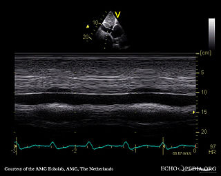
<span class="mw-page-title-main">Eisenmenger syndrome</span> Medical condition

Eisenmenger syndrome or Eisenmenger's syndrome is defined as the process in which a long-standing left-to-right cardiac shunt caused by a congenital heart defect causes pulmonary hypertension and eventual reversal of the shunt into a cyanotic right-to-left shunt. Because of the advent of fetal screening with echocardiography early in life, the incidence of heart defects progressing to Eisenmenger syndrome has decreased.

<span class="mw-page-title-main">Ventricular hypertrophy</span> Medical condition

Ventricular hypertrophy (VH) is thickening of the walls of a ventricle of the heart. Although left ventricular hypertrophy (LVH) is more common, right ventricular hypertrophy (RVH), as well as concurrent hypertrophy of both ventricles can also occur.

<span class="mw-page-title-main">Valvular heart disease</span> Disease in the valves of the heart

Valvular heart disease is any cardiovascular disease process involving one or more of the four valves of the heart. These conditions occur largely as a consequence of aging, but may also be the result of congenital (inborn) abnormalities or specific disease or physiologic processes including rheumatic heart disease and pregnancy.

<span class="mw-page-title-main">Atrioventricular septal defect</span> Medical condition

Atrioventricular septal defect (AVSD) or atrioventricular canal defect (AVCD), also known as "common atrioventricular canal" or "endocardial cushion defect" (ECD), is characterized by a deficiency of the atrioventricular septum of the heart that creates connections between all four of its chambers. It is a very specific combination of 3 defects:

<span class="mw-page-title-main">Cardiomegaly</span> Medical condition

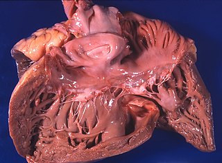
Cardiomegaly is a medical condition in which the heart becomes enlarged. It is more commonly referred to simply as "having an enlarged heart". It is usually the result of underlying conditions that make the heart work harder, such as obesity, heart valve disease, high blood pressure (hypertension), and coronary artery disease. Cardiomyopathy is also associated with cardiomegaly.

A right-to-left shunt is a cardiac shunt which allows blood to flow from the right heart to the left heart. This terminology is used both for the abnormal state in humans and for normal physiological shunts in reptiles.

The following outline is provided as an overview of and topical guide to cardiology, the branch of medicine dealing with disorders of the human heart. The field includes medical diagnosis and treatment of congenital heart defects, coronary artery disease, heart failure, valvular heart disease and electrophysiology. Physicians who specialize in cardiology are called cardiologists.

<span class="mw-page-title-main">Left axis deviation</span> Heart condition

In electrocardiography, left axis deviation (LAD) is a condition wherein the mean electrical axis of ventricular contraction of the heart lies in a frontal plane direction between −30° and −90°. This is reflected by a QRS complex positive in lead I and negative in leads aVF and II.

<span class="mw-page-title-main">Heart failure with preserved ejection fraction</span> Medical condition

Heart failure with preserved ejection fraction (HFpEF) is a form of heart failure in which the ejection fraction – the percentage of the volume of blood ejected from the left ventricle with each heartbeat divided by the volume of blood when the left ventricle is maximally filled – is normal, defined as greater than 50%; this may be measured by echocardiography or cardiac catheterization. Approximately half of people with heart failure have preserved ejection fraction, while the other half have a reduction in ejection fraction, called heart failure with reduced ejection fraction (HFrEF).

<span class="mw-page-title-main">Pathophysiology of heart failure</span>

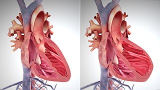
The main pathophysiology of heart failure is a reduction in the efficiency of the heart muscle, through damage or overloading. As such, it can be caused by a wide number of conditions, including myocardial infarction, hypertension and cardiac amyloidosis. Over time these increases in workload will produce changes to the heart itself:

References

  1. "Pulmonary Hypertension". nhs.uk. NHS. 14 March 2017. Retrieved 23 March 2019.
  2. Ibrahim, Bassem (12 December 2016). "Right Ventricular Failure". e-Journal of Cardiology Practice. 14 (32). Retrieved 23 March 2019.
  3. 1 2 3 4 5 6 7 8 9 Bhattacharya, Priyanka; Sharma, Sandeep (15 February 2019). "Right Ventricular Hypertrophy". StatPearls. NCBI. Retrieved 23 March 2019.
  4. Oudiz, Ronald (21 June 2018). "Idiopathic Pulmonary Arterial Hypertension". Medscape. Retrieved 23 March 2019.
  5. 1 2 Johnson, Jon (16 August 2017). "What is right ventricular hypertrophy?". Medical News Today. Retrieved 23 March 2019.
  6. 1 2 3 Nakamura, Michinari; Sadoshima, Junichi (19 April 2018). "Mechanisms of physiological and pathological cardiac hypertrophy". Nature Reviews Cardiology. 15 (7): 387–407. doi:10.1038/s41569-018-0007-y. PMID   29674714. S2CID   4975072.
  7. Braz, Julian; Gregory, Kimberley (15 February 2004). "PKC-α regulates cardiac contractility and propensity toward heart failure". Nature Medicine. 10 (3): 248–254. doi:10.1038/nm1000. PMID   14966518. S2CID   8812202.
  8. Lehtonen, Jari (1988). "Electrocardiographic Criteria for the Diagnosis of Right Ventricular Hypertrophy Verified at Autopsy". Chest. 93 (4): 839–42. doi:10.1378/chest.93.4.839. PMID   2964996.
  9. Hancock, William (2009). "AHA/ACCF/HRS Recommendations for the Standardization and Interpretation of the Electrocardiogram". Journal of the American College of Cardiology. 53 (11): 992–1002. doi: 10.1016/j.jacc.2008.12.015 . PMID   19281932.
  10. Ho, Siew Yen (2006). "Anatomy, echocardiography, and normal right ventricular dimensions". Heart. 92 (Supp 1): i2–i13. doi:10.1136/hrt.2005.077875. PMC   1860731 . PMID   16543598.
  11. 1 2 van der Bruggen, C (2017). "RV pressure overload: from hypertrophy to failure". Cardiovascular Research. 113 (12): 1423–1432. doi: 10.1093/cvr/cvx145 . PMID   28957530.
  12. "Tricuspid Regurgitation". BMJ Best Practice. BMJ. March 2019. Retrieved 23 March 2019.