Treatment depends on the type of cardiomyopathy and the severity of symptoms.[5] Treatments may include lifestyle changes, medications, or surgery.[5] Surgery may include a ventricular assist device or heart transplant.[5] In 2015 cardiomyopathy and myocarditis affected 2.5 million people.[6] In a recent nationwide study of cardiomyopathies between 2004 and 2023, the overall prevalence more than doubled over the two decades, with an age-standardized prevalence of 5 in 1,000 in men, and 3 in 1,000 women in 2023. Dilated cardiomyopathy was the most common subtype (3 in 1,000 for men, 1 in 1,000 in women), followed by hypertrophic cardiomyopathy (about 1 in 1,000 for men, 0.5 in 1,000 for women).[10] In this investigation, cardiomyopathies were associated with a substantial excess mortality compared with the general population, ranging from a 32-fold and 16-fold increase in the youngest men and women with cardiomyopathies, to a two-fold increase in the oldest patients.[10]
Shortness of breath or trouble breathing, especially with physical exertion
Fatigue
Swelling in the ankles, feet, legs, abdomen and veins in the neck
Dizziness
Lightheadedness
Fainting during physical activity
Arrhythmias (abnormal heartbeats)
Chest pain, especially after physical exertion or heavy meals
Heart murmurs (unusual sounds associated with heartbeats)
Causes
Cardiomyopathies can be of genetic (familial) or non-genetic (acquired) origin.[11] Genetic cardiomyopathies usually are caused by sarcomere or cytoskeletal diseases, neuromuscular disorders, inborn errors of metabolism, malformation syndromes and sometimes are unidentified.[12][13] Non-genetic cardiomyopathies can have definitive causes such as viral infections, myocarditis and others.[14][15]
Cardiomyopathies are either confined to the heart or are part of a generalized systemic disorder, both often leading to cardiovascular death or progressive heart failure-related disability. Other diseases that cause heart muscle dysfunction are excluded, such as coronary artery disease, hypertension, or abnormalities of the heart valves.[16] Often, the underlying cause remains unknown, but in many cases the cause may be identifiable.[17] Alcoholism, for example, has been identified as a cause of dilated cardiomyopathy, as has drug toxicity, and certain infections (including Hepatitis C).[18][19][20] Untreated celiac disease can cause cardiomyopathies, which can completely reverse with a timely diagnosis.[21] In addition to acquired causes, molecular biology and genetics have given rise to the recognition of various genetic causes.[19][22]
A more clinical categorization of cardiomyopathy as 'hypertrophied', 'dilated', or 'restrictive',[23] has become difficult to maintain because some of the conditions could fulfill more than one of those three categories at any particular stage of their development.[24]
The current American Heart Association (AHA) definition divides cardiomyopathies into primary, which affect the heart alone, and secondary, which are the result of illness affecting other parts of the body. These categories are further broken down into subgroups which incorporate new genetic and molecular biology knowledge.[25]
Mechanism
The pathophysiology of cardiomyopathies is better understood at the cellular level with advances in molecular techniques. Mutant proteins can disturb cardiac function in the contractile apparatus (or mechanosensitive complexes). Cardiomyocyte alterations and their persistent responses at the cellular level cause changes that are correlated with sudden cardiac death and other cardiac problems.[26]
Cardiomyopathies are generally varied individually. Different factors can cause cardiomyopathies in adults as well as children. For example, dilated cardiomyopathy in adults is associated with ischemic cardiomyopathy, hypertension, valvular diseases, and genetics. In children, neuromuscular diseases such as Becker muscular dystrophy or X-linked genetic disorder, are directly linked with cardiomyopathies.[27]
Diagnosis
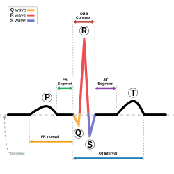
Normal sinus rhythm on EKG
Among the diagnostic procedures done to determine a cardiomyopathy are:[28]
Ischemic cardiomyopathy (not formally included in the classification, due to ischemic cardiomyopathy being a direct result of another cardiac problem)[30]
Treatment may include suggestion of lifestyle changes to better manage the condition. Treatment depends on the type of cardiomyopathy and condition of disease, but may include medication (conservative treatment) or iatrogenic/implanted pacemakers for slow heart rates, defibrillators for those prone to fatal heart rhythms, ventricular assist devices (VADs) for severe heart failure, or catheter ablation for recurring dysrhythmias that cannot be eliminated by medication or mechanical cardioversion. The goal of treatment is often symptom relief, and some patients may eventually require a heart transplant.[28]
Acoramidis (Attruby) was approved for medical use in the United States in November 2024, to treat adults with cardiomyopathy of wild-type or variant (hereditary) transthyretin-mediated amyloidosis (ATTR-CM) to reduce death and hospitalization related to heart problems.[34]
↑Lakdawala NK, Stevenson LW, Loscalzo J (2015). "Chapter 287". In Kasper DL, Fauci AS, Hauser SL, Longo DL, Jameson JL, Loscalzo J (eds.). Harrison's Principles of Internal Medicine (19thed.). McGraw-Hill. p.1553. ISBN978-0-07-180215-4.
↑Lilly, Leonard S., ed. (2011). Pathophysiology of heart disease: a collaborative project of medical students and faculty (5thed.). Baltimore, MD: Wolters Kluwer/Lippincott Williams & Wilkins. ISBN978-1-60547-723-7. OCLC649701807.
12Westphal JG, Rigopoulos AG, Bakogiannis C, Ludwig SE, Mavrogeni S, Bigalke B, etal. (2017). "The MOGE(S) classification for cardiomyopathies: current status and future outlook". Heart Fail Rev (Review). 22 (6): 743–752. doi:10.1007/s10741-017-9641-4. PMID28721466. S2CID36117047.
↑Valentin Fuster, John Willis Hurst (2004). Hurst's the heart. McGraw-Hill Professional. p.1884. ISBN978-0-07-143225-2. Archived from the original on 27 May 2013. Retrieved 11 November 2010.
Maron BJ, Udelson JE, Bonow RO, Nishimura RA, Ackerman MJ, Estes NA, Cooper LT, Link MS, Maron MS (1 December 2015). "Eligibility and Disqualification Recommendations for Competitive Athletes With Cardiovascular Abnormalities: Task Force 3: Hypertrophic Cardiomyopathy, Arrhythmogenic Right Ventricular Cardiomyopathy and Other Cardiomyopathies, and Myocarditis: A Scientific Statement From the American Heart Association and American College of Cardiology". Circulation. 132 (22): e273–280. doi:10.1161/CIR.0000000000000239. ISSN1524-4539. PMID26621644. S2CID207639288.
This page is based on this Wikipedia article Text is available under the CC BY-SA 4.0 license; additional terms may apply. Images, videos and audio are available under their respective licenses.