| Pulmonary valve regurgitation | |
|---|---|
| Other names | Pulmonary insufficiency, pulmonary incompetence |
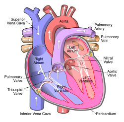 | |
| Diagram of the human heart | |
| Specialty | Cardiology |
| Causes | Pulmonary hypertension, Infective endocarditis [1] |
| Diagnostic method | EKG, Echocardiogram [2] |
| Treatment | Depends on cause [3] (See cause) |
Pulmonary (or pulmonic [4] ) regurgitation (or insufficiency, incompetence) is a condition in which the pulmonary valve is incompetent [5] and allows backflow from the pulmonary artery to the right ventricle of the heart during diastole. [6] While a small amount of backflow may occur ordinarily, it is usually only shown on an echocardiogram and is harmless. More pronounced regurgitation that is noticed through a routine physical examination is a medical sign of disease and warrants further investigation.[ medical citation needed ] If it is secondary to pulmonary hypertension it is referred to as a Graham Steell murmur. [7]
