Related Research Articles

A heart valve is a one-way valve that allows blood to flow in one direction through the chambers of the heart. Four valves are usually present in a mammalian heart and together they determine the pathway of blood flow through the heart. A heart valve opens or closes according to differential blood pressure on each side.

Heart sounds are the noises generated by the beating heart and the resultant flow of blood through it. Specifically, the sounds reflect the turbulence created when the heart valves snap shut. In cardiac auscultation, an examiner may use a stethoscope to listen for these unique and distinct sounds that provide important auditory data regarding the condition of the heart.
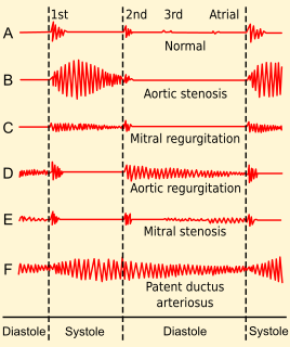
Heart murmurs are heart sounds produced when blood is pumped across a heart valve and creates a sound loud enough to be heard with a stethoscope. Murmurs are of various types and are important in the detection of cardiac and valvular pathologies.

Mitral valve prolapse (MVP) is a valvular heart disease characterized by the displacement of an abnormally thickened mitral valve leaflet into the left atrium during systole. It is the primary form of myxomatous degeneration of the valve. There are various types of MVP, broadly classified as classic and nonclassic. In severe cases of classic MVP, complications include mitral regurgitation, infective endocarditis, congestive heart failure, and, in rare circumstances, cardiac arrest.

Mitral stenosis is a valvular heart disease characterized by the narrowing of the orifice of the mitral valve of the heart. It is almost always caused by rheumatic valvular heart disease. Normally, the mitral valve is about 5 cm2 during diastole. Any decrease in area below 2 cm2 causes mitral stenosis. Early diagnosis of mitral stenosis in pregnancy is very important as the heart cannot tolerate increased cardiac output demand as in the case of exercise and pregnancy. Atrial fibrillation is a common complication of resulting left atrial enlargement, which can lead to systemic thromboembolic complications like stroke.

Aortic insufficiency (AI), also known as aortic regurgitation (AR), is the leaking of the aortic valve of the heart that causes blood to flow in the reverse direction during ventricular diastole, from the aorta into the left ventricle. As a consequence, the cardiac muscle is forced to work harder than normal.

Mitral insufficiency(MI), also known as mitral regurgitation(MR), or mitral incompetence is a form of valvular heart disease in which the mitral valve does not close properly when the heart pumps out blood. It is the abnormal leaking of blood backwards – regurgitation from the left ventricle, through the mitral valve, into the left atrium, when the left ventricle contracts. Mitral insufficiency is the most common form of valvular heart disease.
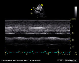
A transthoracic echocardiogram (TTE) is the most common type of echocardiogram, which is a still or moving image of the internal parts of the heart using ultrasound. In this case, the probe is placed on the chest or abdomen of the subject to get various views of the heart. It is used as a non-invasive assessment of the overall health of the heart, including a patient's heart valves and degree of heart muscle contraction. The images are displayed on a monitor for real-time viewing and then recorded.
Valvular heart disease is any cardiovascular disease process involving one or more of the four valves of the heart. These conditions occur largely as a consequence of aging, but may also be the result of congenital (inborn) abnormalities or specific disease or physiologic processes including rheumatic heart disease and pregnancy.

Right ventricular hypertrophy (RVH) is a condition defined by an abnormal enlargement of the cardiac muscle surrounding the right ventricle. The right ventricle is one of the four chambers of the heart. It is located towards the lower-end of the heart and it receives blood from the right atrium and pumps blood into the lungs.
Tricuspid Valve Stenosis is a valvular heart disease that narrows the opening of the heart's tricuspid valve. It is a relatively rare condition that causes stenosis-increased restriction of blood flow through the valve.
Tricuspid insufficiency (TI), more commonly called tricuspid regurgitation (TR), is a type of valvular heart disease in which the tricuspid valve of the heart, located between the right atrium and right ventricle, does not close completely when the right ventricle contracts (systole). TR allows the blood to flow backwards from the right ventricle to the right atrium, which increases the volume and pressure of the blood both in the right atrium and the right ventricle, which may increase central venous volume and pressure if the backward flow is sufficiently severe.

A myxoma is a rare benign tumor of the heart. Myxomata are the most common primary cardiac tumor in adults, and are most commonly found within the left atrium near the valve of the fossa ovalis. Myxomata may also develop in the other heart chambers. The tumor is derived from multipotent mesenchymal cells. Cardiac myxoma can affect adults between 30 and 60 years of age.

Lutembacher's syndrome is a very rare form of congenital heart disease that affects one of the chambers of the heart as well as a valve. It is commonly known as both congenital atrial septal defect (ASD) and acquired mitral stenosis (MS). Congenital atrial septal defect refers to a hole being in the septum or wall that separates the two atria; this condition is usually seen in fetuses and infants. Mitral stenosis refers to mitral valve leaflets sticking to each other making the opening for blood to pass from the atrium to the ventricles very small. With the valve being so small, blood has difficulty passing from the left atrium into the left ventricle. Septal defects that may occur with Lutembacher's syndrome include: Ostium primum atrial septal defect or ostium secundum which is more prevalent.
Pulmonaryinsufficiency is a condition in which the pulmonary valve is incompetent and allows backflow from the pulmonary artery to the right ventricle of the heart during diastole. While a small amount of backflow may occur ordinarily, it is usually only shown on an echocardiogram and is harmless. More pronounced regurgitation that is noticed through a routine physical examination is a medical sign of disease and warrants further investigation. If it is secondary to pulmonary hypertension it is referred to as a Graham Steell murmur.
The third heart sound or S3 is a rare extra heart sound that occurs soon after the normal two "lub-dub" heart sounds (S1 and S2). S3 is associated with heart failure.
Diastolic heart murmurs are heart murmurs heard during diastole, i.e. they start at or after S2 and end before or at S1. Many involve stenosis of the atrioventricular valves or regurgitation of the semilunar valves.
A presystolic murmur, also called presystolic accentuation, is a type of diastolic heart murmur typically associated with the opening snap in mitral valve stenosis. It is heard following the middiastolic rumble of the stenotic valve, during the diastasis phase, making it a "late diastolic" murmur.
Heart murmurs are most frequently organized by timing, into systolic heart murmurs and diastolic heart murmurs. However, continuous murmurs can not be directly placed into either category.
The Carey Coombs murmur or Coombs murmur is a clinical sign which occurs in patients with mitral valvulitis due to acute rheumatic fever. It is described as a short, mid-diastolic rumble best heard at the apex, which disappears as the valvulitis improves. It is often associated with an S3 gallop rhythm, and can be distinguished from the diastolic murmur of mitral stenosis by the absence of an opening snap before the murmur. It is audible at apex. The murmur is caused by increased blood flow across a thickened mitral valve.
References
- ↑ Archives of Diagnosis, Volume 10. University of Chicago. 1918. p. 60. Retrieved 27 October 2017.
- ↑ Heart sounds made incredibly easy!. Ambler, Pa.: Lippincott Williams & Wilkins. 2005. p. 143. ISBN 9781582553580.
- 1 2 Jose, V. Jacob (2017). Cardiology: Clinical Methods. JP Medical Ltd. p. 82. ISBN 9789385999451.
- ↑ Fraser, Alan G.; Weston, Clive F. M. (January 1991). "The Graham Steell Murmur: Eponymous Serendipity?". J R Coll Physicians Lond. 25 (1): 66–70. PMC 5377079 . PMID 2023159.
| | This medical sign article is a stub. You can help Wikipedia by expanding it. |