
A heart valve is a one-way valve that normally allows blood to flow in only one direction through the heart. The four valves are commonly represented in a mammalian heart that determines the pathway of blood flow through the heart. A heart valve opens or closes incumbent on differential blood pressure on each side.

Heart sounds are the noises generated by the beating heart and the resultant flow of blood through it. Specifically, the sounds reflect the turbulence created when the heart valves snap shut. In cardiac auscultation, an examiner may use a stethoscope to listen for these unique and distinct sounds that provide important auditory data regarding the condition of the heart.

The aortic valve is a valve in the human heart between the left ventricle and the aorta. It is one of the two semilunar valves of the heart, the other being the pulmonary valve. The heart has four valves; the other two are the mitral and the tricuspid valves. The aortic valve normally has three cusps or leaflets, although in 1–2% of the population it is found to congenitally have two leaflets. The aortic valve is the last structure in the heart the blood travels through before stopping the flow through the systemic circulation.
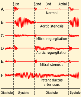
Heart murmurs are heart sounds produced when blood is pumped across a heart valve and creates a sound loud enough to be heard with a stethoscope. Murmurs are of various types and are important in the detection of cardiac and valvular pathologies.

The systole is the part of the cardiac cycle during which some chambers of the heart muscle contract after refilling with blood. The term originates, via New Latin, from Ancient Greek συστολή (sustolē), from συστέλλειν, and is similar to the use of the English term to squeeze.
A ventricle is one of two large chambers toward the bottom of the heart that collect and expel blood received from an atrium towards the peripheral beds within the body and lungs. The atrium primes the pump.
Afterload is the pressure that the heart must work against to eject blood during systole. Afterload is proportional to the average arterial pressure. As aortic and pulmonary pressures increase, the afterload increases on the left and right ventricles respectively. Afterload changes to adapt to the continually changing demands on an animal's cardiovascular system. Afterload is proportional to mean systolic blood pressure and is measured in millimeters of mercury.

Mitral stenosis is a valvular heart disease characterized by the narrowing of the orifice of the mitral valve of the heart. It is almost always caused by rheumatic valvular heart disease. Normally, the mitral valve is about 5 cm2 during diastole. Any decrease in area below 2 cm2 causes mitral stenosis. Early diagnosis of mitral stenosis in pregnancy is very important as the heart cannot tolerate increased cardiac output demand as in the case of exercise and pregnancy. Atrial fibrillation is a common complication of resulting left atrial enlargement, which can lead to systemic thromboembolic complications like stroke.
A stenosis is an abnormal narrowing in a blood vessel or other tubular organ or structure such as foramina and canals. It is also sometimes called a stricture.

Mitral regurgitation (MR), also known as mitral insufficiency, or mitral incompetence is a form of valvular heart disease in which the mitral valve does not close properly when the heart pumps out blood. It is the abnormal leaking of blood backwards from the left ventricle, through the mitral valve, into the left atrium, when the left ventricle contracts, i.e. there is regurgitation of blood back into the left atrium. MR is the most common form of valvular heart disease.
In cardiology, an Austin Flint murmur is a low-pitched rumbling heart murmur which is best heard at the cardiac apex. It can be a mid-diastolic or presystolic murmur It is associated with severe aortic regurgitation, although the role of this sign in clinical practice has been questioned.
A transthoracic echocardiogram (TTE) is the most common type of echocardiogram, which is a still or moving image of the internal parts of the heart using ultrasound. In this case, the probe is placed on the chest or abdomen of the subject to get various views of the heart. It is used as a non-invasive assessment of the overall health of the heart, including a patient's heart valves and degree of heart muscle contraction. The images are displayed on a monitor for real-time viewing and then recorded.
Valvular heart disease is any cardiovascular disease process involving one or more of the four valves of the heart. These conditions occur largely as a consequence of aging, but may also be the result of congenital (inborn) abnormalities or specific disease or physiologic processes including rheumatic heart disease and pregnancy.
Regurgitation is blood flow in the opposite direction from normal, as the backward flowing of blood into the heart or between heart chambers. It is the circulatory equivalent of backflow in engineered systems. It is sometimes called reflux, as in abdominojugular reflux.
The following outline is provided as an overview of and topical guide to cardiology, the branch of medicine dealing with disorders of the human heart. The field includes medical diagnosis and treatment of congenital heart defects, coronary artery disease, heart failure, valvular heart disease and electrophysiology. Physicians who specialize in cardiology are called cardiologists.
Systolic heart murmurs are heart murmurs heard during systole, i.e. they begin and end between S1 and S2. Many involve stenosis of the semilunar valves or regurgitation of the atrioventricular valves.
Cardiac physiology or heart function is the study of healthy, unimpaired function of the heart: involving blood flow; myocardium structure; the electrical conduction system of the heart; the cardiac cycle and cardiac output and how these interact and depend on one another.

In cardiac physiology, isovolumetric contraction is an event occurring in early systole during which the ventricles contract with no corresponding volume change (isovolumetrically). This short-lasting portion of the cardiac cycle takes place while all heart valves are closed.
