Left anterior descending artery

Last updated
Left anterior descending artery
Gray492.png
Sternocostal surface of heart. Anterior descending branch labeled at upper right
Anterior interventricular artery.png
Details
Location Heart
Source left coronary artery
Branchesseptals, diagonals
Suppliesanterolateral myocardium, apex, interventricular septum, 45–55% of the left ventricle (LV)
Identifiers
Latin ramus interventricularis anterior arteriae coronariae sinistrae
Acronym(s)LAD
TA98 A12.2.03.202
TA2 4143
FMA 3862
Anatomical terminology

The left anterior descending artery (also LAD, anterior interventricular branch of left coronary artery, or anterior descending branch) is a branch of the left coronary artery. It supplies the anterior portion of the left ventricle. [1] It provides about half of the arterial supply to the left ventricle and is thus considered the most important vessel supplying the left ventricle.[ citation needed ] Blockage of this artery is often called the widow-maker infarction due to a high risk of death. [2]

Contents

Structure

Course

It first passes at posterior to the pulmonary artery, then passes anteriorward between that pulmonary artery and the left atrium to reach the anterior interventricular sulcus, along which it descends to the notch of cardiac apex. In 78% of cases, it reaches the apex of the heart.[ citation needed ]

Although rare, multiple anomalous courses of the LAD have been described. These include the origin of the artery from the right aortic sinus. [3]

Branches

The LAD gives off two types of branches: septals and diagonals.

Segments

Function

The artery supplies the anterior region of the left ventricle, including: the anterolateral myocardium, apex, anterior interventricular septum, and anterolateral papillary muscle. [7] The LAD typically supplies 45–55% of the left ventricle and is therefore considered the most critical vessel in terms of myocardial blood supply.[ citation needed ]

Left: Critical stenosis (95%) of the proximal LAD in a patient with Wellens' Warning.
Right: The same patient after reperfusion. Wellens' Warning.gif
Left: Critical stenosis (95%) of the proximal LAD in a patient with Wellens' Warning.
Right: The same patient after reperfusion.

Widow maker

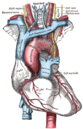
Widow maker is an alternative name for the anterior interventricular branch of the left coronary artery. [8] [2] The name widow maker may also apply to the left coronary artery [9] or severe occlusions to that artery. [10] [11]

This term is used because the left main coronary and/or the left anterior descending supply blood to large areas of the heart. This means that if these arteries are abruptly and completely occluded it will cause a massive heart attack that will likely lead to sudden death. The blockage that kills is made up of platelets streaming to the site of a ruptured cholesterol plaque. Even a small amount of plaque in this area can (for a variety of poorly understood reasons) rupture and cause death; bypassing chronic blockages or trying to open them up with angioplasty does not prevent heart attack but it can restore blood flow in case of a sudden blockage or heart attack and if performed within a rapid time period can minimize the damage done. An example of the devastating results of a complete occlusion of the LAD artery was the sudden death of former NBC News Washington Bureau Chief Tim Russert, [12] as well as the near-death of film director Kevin Smith. [13]

From the minute a widow maker heart attack hits, survival time ranges from minutes to several hours. Rapidly progressing symptoms should signal the need for immediate attention. Symptoms of initial onset may include nausea, shortness of breath, pain in the head, jaw, arms or chest, numbness in fingers, often of a novel but imprecise sensation which builds with irregular heart beat. Early symptoms may be mistaken for food poisoning, flu or general malaise until they intensify. A widow maker cannot kill instantly but induces cardiac arrest which may do so within 10 to 20 minutes of no circulation. A victim with no pulse or breath is still alive, living off oxygen stored in the blood and may be able to be rescued if treatment is begun promptly within this window. [14]

Additional images

Related Research Articles

<span class="mw-page-title-main">Aorta</span> Largest artery in the human body

The aorta is the main and largest artery in the human body, originating from the left ventricle of the heart, branching upwards immediately after, and extending down to the abdomen, where it splits at the aortic bifurcation into two smaller arteries. The aorta distributes oxygenated blood to all parts of the body through the systemic circulation.

<span class="mw-page-title-main">Coronary circulation</span> Circulation of blood in the blood vessels of the heart muscle (myocardium)

Coronary circulation is the circulation of blood in the arteries and veins that supply the heart muscle (myocardium). Coronary arteries supply oxygenated blood to the heart muscle. Cardiac veins then drain away the blood after it has been deoxygenated. Because the rest of the body, and most especially the brain, needs a steady supply of oxygenated blood that is free of all but the slightest interruptions, the heart is required to function continuously. Therefore its circulation is of major importance not only to its own tissues but to the entire body and even the level of consciousness of the brain from moment to moment. Interruptions of coronary circulation quickly cause heart attacks, in which the heart muscle is damaged by oxygen starvation. Such interruptions are usually caused by coronary ischemia linked to coronary artery disease, and sometimes to embolism from other causes like obstruction in blood flow through vessels.

<span class="mw-page-title-main">Coronary artery bypass surgery</span> Surgical procedure to restore normal blood flow to an obstructed coronary artery

Coronary artery bypass surgery, also known as coronary artery bypass graft, is a surgical procedure to treat coronary artery disease (CAD), the buildup of plaques in the arteries of the heart. It can relieve chest pain caused by CAD, slow the progression of CAD, and increase life expectancy. It aims to bypass narrowings in heart arteries by using arteries or veins harvested from other parts of the body, thus restoring adequate blood supply to the previously ischemic heart.

<span class="mw-page-title-main">Papillary muscle</span> Heart ventricle muscles

The papillary muscles are muscles located in the ventricles of the heart. They attach to the cusps of the atrioventricular valves via the chordae tendineae and contract to prevent inversion or prolapse of these valves on systole.

<span class="mw-page-title-main">Coronary arteries</span> Artery of the coronary circulation which transports blood into and out of the cardiac muscle

The coronary arteries are the arterial blood vessels of coronary circulation, which transport oxygenated blood to the heart muscle. The heart requires a continuous supply of oxygen to function and survive, much like any other tissue or organ of the body.

<span class="mw-page-title-main">Atrioventricular node</span> Part of the electrical conduction system of the heart

The atrioventricular node or AV node electrically connects the heart's atria and ventricles to coordinate beating in the top of the heart; it is part of the electrical conduction system of the heart. The AV node lies at the lower back section of the interatrial septum near the opening of the coronary sinus, and conducts the normal electrical impulse from the atria to the ventricles. The AV node is quite compact.

<span class="mw-page-title-main">Cardiac catheterization</span> Insertion of a catheter into a chamber or vessel of the heart

Cardiac catheterization is the insertion of a catheter into a chamber or vessel of the heart. This is done both for diagnostic and interventional purposes.

<span class="mw-page-title-main">Atrium (heart)</span> Part of the human heart

The atrium is one of the two upper chambers in the heart that receives blood from the circulatory system. The blood in the atria is pumped into the heart ventricles through the atrioventricular mitral and tricuspid heart valves.

<span class="mw-page-title-main">Left coronary artery</span> Artery supplying blood to the left side of the heart muscle

The left coronary artery is a coronary artery that arises from the aorta above the left cusp of the aortic valve, and supplies blood to the left side of the heart muscle. The left coronary artery typically runs for 10–25 mm, then bifurcates into the left anterior descending artery, and the left circumflex artery.

<span class="mw-page-title-main">Right coronary artery</span> Blood vessel supplying the human heart

In the blood supply of the heart, the right coronary artery (RCA) is an artery originating above the right cusp of the aortic valve, at the right aortic sinus in the heart. It travels down the right coronary sulcus, towards the crux of the heart. It gives off many branches, including the sinoatrial nodal artery, right marginal artery, posterior interventricular artery, conus artery, and atrioventricular nodal branch. It contributes the right side of the heart, and parts of the interventricular septum.

<span class="mw-page-title-main">Interventricular septum</span> Wall of tissue separating ventricles of human heart

The interventricular septum is the stout wall separating the ventricles, the lower chambers of the heart, from one another.

<span class="mw-page-title-main">Coronary sinus</span> Set of veins which drain blood from the myocardium (heart muscle)

The coronary sinus is the largest vein of the heart. It drains over half of the deoxygenated blood from the heart muscle into the right atrium. It begins on the backside of the heart, in between the left atrium, and left ventricle; it begins at the junction of the great cardiac vein, and oblique vein of the left atrium. It receives multiple tributaries. It passes across the backside of the heart along a groove between left atrium and left ventricle, then drains into the right atrium at the orifice of the coronary sinus.

<span class="mw-page-title-main">Great cardiac vein</span>

The great cardiac vein is a vein of the heart. It begins at the apex of the heart and ascends along the anterior interventricular sulcus before joining the oblique vein of the left atrium to form the coronary sinus upon the posterior surface of the heart.

<span class="mw-page-title-main">Coronary sulcus</span> Groove on the surface of the heart that separates the atria from the ventricles

The coronary sulcus is a groove on the surface of the heart at the base of right auricle that separates the atria from the ventricles. The structure contains the trunks of the nutrient vessels of the heart, and is deficient in front, where it is crossed by the root of the pulmonary trunk. On the posterior surface of the heart, the coronary sulcus contains the coronary sinus. The right coronary artery, circumflex branch of left coronary artery, and small cardiac vein all travel along parts of the coronary sulcus.

<span class="mw-page-title-main">Posterior descending artery</span> Artery supplying the heart

In the coronary circulation, the posterior descending artery (PDA), also called the posterior interventricular artery, is an artery running in the posterior interventricular sulcus to the apex of the heart where it meets with the left anterior descending artery also known as the anterior interventricular artery. The PDA supplies the posterior third of the interventricular septum. The remaining anterior two-thirds is supplied by the left anterior descending artery, which is a branch of left coronary artery.

<span class="mw-page-title-main">Circumflex branch of left coronary artery</span> Artery of heart

The circumflex branch of left coronary artery is a branch of the left coronary artery. It winds around the left side of the heart along the atrioventricular groove. It supplies the posterolateral portion of the left ventricle.

<span class="mw-page-title-main">Anterior interventricular sulcus</span> Groove separating the hearts ventricles

The anterior interventricular sulcus is one of two grooves separating the ventricles of the heart. They can also be known as paraconal interventricular groove or subsinosal interventricular groove respectively. It is situated on the sternocostal surface of the heart, close to the left margin of the heart. It extends between the coronary sulcus, and the apex of the heart; upon reaching the diaphragmatic surface of the heart, it ends at the notch of cardiac apex. It contains the anterior interventricular branch of the left coronary artery, and great cardiac vein.

Coronary artery anomalies are variations of the coronary circulation, affecting <1% of the general population. Symptoms include chest pain, shortness of breath and syncope, although cardiac arrest may be the first clinical presentation. Several varieties are identified, with a different potential to cause sudden cardiac death.

Crista supraventricularis is a muscular ridge within the right ventricle of the heart. It is located between the tricuspid and pulmonic valves, at the junction of the right ventricular anterior (free) wall and the interventricular septum. It has a "U-shaped" morphology, which serves as a "trough" for the proximal right coronary artery.

The heart is a muscular organ situated in the mediastinum. It consists of four chambers, four valves, two main arteries, and the conduction system. The left and right sides of the heart have different functions: the right side receives de-oxygenated blood through the superior and inferior venae cavae and pumps blood to the lungs through the pulmonary artery, and the left side receives saturated blood from the lungs.

References

PD-icon.svgThis article incorporates text in the public domain from page 547 of the 20th edition of Gray's Anatomy (1918)

  1. Wilson, Alexander; Bhutta, Beenish S. (2022), "Anatomy, Thorax, Coronary Sinus", StatPearls, Treasure Island (FL): StatPearls Publishing, PMID   32491498 , retrieved 2023-01-05
  2. 1 2 Topol, Eric J.; Califf, Robert M. (2007). Textbook of Cardiovascular Medicine. Lippincott Williams & Wilkins. p. 283. ISBN   9780781770125 . Retrieved 6 November 2014. The most worrisome type is the proximal left anterior descending (LAD) MI, often referred to as the widow-maker infarction, which carries a high mortality and is attributed to an occlusion of the LAD before or at the first septal perforator.
  3. Ropers, D. (12 February 2002). "Anomalous Course of the Left Main or Left Anterior Descending Coronary Artery Originating From the Right Sinus of Valsalva: Identification of Four Common Variations by Electron Beam Tomography". Circulation. 105 (6): 42e–43. doi: 10.1161/hc0602.102020 . PMID   11839639.
  4. 1 2 3 Villa, AD; Sammut, E; Nair, A; Rajani, R; Bonamini, R; Chiribiri, A (28 June 2016). "Coronary artery anomalies overview: The normal and the abnormal". World Journal of Radiology. 8 (6): 537–55. doi: 10.4329/wjr.v8.i6.537 . PMC   4919754 . PMID   27358682.
  5. 1 2 3 Weber, C; Brown, KN; Borger, J (January 2020). "Anatomy, Thorax, Heart Anomalous Left Anterior Descending (LAD) Artery". PMID   30844189.{{cite journal}}: Cite journal requires |journal= (help)
  6. 1 2 Kini, S; Bis, KG; Weaver, L (June 2007). "Normal and variant coronary arterial and venous anatomy on high-resolution CT angiography". AJR. American Journal of Roentgenology. 188 (6): 1665–74. doi:10.2214/AJR.06.1295. PMID   17515392.
  7. Wilson, Alexander; Bhutta, Beenish S. (2022), "Anatomy, Thorax, Coronary Sinus", StatPearls, Treasure Island (FL): StatPearls Publishing, PMID   32491498 , retrieved 2023-01-05
  8. Lewis, Kathryn (1 December 2009). Multiple Lead ECGs: A Practical Analysis of Arrhythmias. Cengage Learning. p. 10. ISBN   9781435441248 . Retrieved 6 November 2014. The LAD is frequently implicated in sudden cardiac death, predominantly in adult males. Clinicians often refer to the LAD as the widow maker because obstruction here predisposes to a high incidence of sudden death.
  9. Barbara J. Aehlert; Robert Vroman (2011). "22". Paramedic Practice Today: Above and Beyond (1st ed.). Jones & Bartlett Publishers. p. 819. ISBN   978-0-323-08535-9 . Retrieved 2 March 2015.
  10. Richard Beebe; Jeff Myers (11 December 2009). "34". Professional Paramedic, Volume I: Foundations of Paramedic Care. Cengage Learning. p. 764. ISBN   978-1-4283-2345-2 . Retrieved 2 March 2015.
  11. Carlos S Restrepo; Dianna M. E. Bardo (1 January 2011). Cardiac Imaging. Thieme. p. 188. ISBN   978-1-60406-186-4 . Retrieved 2 March 2015.
  12. Morgan, David (June 13, 2008). "TV newsman Tim Russert dies of heart attack". Reuters. Archived from the original on June 24, 2008. Retrieved June 13, 2008.
  13. "How Kevin Smith Survived a 'Widow-Maker' Heart Attack". Live Science . 26 February 2018.
  14. Kearl, Mary (June 2009). "Surviving a Widow-Maker Heart Attack". AOL Health. Retrieved June 22, 2009.