Related Research Articles

Coronary circulation is the circulation of blood in the blood vessels that supply the heart muscle (myocardium). Coronary arteries supply oxygenated blood to the heart muscle, and cardiac veins drain away the blood once it has been deoxygenated. Because the rest of the body, and most especially the brain, needs a steady supply of oxygenated blood that is free of all but the slightest interruptions, the heart is required to function continuously. Therefore its circulation is of major importance not only to its own tissues but to the entire body and even the level of consciousness of the brain from moment to moment. Interruptions of coronary circulation quickly cause heart attacks, in which the heart muscle is damaged by oxygen starvation. Such interruptions are usually caused by coronary ischemia linked to coronary artery disease, and sometimes to embolism from other causes like obstruction in blood flow through vessels.
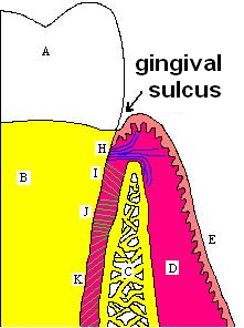
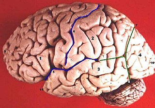
In biological morphology and anatomy, a sulcus is a furrow or fissure. It may be a groove in the surface of a limb or an organ, notably in the surface of the brain, but also in the lungs, certain muscles, as well as in bones, and elsewhere. Many sulci are the product of a surface fold or junction, such as in the gums, where they fold around the neck of the tooth.
In the blood supply of the heart, the right coronary artery (RCA) is an artery originating above the right cusp of the aortic valve, at the right aortic sinus in the heart. It travels down the right coronary sulcus, towards the crux of the heart. It supplies the right side of the heart, and the interventricular septum.

The petrous part of the temporal bone is pyramid-shaped and is wedged in at the base of the skull between the sphenoid and occipital bones. Directed medially, forward, and a little upward, it presents a base, an apex, three surfaces, and three angles, and houses in its interior, the components of the inner ear. The petrous portion is among the most basal elements of the skull and forms part of the endocranium. Petrous comes from the Latin word petrosus, meaning "stone-like, hard". It is one of the densest bones in the body.

There are two surfaces of the squamous part of the frontal bone: the external surface, and the internal surface.

The coronary sulcus is a groove on the surface of the heart that separates the atria from the ventricles. The structure contains the trunks of the nutrient vessels of the heart, and is deficient in front, where it is crossed by the root of the pulmonary trunk. On the posterior surface of the heart, the coronary sulcus contains the coronary sinus.

In the coronary circulation, the posterior interventricular artery, most often called the posterior descending artery (PDA), is an artery running in the posterior interventricular sulcus to the apex of the heart where it meets with the anterior interventricular artery or also known as Left Anterior Descending artery. It supplies the posterior third of the interventricular septum. The remaining anterior two-thirds is supplied by the anterior interventricular artery which is a septal branch of the left anterior descending artery, which is a branch of left coronary artery.
The "LCX", or left circumflex artery is an artery of the heart.

The hypothalamic sulcus is a groove in the lateral wall of the third ventricle, marking the boundary between the thalamus and hypothalamus. The upper and lower portions of the lateral wall of the third ventricle correspond to the alar lamina and basal lamina, respectively, of the lateral wall of the fore-brain vesicle and are separated from each other by a furrow, the hypothalamic sulcus, which extends from the interventricular foramen to the cerebral aqueduct.
The anterior interventricular sulcus is one of two grooves that separates the ventricles of the heart, the other being the posterior interventricular sulcus.
The posterior interventricular sulcus or posterior longitudinal sulcus is one of the two grooves that separates the ventricles of the heart and is on the diaphragmatic surface of the heart near the right margin. The other groove is the anterior interventricular sulcus, situated on the sternocostal surface of the heart, close to its left margin.
The right marginal branch of right coronary artery is the largest marginal branch of the right coronary artery. It follows the acute margin of the heart. It supplies blood to both surfaces of the right ventricle.

The olfactory trigone is a small triangular area in front of the anterior perforated substance.
The left anterior descending artery is a branch of the left coronary artery. Blockage of this artery is often called the widow-maker infarction due to a high death risk.
The left marginal artery is a branch of the circumflex artery, originating at the left atrioventricular sulcus, traveling along the left margin of heart towards the apex of the heart.
Interventricular sulcus may refer to:

The curvatures of the stomach refer to the greater and lesser curvatures. The greater curvature of the stomach is four or five times as long as the lesser curvature.

The intercondylar fossa of femur is a deep notch between the rear surfaces of the medial and lateral epicondyle of the femur, two protrusions on the distal end of the femur that joins the knee. On the front of the femur, the condyles are but much less prominent and are separated from one another by a smooth shallow articular depression called the patellar surface because it articulates with the posterior surface of the patella (kneecap).
About 5 centimetres (2.0 in) in front of the occipital pole of the human brain, on the infero-lateral border is an indentation or notch, named the preoccipital notch. It is considered a landmark because the occipital lobe is located just behind the line that connects that notch with the parietoccipital sulcus.
Heart is a muscular organ sited in the mediastinum. It consist of four chambers, four valves, arteries, and the conduction system. Heart functionally can be separated in left and right side. Right heart receives blood coming from the body through superior and inferior vena cava. It pumps blood to the lungs through the pulmonary artery and left heart receives saturated blood from the lungs.
References
This article incorporates text in the public domain from page 527 of the 20th edition of Gray's Anatomy (1918)