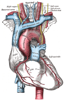
The superior vena cava (SVC) is the superior of the two venae cavae, the great venous trunks that return deoxygenated blood from the systemic circulation to the right atrium of the heart. It is a large-diameter (24 mm) short length vein that receives venous return from the upper half of the body, above the diaphragm. Venous return from the lower half, below the diaphragm, flows through the inferior vena cava. The SVC is located in the anterior right superior mediastinum. It is the typical site of central venous access via a central venous catheter or a peripherally inserted central catheter. Mentions of "the cava" without further specification usually refer to the SVC.

Cardiopulmonary bypass (CPB) is a technique in which a machine temporarily takes over the function of the heart and lungs during surgery, maintaining the circulation of blood and the oxygen content of the patient's body. The CPB pump itself is often referred to as a heart–lung machine or "the pump". Cardiopulmonary bypass pumps are operated by perfusionists. CPB is a form of extracorporeal circulation. Extracorporeal membrane oxygenation is generally used for longer-term treatment.
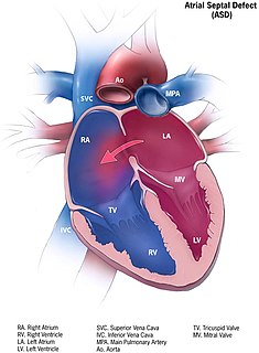
Atrial septal defect (ASD) is a congenital heart defect in which blood flows between the atria of the heart. Some flow is a normal condition both pre-birth and immediately post-birth via the foramen ovale; however, when this does not naturally close after birth it is referred to as a patent (open) foramen ovale (PFO). It is common in patients with a congenital atrial septal aneurysm (ASA).
Situs ambiguus is a rare congenital defect in which the major visceral organs are distributed abnormally within the chest and abdomen. Heterotaxy in general refers to any defect of left-right laterality and arrangement of the visceral organs. This does not include the congenital defect situs inversus, which results when arrangement of all the organs in the abdomen and chest are mirrored, so the positions are opposite the normal placement. Situs inversus is the mirror image of situs solitus, which is normal asymmetric distribution of the abdominothoracic visceral organs. Situs ambiguus can also be subdivided into left-isomerism and right isomerism based on the defects observed in the spleen, lungs and atria of the heart.

The jugular veins are veins that take deoxygenated blood from the head back to the heart via the superior vena cava.

The atrium or auricle is the upper chamber through which blood enters the ventricles of the heart. There are two atria in the human heart – the left atrium receives blood from the pulmonary (lung) circulation, and the right atrium receives blood from the venae cavae. The atria receive blood while relaxed (diastole), then contract (systole) to move blood to the ventricles. All animals with a closed circulatory system have at least one atrium. Humans have two atria.

The chordae tendineae , colloquially known as the heart strings, are inelastic cords of fibrous connective tissue that connect the papillary muscles to the tricuspid valve and the mitral valve in the heart.
A transthoracic echocardiogram (TTE) is the most common type of echocardiogram, which is a still or moving image of the internal parts of the heart using ultrasound. In this case, the probe is placed on the chest or abdomen of the subject to get various views of the heart. It is used as a non-invasive assessment of the overall health of the heart, including a patient's heart valves and degree of heart muscle contraction. The images are displayed on a monitor for real-time viewing and then recorded.

The hepatic veins are the veins that drain de-oxygenated blood from the liver into the inferior vena cava. There are usually three upper hepatic veins draining from the left, middle, and right parts of the liver. These are larger than the group of lower hepatic veins that can number from six to twenty. All of the hepatic veins drain into the inferior vena cava.

Tricuspid atresia is a form of congenital heart disease whereby there is a complete absence of the tricuspid valve. Therefore, there is an absence of right atrioventricular connection. This leads to a hypoplastic (undersized) or absent right ventricle. This defect is contracted during prenatal development, when the heart does not finish developing. It causes the systemic circulation to be filled with relatively deoxygenated blood. Because of this, hypoxia occurs, so other defects must occur to maintain blood flow. Because of the lack of an atrioventricular connection, an atrial septal defect (ASD) must be present to fill the left atrium and the left ventricle with blood. Since there is a lack of a right ventricle, there must be a way to pump blood into the pulmonary arteries, and this is accomplished by a ventricular septal defect (VSD). The causes of tricupsid atresia are unknown.
Cor triatriatum is a congenital heart defect where the left atrium or right atrium is subdivided by a thin membrane, resulting in three atrial chambers.

In the heart's conduction system, Bachmann's bundle is a branch of the anterior internodal tract that resides on the inner wall of the left atrium. It is a broad band of cardiac muscle that passes from the right atrium, between the superior vena cava and the ascending aorta. Bachmann's bundle is, during normal sinus rhythm, the preferential path for electrical activation of the left atrium. It is therefore considered to be part of the "atrial conduction system" of the heart.
In anatomy, a persistent left superior vena cava is the most common variation of the thoracic venous system. It is present in between 0.3% and 0.5% of the population, and is an embryologic remnant that results from a failure to involute.
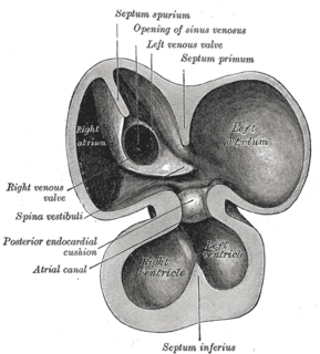
The sinus venosus is a large quadrangular cavity which precedes the atrium on the venous side of the chordate heart. In mammals, it exists distinctly only in the embryonic heart, where it is found between the two venae cavae. However, the sinus venosus persists in the adult. In the adult, it is incorporated into the wall of the right atrium to form a smooth part called the sinus venarum, which is separated from the rest of the atrium by a ridge of fibres called the crista terminalis. The sinus venosus also forms the sinoatrial node and the coronary sinus; in (most) mammals only.
The terminal sulcus is a groove in the right atrium of the heart. The terminal sulcus marks the separation of the right atrial pectinate muscles from the sinus venarum. The terminal sulcus extends from the front of the superior vena cava to the front of the inferior vena cava, and represents the line of union of the sinus venosus of the embryo with the primitive atrium. On the internal aspect of the right atrium, corresponding to the terminal sulcus is the crista terminalis.
The heart is the first functional organ in a vertebrate embryo. There are 5 stages to heart development.

Anomalous pulmonary venous connection is a congenital defect of the pulmonary veins.
The Senning procedure is an atrial switch heart operation performed to treat transposition of the great arteries. It is named after its inventor, the Swedish cardiac surgeon Åke Senning (1915–2000), also known for implanting the first permanent cardiac pacemaker in 1958.
Stella Van Praagh was a pediatric cardiologist and pathologist at Children's Hospital Boston. She was internationally known for her contributions to the pathology of congenital heart disease.
