Uhl anomaly

Last updated
Uhl anomaly
Autosomal dominant - en.svg
Uhl anomaly is inherited via autosomal dominant manner
Specialty Medical genetics   OOjs UI icon edit-ltr-progressive.svg

Uhl anomaly is a rare cardiac malformation that was first identified by Dr. Henry Uhl in 1952. It is characterized by the absence of the right ventricle (RV) myocardium, either entirely or partially, and the replacement of the RV myocardium by nonfunctional fibroelastic tissue that resembles parchment. [1] As of 2010 less than 100 cases have been reported in literature. [2]

Contents

Patients will typically present as infants with right-sided heart failure. [3] Atrial right-to-left shunting is frequently observed as the cause of cyanosis. [4]

Typically, magnetic resonance imaging, computed tomography, and echocardiography are used to make the diagnosis. [5]

Signs and symptoms

Infants may exhibit severe cyanosis and right heart failure at birth, though these conditions may get better as pulmonary vascular resistance decreases. The main clinical features in older patients are right heart failure symptoms and signs. [6]

Causes

Although the precise cause of Uhl's anomaly is unknown, there have been reports of primary nondevelopment of myocytes, selective apoptosis, and cardiomyocyte overexpression of vascular endothelial growth factor. [7] [8] A sporadic mutation could indicate that genetics is the underlying cause. [9]

Mechanism

The pathophysiological outcome of Uhl anomaly involves compromised diastolic filling and right ventricular contraction. Systemic venous congestion and elevated right atrial and systemic venous pressure are the results of right ventricular failure. High pulmonary vascular resistance newborns may develop functional pulmonary atresia (a result of ineffective right ventricular forward flow). Cyanosis is caused by a right-to-left shunting of blood through the patent oval foramen. [6]

Diagnosis

Myocardial biopsies can confirm the diagnosis, which is often established by imaging tests like cardiac magnetic resonance imaging or echocardiogram. [10]

Right atrial and right ventricular dilatation is the cause of the cardiomegaly seen on the chest radiograph. The lung fields of newborns with functional pulmonary atresia and high pulmonary vascular resistance appear oligaemic. [6]

Right ventricular and right atrial dilatation is evident on the electrocardiogram. One can observe an epsilon (ε) wave in the right praecordial leads. [6]

An echocardiographic evaluation reveals a thin free wall of the right ventricle without any myocardium, reduced contractility and restricted ventricle filling, dilation of the right atrium, and normal tricuspid valve attachment to the right atrioventricular junction. [6]

Global dyskinesia of a thin-walled right ventricle, absence of myocardium at the right ventricular free wall, lack of fibrofatty infiltration, normal tricuspid valve attachment to the right atrioventricular junction, and normal left ventricular myocardium are all indicative of cardiac magnetic resonance imaging findings. [6]

Histopathologically, nonfunctioning fibroelastic tissue replaces the myocardial layer, giving the right ventricular free wall a parchment-like appearance. [11]

Other anomalies that can result in RV dilatation, such as Ebstein anomaly, RV arrhythmogenic dysplasia, pulmonary atresia, and anomalous pulmonary venous return, are included in the differential diagnosis of Uhl anomaly. [12]

Treatment

When the pulmonary vascular resistance has become high and the newborn has severe cyanosis soon after birth, a brief intravenous prostaglandin E infusion is recommended. In most cases, diuretics are necessary for congestive right heart failure. Various surgical techniques have been used, such as: (1) one-and-a-half ventricular repair alongside partial right ventriculectomy and bidirectional Glenn shunt; (2) right ventricular exclusion alongside atrial septectomy as well as a bidirectional Glenn shunt (superior cavopulmonary anastomosis); and (3) cardiac transplantation. [6]

Related Research Articles

<span class="mw-page-title-main">Cardiology</span> Branch of medicine dealing with the heart

Cardiology is the study of the heart. Cardiology is a branch of medicine that deals with disorders of the heart and the cardiovascular system. The field includes medical diagnosis and treatment of congenital heart defects, coronary artery disease, heart failure, valvular heart disease, and electrophysiology. Physicians who specialize in this field of medicine are called cardiologists, a specialty of internal medicine. Pediatric cardiologists are pediatricians who specialize in cardiology. Physicians who specialize in cardiac surgery are called cardiothoracic surgeons or cardiac surgeons, a specialty of general surgery.

<span class="mw-page-title-main">Tetralogy of Fallot</span> Type of congenital heart defect

Tetralogy of Fallot (TOF), formerly known as Steno-Fallot tetralogy, is a congenital heart defect characterized by four specific cardiac defects. Classically, the four defects are:

<span class="mw-page-title-main">Pulmonary heart disease</span> Medical condition

Pulmonary heart disease, also known as cor pulmonale, is the enlargement and failure of the right ventricle of the heart as a response to increased vascular resistance or high blood pressure in the lungs.

<span class="mw-page-title-main">Arrhythmogenic cardiomyopathy</span> Medical condition

Arrhythmogenic cardiomyopathy (ACM), arrhythmogenic right ventricular dysplasia (ARVD), or arrhythmogenic right ventricular cardiomyopathy (ARVC), most commonly is an inherited heart disease.

<span class="mw-page-title-main">Atrial septal defect</span> Human heart defect present at birth

Atrial septal defect (ASD) is a congenital heart defect in which blood flows between the atria of the heart. Some flow is a normal condition both pre-birth and immediately post-birth via the foramen ovale; however, when this does not naturally close after birth it is referred to as a patent (open) foramen ovale (PFO). It is common in patients with a congenital atrial septal aneurysm (ASA).

<span class="mw-page-title-main">Congenital heart defect</span> Defect in the structure of the heart that is present at birth

A congenital heart defect (CHD), also known as a congenital heart anomaly, congenital cardiovascular malformation, and congenital heart disease, is a defect in the structure of the heart or great vessels that is present at birth. A congenital heart defect is classed as a cardiovascular disease. Signs and symptoms depend on the specific type of defect. Symptoms can vary from none to life-threatening. When present, symptoms are variable and may include rapid breathing, bluish skin (cyanosis), poor weight gain, and feeling tired. CHD does not cause chest pain. Most congenital heart defects are not associated with other diseases. A complication of CHD is heart failure.

<span class="mw-page-title-main">Ventricular septal defect</span> Medical condition

A ventricular septal defect (VSD) is a defect in the ventricular septum, the wall dividing the left and right ventricles of the heart. The extent of the opening may vary from pin size to complete absence of the ventricular septum, creating one common ventricle. The ventricular septum consists of an inferior muscular and superior membranous portion and is extensively innervated with conducting cardiomyocytes.

<span class="mw-page-title-main">Fontan procedure</span> Surgical procedure used in children with univentricular hearts

The Fontan procedure or Fontan–Kreutzer procedure is a palliative surgical procedure used in children with univentricular hearts. It involves diverting the venous blood from the inferior vena cava (IVC) and superior vena cava (SVC) to the pulmonary arteries. The procedure varies for differing congenital heart pathologies. For example in tricuspid atresia, the procedure can be done where the blood does not pass through the morphologic right ventricle; i.e., the systemic and pulmonary circulations are placed in series with the functional single ventricle. Whereas in hypoplastic left heart syndrome, the heart is more reliant on the more functional right ventricle to provide blood flow to the systemic circulation. The procedure was initially performed in 1968 by Francis Fontan and Eugene Baudet from Bordeaux, France, published in 1971, simultaneously described in 1971 by Guillermo Kreutzer from Buenos Aires, Argentina, and finally published in 1973.

Situs ambiguus is a rare congenital defect in which the major visceral organs are distributed abnormally within the chest and abdomen. Clinically heterotaxy spectrum generally refers to any defect of Left-right asymmetry and arrangement of the visceral organs; however, classical heterotaxy requires multiple organs to be affected. This does not include the congenital defect situs inversus, which results when arrangement of all the organs in the abdomen and chest are mirrored, so the positions are opposite the normal placement. Situs inversus is the mirror image of situs solitus, which is normal asymmetric distribution of the abdominothoracic visceral organs. Situs ambiguus can also be subdivided into left-isomerism and right isomerism based on the defects observed in the spleen, lungs and atria of the heart.

<span class="mw-page-title-main">Hypoplastic left heart syndrome</span> Type of congenital heart defect

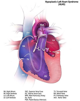
Hypoplastic left heart syndrome (HLHS) is a rare congenital heart defect in which the left side of the heart is severely underdeveloped and incapable of supporting the systemic circulation. It is estimated to account for 2-3% of all congenital heart disease. Early signs and symptoms include poor feeding, cyanosis, and diminished pulse in the extremities. The etiology is believed to be multifactorial resulting from a combination of genetic mutations and defects resulting in altered blood flow in the heart. Several structures can be affected including the left ventricle, aorta, aortic valve, or mitral valve all resulting in decreased systemic blood flow.

<span class="mw-page-title-main">Pulmonary atresia</span> Medical condition

Pulmonary atresia is a congenital malformation of the pulmonary valve in which the valve orifice fails to develop. The valve is completely closed thereby obstructing the outflow of blood from the heart to the lungs. The pulmonary valve is located on the right side of the heart between the right ventricle and pulmonary artery. In a normal functioning heart, the opening to the pulmonary valve has three flaps that open and close.

<span class="mw-page-title-main">Tricuspid atresia</span> Medical condition

Tricuspid atresia is a form of congenital heart disease whereby there is a complete absence of the tricuspid valve. Therefore, there is an absence of right atrioventricular connection. This leads to a hypoplastic (undersized) or absent right ventricle. This defect is contracted during prenatal development, when the heart does not finish developing. It causes the systemic circulation to be filled with relatively deoxygenated blood. The causes of tricuspid atresia are unknown.

A right-to-left shunt is a cardiac shunt which allows blood to flow from the right heart to the left heart. This terminology is used both for the abnormal state in humans and for normal physiological shunts in reptiles.

<span class="mw-page-title-main">Tricuspid regurgitation</span> Type of valvular heart disease

Tricuspid regurgitation (TR), also called tricuspid insufficiency, is a type of valvular heart disease in which the tricuspid valve of the heart, located between the right atrium and right ventricle, does not close completely when the right ventricle contracts (systole). TR allows the blood to flow backwards from the right ventricle to the right atrium, which increases the volume and pressure of the blood both in the right atrium and the right ventricle, which may increase central venous volume and pressure if the backward flow is sufficiently severe.

<span class="mw-page-title-main">Smallest cardiac veins</span> Small veins in the walls of all four heart chambers

The smallest cardiac veins are small, valveless veins in the walls of all four heart chambers that drain venous blood from the myocardium directly into any of the heart chambers.

<span class="mw-page-title-main">Double inlet left ventricle</span> Medical condition

A double inlet left ventricle (DILV) or "single ventricle", is a congenital heart defect appearing in 5 in 100,000 newborns, where both the left atrium and the right atrium feed into the left ventricle. The right ventricle is hypoplastic or does not exist.

<span class="mw-page-title-main">Bidirectional Glenn procedure</span>

The bidirectional Glenn (BDG) shunt, or bidirectional cavopulmonary anastomosis, is a surgical technique used in pediatric cardiac surgery procedure used to temporarily improve blood oxygenation for patients with a congenital cardiac defect resulting in a single functional ventricle. Creation of a bidirectional shunt reduces the amount of blood volume that the heart needs to pump at the time of surgical repair with the Fontan procedure.

<span class="mw-page-title-main">Anomalous pulmonary venous connection</span> Medical condition

Anomalous pulmonary venous connection is a congenital defect of the pulmonary veins.

<span class="mw-page-title-main">Hypoplastic right heart syndrome</span> Type of congenital heart disease

Hypoplastic right heart syndrome or HRHS is a congenital heart defect in which the structures on the right side of the heart, particularly the right ventricle, are underdeveloped. This defect causes inadequate blood flow to the lungs, and thus a cyanotic infant.

References

  1. Uhl, Henry (September 1952). "A previously undescribed congenital malformation of the heart: almost total absence of the myocardium of the right ventricle". Bulletin of the Johns Hopkins Hospital. 91 (3): 197–209. PMID   12978573.
  2. Hoschtitzky, Andreas; Rowlands, Helen; Ilina, Maria; Khambadkone, Sachin; Elliott, Martin J. (2010). "Single Ventricle Strategy for Uhl's Anomaly of the Right Ventricle". The Annals of Thoracic Surgery. Elsevier BV. 90 (6): 2076–2078. doi:10.1016/j.athoracsur.2009.12.084. ISSN   0003-4975.
  3. Richardson, J.D.; Teo, K.S.L.; Bertaso, A.G.; Wong., D.T.L.; Disney, P.; Worthley, S.G. (2012). "Uhl's anomaly". International Journal of Cardiology. Elsevier BV. 154 (2): e36–e37. doi:10.1016/j.ijcard.2011.05.005. ISSN   0167-5273.
  4. Azhari, Nawal; Assaqqat, Mervat; Bulbul, Ziad (2002). "Successful surgical repair of Uhl's anomaly". Cardiology in the Young. Cambridge University Press (CUP). 12 (2): 192–195. doi:10.1017/s1047951102000410. ISSN   1047-9511.
  5. Cammalleri, Valeria; Forcina, Marco; Pugliese, Luca; Romeo, Francesco; Floris, Roberto; Chiocchi, Marcello (2019). "Uhl Anomaly in Asymptomatic Adult Woman". Circulation: Cardiovascular Imaging. Ovid Technologies (Wolters Kluwer Health). 12 (2). doi:10.1161/circimaging.118.008277. ISSN   1941-9651.
  6. 1 2 3 4 5 6 7 Cheung, Yiu-fai (2023). "Uhl Anomaly". Congenital and Paediatric Acquired Heart Disease in Practice. Singapore: Springer Nature Singapore. p. 271–273. doi:10.1007/978-981-99-2862-0_26. ISBN   978-981-99-2861-3.
  7. Uhl, Henry S.M. (April 15, 1996). "Uhl's Anomaly Revisited". Circulation. Ovid Technologies (Wolters Kluwer Health). 93 (8): 1483–1484. doi:10.1161/01.cir.93.8.1483. ISSN   0009-7322.
  8. Feucht, M.; Christ, B.; Wilting, J. "VEGF induces cardiovascular malformation and embryonic lethality". The American Journal of Pathology. American Society for Investigative Pathology. 151 (5). PMID   9358767 . Retrieved January 22, 2024.
  9. Gerlis, L M; Schmidt-Ott, S C; Ho, S Y; Anderson, R H (February 1, 1993). "Dysplastic conditions of the right ventricular myocardium: Uhl's anomaly vs arrhythmogenic right ventricular dysplasia". Heart. BMJ. 69 (2): 142–150. doi:10.1136/hrt.69.2.142. ISSN   1355-6037. PMC   1024941 .
  10. Ganczar, Joanna; English, Robert (2015). "Uhl′s anomaly: Absence of the right ventricular myocardium". Annals of Pediatric Cardiology. Medknow. 8 (1): 71. doi: 10.4103/0974-2069.149529 . ISSN   0974-2069. PMC   4322408 .
  11. Chounoune, Reginald; Lowry, Adam; Ramakrishnan, Karthik; Pearson, Gail D.; Moak, Jeffrey P.; Nath, Dilip S. (2018). "Uhl's anomaly: A one and a half ventricular repair in a patient presenting with cardiac arrest". Journal of the Saudi Heart Association. Saudi Heart Association. 30 (1): 52–54. doi:10.1016/j.jsha.2017.03.011. ISSN   1016-7315. PMC   5744023 .
  12. Caruso, Settimo; Cannataci, Christine; Romano, Giuseppe (2021). "Case 288: Uhl Anomaly". Radiology. 299 (1): 237–241. doi:10.1148/radiol.2021192475. ISSN   0033-8419.

Further reading