Pulmonary atresia

Last updated
Pulmonary atresia
Other namesPulmonary atresia - intact ventricular septum [1]
Gray494.png
Pulmonary valve (center left)
Specialty Cardiology   OOjs UI icon edit-ltr-progressive.svg
Symptoms Cyanosis [1]
CausesNo known cause for pulmonary atresia [1]
Diagnostic method Chest x-ray, Echocardiogram [1]
TreatmentProstaglandin E1, Heart catheterization [1]

Pulmonary atresia is a congenital malformation of the pulmonary valve in which the valve orifice fails to develop. The valve is completely closed thereby obstructing the outflow of blood from the heart to the lungs. [2] The pulmonary valve is located on the right side of the heart between the right ventricle and pulmonary artery. In a normal functioning heart, the opening to the pulmonary valve has three flaps that open and close. [3]

Contents

In congenital heart defects such as pulmonary atresia, structural abnormalities can include the valves of the heart, and the walls and arteries/veins near the heart muscle. Consequently, blood flow due to the aforementioned structural abnormalities is affected, either by blocking or altering the flow of blood through the human cardiac muscle. [4]

Signs and symptoms

The symptoms/signs of pulmonary atresia that will occur in babies are consistent with cyanosis, some fatigue and some shortness of breath (eating may be a problem as well). [1]

In the case of pulmonary atresia with ventricular septal defect, decreased pulmonary blood flow may cause associated defects such as: [5]

Cause

Smoking Papierosa 1 ubt 0069.jpeg
Smoking

In terms of the cause of pulmonary atresia, there is uncertainty as to what instigates this congenital heart defect. [2] Potential risk factors that can cause this congenital heart defect are those the pregnant mother may come in contact with, such as: [2] [6]

Diagnosis

In regards to the diagnosis of pulmonary atresia the body requires oxygenated blood for survival. pulmonary atresia is not threatening to a developing fetus however, because the mother's placenta provides the needed oxygen since the baby's lungs are not yet functional. Once the baby is born its lungs must now provide the oxygen needed for survival, but with pulmonary atresia there is no opening on the pulmonary valve for blood to get to the lungs and become oxygenated. Due to this, the newborn baby is blue in color and pulmonary atresia can usually be diagnosed within hours or minutes after birth.[ medical citation needed ]

The diagnosis of pulmonary atresia can be done via the following exams/methods: an echocardiogram, chest x-ray, EKG and an exam to measure the amount of O
2
in the body. [1]

Classification

Ventricular septal defect via echocardiogram Ventricular Septal Defect.jpg
Ventricular septal defect via echocardiogram

There are two types of pulmonary atresia.

Treatment

Prostaglandin E1 Prostaglandin E1.svg
Prostaglandin E1

The treatment of pulmonary atresia consists of: an IV medication called prostaglandin E1, which is used for treatment of pulmonary atresia, as it stops the ductus arteriosus from closing, allowing mixing of the pulmonary and systemic circulations, but prostaglandin E1 can be dangerous as it can cause apnea. Another example of preliminary treatment is heart catheterization to evaluate the defect or defects of the heart; this procedure is much more invasive. Ultimately, however, the individual will need to have a series of surgeries to improve the blood flow permanently. The first surgery will likely be performed shortly after birth. A shunt can be created between the aorta and the pulmonary artery to help increase blood flow to the lungs. As the child grows, so does the heart and the shunt may need to be revised in order to meet the body's requirements. [5] [8] [9]

Fontan procedure Fontan procedure.svg
Fontan procedure

The type of surgery recommended depends on the size of the right ventricle and the pulmonary artery, if the right ventricle is small and unable to act as a pump, the surgery performed would be the Fontan procedure. In this three-stage procedure, the right atrium is disconnected from the pulmonary circulation. The systemic venous return goes directly to the lungs, by-passing the heart. Very young children with elevated pulmonary vascular resistance may not able to undergo the Fontan procedure. Cardiac catheterization may be done to determine the resistance before going ahead with the surgery. [10] [11] [12]

Prognosis

The prognosis for pulmonary atresia varies for every child, if the condition is left uncorrected it may be fatal, but the prognosis has greatly improved over the years for those with pulmonary atresia. Some factors that affect how well the child does include how well the heart is beating, and the condition of the blood vessels that supply the heart. Most cases of pulmonary atresia can be helped with surgery, if the patient's right ventricle is exceptionally small, many surgeries will be needed in order to help stimulate normal circulation of blood to the heart. If uncorrected, babies with this type of congenital heart disease may only survive for the first few days of life. Many children with pulmonary atresia will go on to lead normal lives, though complications such as endocarditis, stroke and seizures are possible. [1] [8] [13]

See also

Related Research Articles

<span class="mw-page-title-main">Cardiology</span> Branch of medicine dealing with the heart

Cardiology is the study of the heart. Cardiology is a branch of medicine that deals with disorders of the heart and the cardiovascular system. The field includes medical diagnosis and treatment of congenital heart defects, coronary artery disease, heart failure, valvular heart disease, and electrophysiology. Physicians who specialize in this field of medicine are called cardiologists, a specialty of internal medicine. Pediatric cardiologists are pediatricians who specialize in cardiology. Physicians who specialize in cardiac surgery are called cardiothoracic surgeons or cardiac surgeons, a specialty of general surgery.

<span class="mw-page-title-main">Tetralogy of Fallot</span> Type of congenital heart defect

Tetralogy of Fallot (TOF), formerly known as Steno-Fallot tetralogy, is a congenital heart defect characterized by four specific cardiac defects. Classically, the four defects are:

<span class="mw-page-title-main">Atrial septal defect</span> Human heart defect present at birth

Atrial septal defect (ASD) is a congenital heart defect in which blood flows between the atria of the heart. Some flow is a normal condition both pre-birth and immediately post-birth via the foramen ovale; however, when this does not naturally close after birth it is referred to as a patent (open) foramen ovale (PFO). It is common in patients with a congenital atrial septal aneurysm (ASA).

<span class="mw-page-title-main">Congenital heart defect</span> Defect in the structure of the heart that is present at birth

A congenital heart defect (CHD), also known as a congenital heart anomaly, congenital cardiovascular malformation, and congenital heart disease, is a defect in the structure of the heart or great vessels that is present at birth. A congenital heart defect is classed as a cardiovascular disease. Signs and symptoms depend on the specific type of defect. Symptoms can vary from none to life-threatening. When present, symptoms are variable and may include rapid breathing, bluish skin (cyanosis), poor weight gain, and feeling tired. CHD does not cause chest pain. Most congenital heart defects are not associated with other diseases. A complication of CHD is heart failure.

<span class="mw-page-title-main">Ventricular septal defect</span> Medical condition

A ventricular septal defect (VSD) is a defect in the ventricular septum, the wall dividing the left and right ventricles of the heart. The extent of the opening may vary from pin size to complete absence of the ventricular septum, creating one common ventricle. The ventricular septum consists of an inferior muscular and superior membranous portion and is extensively innervated with conducting cardiomyocytes.

<span class="mw-page-title-main">Fontan procedure</span> Surgical procedure used in children with univentricular hearts

The Fontan procedure or Fontan–Kreutzer procedure is a palliative surgical procedure used in children with univentricular hearts. It involves diverting the venous blood from the inferior vena cava (IVC) and superior vena cava (SVC) to the pulmonary arteries. The procedure varies for differing congenital heart pathologies. For example in tricuspid atresia, the procedure can be done where the blood does not pass through the morphologic right ventricle; i.e., the systemic and pulmonary circulations are placed in series with the functional single ventricle. Whereas in hypoplastic left heart syndrome, the heart is more reliant on the more functional right ventricle to provide blood flow to the systemic circulation. The procedure was initially performed in 1968 by Francis Fontan and Eugene Baudet from Bordeaux, France, published in 1971, simultaneously described in 1971 by Guillermo Kreutzer from Buenos Aires, Argentina, and finally published in 1973.

<span class="mw-page-title-main">Cardiac catheterization</span> Insertion of a catheter into a chamber or vessel of the heart

Cardiac catheterization is the insertion of a catheter into a chamber or vessel of the heart. This is done both for diagnostic and interventional purposes.

The Rastelli procedure is an open heart surgical procedure developed by Italian physician and cardiac surgery researcher, Giancarlo Rastelli, in 1967 at the Mayo Clinic, and involves using a pulmonary or aortic homograft conduit to relieve pulmonary obstruction in double outlet right ventricle with pulmonary stenosis.

<span class="mw-page-title-main">Hypoplastic left heart syndrome</span> Type of congenital heart defect

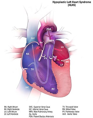
Hypoplastic left heart syndrome (HLHS) is a rare congenital heart defect in which the left side of the heart is severely underdeveloped and incapable of supporting the systemic circulation. It is estimated to account for 2-3% of all congenital heart disease. Early signs and symptoms include poor feeding, cyanosis, and diminished pulse in the extremities. The etiology is believed to be multifactorial resulting from a combination of genetic mutations and defects resulting in altered blood flow in the heart. Several structures can be affected including the left ventricle, aorta, aortic valve, or mitral valve all resulting in decreased systemic blood flow.

<span class="mw-page-title-main">Transposition of the great vessels</span> Group of congenital heart defects

Transposition of the great vessels (TGV) is a group of congenital heart defects involving an abnormal spatial arrangement of any of the great vessels: superior and/or inferior venae cavae, pulmonary artery, pulmonary veins, and aorta. Congenital heart diseases involving only the primary arteries belong to a sub-group called transposition of the great arteries (TGA), which is considered the most common congenital heart lesion that presents in neonates.

<span class="mw-page-title-main">Tricuspid atresia</span> Medical condition

Tricuspid atresia is a form of congenital heart disease whereby there is a complete absence of the tricuspid valve. Therefore, there is an absence of right atrioventricular connection. This leads to a hypoplastic (undersized) or absent right ventricle. This defect is contracted during prenatal development, when the heart does not finish developing. It causes the systemic circulation to be filled with relatively deoxygenated blood. The causes of tricuspid atresia are unknown.

<span class="mw-page-title-main">Atrioventricular septal defect</span> Medical condition

Atrioventricular septal defect (AVSD) or atrioventricular canal defect (AVCD), also known as "common atrioventricular canal" or "endocardial cushion defect" (ECD), is characterized by a deficiency of the atrioventricular septum of the heart that creates connections between all four of its chambers. It is a very specific combination of 3 defects:

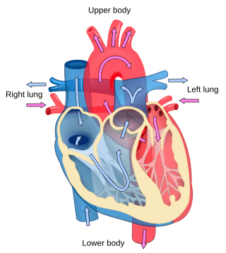
A right-to-left shunt is a cardiac shunt which allows blood to flow from the right heart to the left heart. This terminology is used both for the abnormal state in humans and for normal physiological shunts in reptiles.

<span class="mw-page-title-main">Lutembacher's syndrome</span> Medical condition

Lutembacher's syndrome is a very rare form of congenital heart disease that affects one of the chambers of the heart as well as a valve. It is commonly known as both congenital atrial septal defect (ASD) and acquired mitral stenosis (MS). Congenital atrial septal defect refers to a hole being in the septum or wall that separates the two atria; this condition is usually seen in fetuses and infants. Mitral stenosis refers to mitral valve leaflets sticking to each other making the opening for blood to pass from the atrium to the ventricles very small. With the valve being so small, blood has difficulty passing from the left atrium into the left ventricle. Septal defects that may occur with Lutembacher's syndrome include: Ostium primum atrial septal defect or ostium secundum which is more prevalent.

<span class="mw-page-title-main">Double inlet left ventricle</span> Medical condition

A double inlet left ventricle (DILV) or "single ventricle", is a congenital heart defect appearing in 5 in 100,000 newborns, where both the left atrium and the right atrium feed into the left ventricle. The right ventricle is hypoplastic or does not exist.

<span class="mw-page-title-main">Bidirectional Glenn procedure</span>

The bidirectional Glenn (BDG) shunt, or bidirectional cavopulmonary anastomosis, is a surgical technique used in pediatric cardiac surgery procedure used to temporarily improve blood oxygenation for patients with a congenital cardiac defect resulting in a single functional ventricle. Creation of a bidirectional shunt reduces the amount of blood volume that the heart needs to pump at the time of surgical repair with the Fontan procedure.

<span class="mw-page-title-main">Hypoplastic right heart syndrome</span> Type of congenital heart disease

Hypoplastic right heart syndrome is a congenital heart defect in which the structures on the right side of the heart, particularly the right ventricle, are underdeveloped. This defect causes inadequate blood flow to the lungs, and thus a cyanotic infant.

<span class="mw-page-title-main">Ventricular outflow tract obstruction</span> Medical condition

A ventricular outflow tract obstruction is a heart condition in which either the right or left ventricular outflow tract is blocked or obstructed. These obstructions represent a spectrum of disorders. Majority of these cases are congenital, but some are acquired throughout life.

The Senning procedure is an atrial switch heart operation performed to treat transposition of the great arteries. It is named after its inventor, the Swedish cardiac surgeon Åke Senning (1915–2000), also known for implanting the first permanent cardiac pacemaker in 1958.

<span class="mw-page-title-main">Pulmonary atresia with ventricular septal defect</span> Type of congenital heart defect

Pulmonary atresia with ventricular septal defect is a rare birth defect characterized by pulmonary valve atresia occurring alongside a defect on the right ventricular outflow tract.

References

  1. 1 2 3 4 5 6 7 8 "Pulmonary atresia: MedlinePlus Medical Encyclopedia". www.nlm.nih.gov. Retrieved 2016-05-27.
  2. 1 2 3 4 5 "Facts about Pulmonary Atresia | Congenital Heart Defects | NCBDDD | CDC". www.cdc.gov. Retrieved 2016-06-24.
  3. "Pulmonic Valve Anatomy: Overview, Pathophysiologic Variants". 2017-09-14.{{cite journal}}: Cite journal requires |journal= (help)
  4. "Congenital Heart Defects: MedlinePlus" . Retrieved 2016-06-24.
  5. 1 2 3 "Pulmonary Atresia With Ventricular Septal Defect: Background, Epidemiology, Etiology". August 2018.{{cite journal}}: Cite journal requires |journal= (help)
  6. "Research | Congenital Heart Defects | NCBDDD | CDC". www.cdc.gov. Retrieved 24 June 2016.
  7. "Pulmonary Atresia With Intact Ventricular Septum: Background, Pathophysiology, Etiology". 2018-06-07.{{cite journal}}: Cite journal requires |journal= (help)
  8. 1 2 Murthy, KS; Reddy, K Pramod; Nagarajan, R; Goutami, V; Cherian, KM (2010-01-01). "Management of ventricular septal defect with pulmonary atresia and major aorto pulmonary collateral arteries: Challenges and controversies". Annals of Pediatric Cardiology. 3 (2): 127–135. doi: 10.4103/0974-2069.74040 . ISSN   0974-2069. PMC   3017916 . PMID   21234191.
  9. "Alprostadil (PGE1) for Maintaining Ductal Patency".
  10. Fredenburg, Tyler B.; Johnson, Tiffanie R.; Cohen, Mervyn D. (2011-03-01). "The Fontan Procedure: Anatomy, Complications, and Manifestations of Failure". RadioGraphics. 31 (2): 453–463. doi:10.1148/rg.312105027. ISSN   0271-5333. PMID   21415190.
  11. White, Charles S.; Haramati, Linda B.; Chen, Joseph Jen-Sho; Levsky, Jeffrey M. (2014-06-03). Cardiac Imaging. Oxford University Press. p. 477. ISBN   9780199829484.
  12. Feltes, Timothy F.; Bacha, Emile; Beekman, Robert H.; Cheatham, John P.; Feinstein, Jeffrey A.; Gomes, Antoinette S.; Hijazi, Ziyad M.; Ing, Frank F.; Moor, Michael de (2011-06-07). "Indications for Cardiac Catheterization and Intervention in Pediatric Cardiac Disease A Scientific Statement From the American Heart Association". Circulation. 123 (22): 2607–2652. doi: 10.1161/CIR.0b013e31821b1f10 . ISSN   0009-7322. PMID   21536996.
  13. Abuhamad, Alfred Z.; Chaoui, Rabih (2012-03-28). A Practical Guide to Fetal Echocardiography: Normal and Abnormal Hearts. Lippincott Williams & Wilkins. pp. 185–86. ISBN   9781451147803.

Further reading