Double aortic arch

Last updated
Double aortic arch
Other namesDAA

Double aortic arch is a relatively rare congenital cardiovascular malformation. DAA is an anomaly of the aortic arch in which two aortic arches form a complete vascular ring that can compress the trachea and/or esophagus. [1] [2] Most commonly there is a larger (dominant) right arch behind and a smaller (hypoplastic) left aortic arch in front of the trachea/esophagus. The two arches join to form the descending aorta which is usually on the left side (but may be right-sided or in the midline). In some cases the end of the smaller left aortic arch closes (left atretic arch) and the vascular tissue becomes a fibrous cord. Although in these cases a complete ring of two patent aortic arches is not present, the term ‘vascular ring’ is the accepted generic term even in these anomalies.

Contents

The symptoms are related to the compression of the trachea, esophagus or both by the complete vascular ring. Diagnosis can often be suspected or made by chest x-ray, barium esophagram, or echocardiography. Computed tomography (CT) or magnetic resonance imaging (MRI) show the relationship of the aortic arches to the trachea and esophagus and also the degree of tracheal narrowing. Bronchoscopy can be useful in internally assessing the degree of tracheomalacia. Treatment is surgical and is indicated in all symptomatic patients. In the current era the risk of mortality or significant morbidity after surgical division of the lesser arch is low. However, the preoperative degree of tracheomalacia has an important impact on postoperative recovery. In certain patients it may take several months (up to 1–2 years) for the obstructive respiratory symptoms (wheezing) to disappear.

Signs and symptoms

Symptoms are caused by vascular compression of the airway, esophagus or both. Presentation is often within the first month (neonatal period) and usually within the first 6 months of life. Starting at birth an inspiratory and expiratory stridor (high pitch noise from turbulent airflow in trachea) may be present often in combination with an expiratory wheeze. The severity of the stridor may depend on the patient’s body position. It can be worse when the baby is lying on their back rather than their side. Sometimes the stridor can be relieved by extending the neck (lifting the chin up). Parents may notice that the baby’s cry is hoarse and the breathing noisy. Frequently a persistent cough is present. When the airway obstruction is significant there may be episodes of severe cyanosis (“blue baby”) that can lead to unconsciousness. Recurrent respiratory infections are common and secondary pulmonary secretions can further increase the airway obstruction.[ citation needed ]

Secondary to compression of the esophagus babies often feed poorly. They may have difficulties in swallowing liquids with choking or regurgitating and increased respiratory obstruction during feeding. Older patients might refuse to take solid food, although most infants with severe symptoms nowadays are operated upon before they are offered solid food. Occasionally patients with double aortic arches present late (during later childhood or adulthood). Symptoms may mimic asthma.[ citation needed ]

Causes

Little is known regarding the exact causes of aortic arch anomalies. However, the association with chromosome 22q11 deletion (DiGeorge Syndrome) implies that a genetic component is likely in certain cases. [3] Esophageal atresia also occurs in some patients with double aortic arch. [4]

Diagnosis

Prenatal diagnosis (fetal ultrasound): Today the diagnosis of double aortic arch can be obtained in-utero in experienced centers. [5] Scheduled repair soon after birth in symptomatic patients can relieve tracheal compression early and therefore potentially prevent the development of severe tracheomalacia.

Chest X-ray: Plain chest x-rays of patients with double aortic arch may appear normal (often) or show a dominant right aortic arch or two aortic arches . There might be evidence of tracheal deviation and/or compression. Sometimes patients present with radiologic findings of pneumonia.

Barium swallow (esophagraphy): Historically the esophagram used to be the gold standard for diagnosis of double aortic arch. In patients with double aortic arch the esophagus shows left- and right-sided indentations from the vascular compression. Due to the blood-pressure related movement of the aorta and the two arches, moving images of the barium-filled esophagus can demonstrate the typical pulsatile nature of the obstruction. The indentation from a dominant right arch is usually deeper and higher compared to the dent from the left arch.

Bronchoscopy: Although bronchoscopy is not routinely done in patients with suspected or confirmed double aortic arch, it can visualize sites and severity of pulsatile tracheal compression.

Echocardiography: In babies under the age of 12 months, echocardiography is considered to be sensitive and specific in making the diagnosis of double aortic arch when both arches are open. Non-perfused elements of other types of vascular rings (e.g. left arch with atretic (closed) end) or the ligamentum arteriosum might be difficult to visualize by echocardiography.

Computed tomography (CT): Computed tomography after application of contrast media is usually diagnostically accurate. It shows the relationship of the arches to the trachea and bronchi.

Magnetic resonance imaging (MRI): Magnetic resonance imaging provides excellent images of the trachea and surrounding vascular structures and has the advantage of not using radiation for imaging compared to Computed tomography.

Cardiac catherization/aortography: Today patients with double aortic arch usually only undergo cardiac catherization to evaluate the hemodynamics and anatomy of associated congenital cardiac defects. Through a catheter in the ascending aorta contrast media is injected and the resulting aortography may be used to delineate the anatomy of the double aortic arch including sites of narrowing in the left aortic arch. Aortography can also be used to visualize the origin of all head and arm vessels originating from the two arches.

Classification

Double aortic arch is a subtype of complete vascular ring. There are three types of double aortic arch:

Double aortic arch with right dominant arch: Normally there is only one (left) aortic arch. In patients with double aortic arch the ascending aorta arises normally from the left ventricle but then divides into two arches, a left and a right aortic arch which join posteriorly to become the descending aorta.

The smaller left arch passes anteriorly and to the left of the trachea in the usual position. It is often joined by the ligamentum arteriosum (or patent ductus arteriosus) where it forms the descending aorta. The left arch gives first origin to the left common carotid artery and then the left subclavian artery.

The right aortic arch is completing the vascular ring by passing to the right and then behind the esophagus and trachea to join the usually left-sided descending aorta. The first vessel coming off the right arch is usually the right common carotid artery followed by the right subclavian artery.

Double aortic arch with left dominant arch: In this less common condition, as the name indicates, the left arch is the larger of the two aortic arches. The origins of the arm and head vessels are identical to the anatomy of double aortic arch with right dominant arch

Balanced or codominant double aortic arch: In this rare condition both aortic arches are of the same diameter.

Treatment

Surgical correction is indicated in all double aortic arch patients with obstructive symptoms (stridor, wheezing, pulmonary infections, poor feeding with choking). If symptoms are absent a conservative approach (watchful waiting) can be reasonable. Children with very mild symptoms may outgrow their symptoms but need regular follow-up.[ citation needed ]

Anesthesia and intraoperative monitoring

The procedure is performed in general anesthesia. It is useful to place pulse oximeter probes on both hands and one foot so that test occlusion of one arch or its branches will allow confirmation of the anatomy. In addition blood pressure cuffs should also be placed on one leg and both arms to confirm the absence of a pressure gradient when the intended point of division of the lesser arch is temporarily occluded with forceps.[ citation needed ]

Open division of vascular ring

Isolated double aortic arch without associated intracardiac defects is a vascular anomaly that can be corrected without the support of cardiopulmonary bypass.[ citation needed ]

For surgical division of the narrower left aortic arch in a typical double arch patient with a dominant right arch, the patient is placed on the right side. After prepping and draping of the left chest a posterolateral thoracotomy is performed. The chest cavity is entered between the fourth and fifth rib (fourth intercostal space, as in the operation for patent ductus arteriosus or coarctation). After pulling the left lung aside, the layer (mediastinal pleura) above the left arch is incised and the left arch and the ligamentum arteriosum are dissected out and separated from the surrounding structures. The ligamentum is divided and two vascular clamps are placed on the junction of the left arch with the descending aorta. After division the two aortic ends are oversewn with 2 running layers of non-absorbable sutures. The end of the left arch is now further dissected from the mediastinal tissues for relief of any remaining constricting mechanism. The medial surface of the descending aorta and the distal end are also carefully dissected away from the esophagus. Additional relief can be obtained by stitching the lateral wall of the aorta to the adjacent rib to pull it away from the esophagus.[ citation needed ]

After insertion of a chest tube to prevent hemothorax and/or pneumothorax, the fourth and fifth rib are approximated by an absorbable suture. The surgery is completed by closure of the left thoracotomy wound in layers.[ citation needed ]

In most centers, the mortality risk for surgery is between zero and 2%.[ citation needed ] A specific risk of open surgical repair of double aortic arch is injury to the recurrent laryngeal nerve, which can cause vocal cord paralysis. Injury to the lymphatic system can lead to postoperative chylothorax. Additional risks include lung injury, bleeding with the need for blood transfusions and wound infection.[ citation needed ]

Postoperative care

After the surgery, some patients require intubation and mechanical ventilation for several days to allow adequate tracheal toilet, but most patients can have the tubes removed soon after the surgery. The obstructive airway symptoms may be worse in the first postoperative weeks. Only a few patients have immediate relief of stridor, but many obtain immediate relief of problems with swallowing (dysphagia). After extubation, it might be necessary to maintain positive airway pressure by appropriate flows of a humidified oxygen/air mixture.[ citation needed ]

Epidemiology

Complete vascular rings represent about 0.5-1% of all congenital cardiovascular malformations. The majority of these are double aortic arches. There is no known gender preference, i.e. males and females are about equally affected. There is also no known ethnic or geographic disposition.[ citation needed ]

Associated cardiovascular anomalies are found in 10-15% of patients. These include:[ citation needed ]

History

Congenital abnormalities of the aortic arch have been known for a long time. The first postmortem description of double aortic arch was in 1737 by Hommell. [6] In 1837 von Siebold published a case report in the German medical literature entitled "Ringfoermiger Aortenbogen bei einem neugeborenen blausuechtigen Kinde" (Ring-shaped aortic arch in a cyanotic neonate). [7]

With the use of barium esophagography it became possible to diagnose aortic arch anomalies during life in the 1930s. The first open surgical correction via thoracotomy was performed by Robert E. Gross at Children’s Hospital Boston in 1945. [8] Gross is one of the pioneers of cardiovascular surgery, who also performed the first successful ligation of a patent ductus arteriosus 7 years earlier. The basis for the radiologic diagnosis by barium swallow (esophagram) of double aortic arch (and other forms of vascular rings) was described in 1946 by Neuhauser from the same institution. [9]

Certain types of double aortic arch with a left arch that is small in diameter (less than 2 or 3mm patent) or atretic might be suitable for a so-called minimally invasive video-assisted thoracoscopic surgery (VATS) approach. [10]

Related Research Articles

<span class="mw-page-title-main">Esophageal atresia</span> Congenital discontinuity of the oesophagus

Esophageal atresia is a congenital medical condition that affects the alimentary tract. It causes the esophagus to end in a blind-ended pouch rather than connecting normally to the stomach. It comprises a variety of congenital anatomic defects that are caused by an abnormal embryological development of the esophagus. It is characterized anatomically by a congenital obstruction of the esophagus with interruption of the continuity of the esophageal wall.

<span class="mw-page-title-main">Brachiocephalic artery</span>

The brachiocephalic artery is an artery of the mediastinum that supplies blood to the right arm and the head and neck. It was previously known as the innominate artery, meaning unnamed artery.

<span class="mw-page-title-main">Aortic dissection</span> Injury to the innermost layer of the aorta

Aortic dissection (AD) occurs when an injury to the innermost layer of the aorta allows blood to flow between the layers of the aortic wall, forcing the layers apart. In most cases, this is associated with a sudden onset of severe chest or back pain, often described as "tearing" in character. Also, vomiting, sweating, and lightheadedness may occur. Other symptoms may result from decreased blood supply to other organs, such as stroke, lower extremity ischemia, or mesenteric ischemia. Aortic dissection can quickly lead to death from insufficient blood flow to the heart or complete rupture of the aorta.

<span class="mw-page-title-main">Ligamentum arteriosum</span>

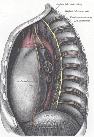
The ligamentum arteriosum, also known as the Ligament of Botallo or Harvey's ligament, is a small ligament attaching the aorta to the pulmonary artery. It serves no function in adults but is the remnant of the ductus arteriosus formed within three weeks after birth.

dextro-Transposition of the great arteries Medical condition

dextro-Transposition of the great arteries is a potentially life-threatening birth defect in the large arteries of the heart. The primary arteries are transposed.

<span class="mw-page-title-main">Ductus arteriosus</span> Blood vessel connecting the pulmonary artery to the proximal descending aorta

The ductus arteriosus, also called the ductus Botalli, named after the Italian physiologist Leonardo Botallo, is a blood vessel in the developing fetus connecting the trunk of the pulmonary artery to the proximal descending aorta. It allows most of the blood from the right ventricle to bypass the fetus's fluid-filled non-functioning lungs. Upon closure at birth, it becomes the ligamentum arteriosum.

<span class="mw-page-title-main">Aortic aneurysm</span> Notable enlargement of the aorta (heart artery)

An aortic aneurysm is an enlargement (dilatation) of the aorta to greater than 1.5 times normal size. They usually cause no symptoms except when ruptured. Occasionally, there may be abdominal, back, or leg pain. The prevalence of abdominal aortic aneurysm ("AAA") has been reported to range from 2 to 12% and is found in about 8% of men more than 65 years of age. The mortality rate attributable to AAA is about 15,000 per year in the United States and 6,000 to 8,000 per year in the United Kingdom and Ireland. Between 2001 and 2006, there were approximately 230,000 AAA surgical repairs performed on Medicare patients in the United States.

<span class="mw-page-title-main">Coarctation of the aorta</span> Medical condition

Coarctation of the aorta, also called aortic narrowing, is a congenital condition whereby the aorta is narrow, usually in the area where the ductus arteriosus inserts. The word coarctation means "pressing or drawing together; narrowing". Coarctations are most common in the aortic arch. The arch may be small in babies with coarctations. Other heart defects may also occur when coarctation is present, typically occurring on the left side of the heart. When a patient has a coarctation, the left ventricle has to work harder. Since the aorta is narrowed, the left ventricle must generate a much higher pressure than normal in order to force enough blood through the aorta to deliver blood to the lower part of the body. If the narrowing is severe enough, the left ventricle may not be strong enough to push blood through the coarctation, thus resulting in a lack of blood to the lower half of the body. Physiologically its complete form is manifested as interrupted aortic arch.

<span class="mw-page-title-main">Recurrent laryngeal nerve</span> Nerve in the human body

The recurrent laryngeal nerve (RLN) is a branch of the vagus nerve that supplies all the intrinsic muscles of the larynx, with the exception of the cricothyroid muscles. There are two recurrent laryngeal nerves, right and left. The right and left nerves are not symmetrical, with the left nerve looping under the aortic arch, and the right nerve looping under the right subclavian artery then traveling upwards. They both travel alongside the trachea. Additionally, the nerves are among the few nerves that follow a recurrent course, moving in the opposite direction to the nerve they branch from, a fact from which they gain their name.

<span class="mw-page-title-main">Persistent truncus arteriosus</span> Medical condition

Persistent truncus arteriosus (PTA), often referred to simply as truncus arteriosus, is a rare form of congenital heart disease that presents at birth. In this condition, the embryological structure known as the truncus arteriosus fails to properly divide into the pulmonary trunk and aorta. This results in one arterial trunk arising from the heart and providing mixed blood to the coronary arteries, pulmonary arteries, and systemic circulation. For the International Classification of Diseases (ICD-11), the International Paediatric and Congenital Cardiac Code (IPCCC) was developed to standardize the nomenclature of congenital heart disease. Under this system, English is now the official language, and persistent truncus arteriosus should properly be termed common arterial trunk.

<span class="mw-page-title-main">Aortic arch</span>

The aortic arch, arch of the aorta, or transverse aortic arch is the part of the aorta between the ascending and descending aorta. The arch travels backward, so that it ultimately runs to the left of the trachea.

<span class="mw-page-title-main">Aortic arches</span>

The aortic arches or pharyngeal arch arteries are a series of six paired embryological vascular structures which give rise to the great arteries of the neck and head. They are ventral to the dorsal aorta and arise from the aortic sac.

<span class="mw-page-title-main">Aberrant subclavian artery</span> Medical condition

Aberrant subclavian artery, or aberrant subclavian artery syndrome, is a rare anatomical variant of the origin of the right or left subclavian artery. This abnormality is the most common congenital vascular anomaly of the aortic arch, occurring in approximately 1% of individuals.

<span class="mw-page-title-main">Ortner's syndrome</span> Medical condition

Ortner's syndrome is a rare cardiovocal syndrome and refers to recurrent laryngeal nerve palsy from cardiovascular disease. It was first described by Norbert Ortner (1865–1935), an Austrian physician, in 1897.

Interrupted aortic arch is a very rare heart defect in which the aorta is not completely developed. There is a gap between the ascending and descending thoracic aorta. In a sense it is the complete form of a coarctation of the aorta. Almost all patients also have other cardiac anomalies, including a ventricular septal defect (VSD), aorto-pulmonary window, and truncus arteriosus. There are three types of interrupted aortic arch, with type B being the most common. Interrupted aortic arch is often associated with DiGeorge syndrome.

A vascular ring is a congenital defect in which there is an abnormal formation of the aorta and/or its surrounding blood vessels. The trachea and esophagus are completely encircled and sometimes compressed by a "ring" formed by these vessels, which can lead to breathing and digestive difficulties.
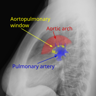
<span class="mw-page-title-main">Traumatic aortic rupture</span> Medical condition

Traumatic aortic rupture, also called traumatic aortic disruption or transection, is a condition in which the aorta, the largest artery in the body, is torn or ruptured as a result of trauma to the body. The condition is frequently fatal due to the profuse bleeding that results from the rupture. Since the aorta branches directly from the heart to supply blood to the rest of the body, the pressure within it is very great, and blood may be pumped out of a tear in the blood vessel very rapidly. This can quickly result in shock and death. Thus traumatic aortic rupture is a common killer in automotive accidents and other traumas, with up to 18% of deaths that occur in automobile collisions being related to the injury. In fact, aortic disruption due to blunt chest trauma is the second leading cause of injury death behind traumatic brain injury.

<span class="mw-page-title-main">Thoracic aorta injury</span> Medical condition

Injury of the thoracic aorta refers to any injury which affects the portion of the aorta which lies within the chest cavity. Injuries of the thoracic aorta are usually the result of physical trauma; however, they can also be the result of a pathological process. The main causes of this injury are deceleration and crush injuries. There are different grades to injuries to the aorta depending on the extent of injury, and the treatment whether surgical or medical depends on that grade. It is difficult to determine if a patient has a thoracic injury just by their symptoms, but through imaging and a physical exam the extent of injury can be determined. All patients with a thoracic aortic injury need to be treated either surgically with endovascular repair or open surgical repair or with medicine to keep their blood pressure and heart rate in the appropriate range. However, most patients that have a thoracic aortic injury do not live for 24 hours.

<span class="mw-page-title-main">Right-sided aortic arch</span> Medical condition

Right-sided aortic arch is a rare anatomical variant in which the aortic arch is on the right side rather than on the left. During normal embryonic development, the aortic arch is formed by the left fourth aortic arch and the left dorsal aorta. In people with a right-sided aortic arch, instead the right dorsal aorta persists and the distal left aorta disappears.

<span class="mw-page-title-main">Tracheoinnominate fistula</span> Medical condition

Tracheoinnominate fistula is an abnormal connection (fistula) between the innominate artery and the trachea. A TIF is a rare but life-threatening iatrogenic injury, usually the sequela of a tracheotomy.

References

  1. Jonas RA. Comprehensive Surgical Management of Congenital Heart Disease, London, Arnold, 2004, p. 497
  2. Kirklin JW, Barratt-Boyes BG. Cardiac Surgery, 3rd ed., New York, Churchill Livingstone, 2003, p. 1415
  3. Momma K, Matsuoka R, Takao A. Aortic arch anomalies associated with chromosome 22q11 deletion. Pediatr Cardiol. 1999 Mar-Apr;20(2):97-102
  4. Hartenberg, M. A.; Salzberg, A. M.; Krummel, T. M.; Bush, J. J. (1989-05-01). "Double aortic arch associated with esophageal atresia and tracheoesophageal fistula". Journal of Pediatric Surgery. 24 (5): 488–490. doi:10.1016/s0022-3468(89)80409-9. ISSN   0022-3468. PMID   2738813.
  5. Tuo G, Volpe P, Bava GL, Bondanza S, De Robertis V, Pongiglione G, Marasini M. Prenatal diagnosis and outcome of isolated vascular rings. Am J Cardiol. 2009 Feb 1;103(3):416-9
  6. Hommell L. Observationes anatomicae de arcu aortae bifido du dueto thoracico duplica, et de carstidum atque subclaviarum. Holdomas 1737;21:161
  7. von Siebold CT. Ringfoermiger Aortenbogen bei einem neugeborenen blausuechtigen Kinde. J Geburtsh Frauenzimmer-Kinderkrank 1837;16:294
  8. Gross RE. Surgical relief for tracheal obstruction from a vascular ring. N Engl J Med 1945;233:586
  9. Neuhauser EB. The roentgen diagnosis of double aortic arch and other anomalies of the great vessels. Am J Roentgenol Radium Ther Nucl Med 1946;56:1
  10. Burke RP, Rosenfeld HM, Wernovsky G, Jonas RA.Video-assisted thoracoscopic vascular ring division in infants and children. J Am Coll Cardiol. 1995;25:943