Airway obstruction

Last updated
Airway obstruction
Specialty Pulmonology   OOjs UI icon edit-ltr-progressive.svg

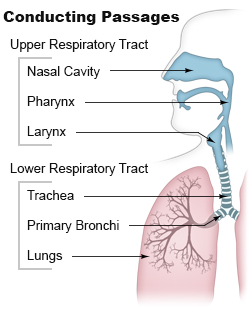
Airway obstruction is a blockage of respiration in the airway that hinders the free flow of air. It can be broadly classified into being either in the upper airway (UPA) or lower airway (LOA).

Contents

Airway obstruction is a life-threatening condition and requires urgent attention, and assistance when it is needed. [1] The assistance to clear an upper airway obstruction would begin employing first-aid anti-choking techniques.

Upper airway obstruction

Causes of upper airway obstruction include foreign body aspiration, blunt laryngotracheal trauma, penetrating laryngotracheal trauma, tonsillar hypertrophy, paralysis of the vocal cord or vocal fold, acute laryngotracheitis such as viral croup, bacterial tracheitis, epiglottitis, peritonsillar abscess, pertussis, retropharyngeal abscess, spasmodic croup. [2] In basic and advanced life support airway obstructions are often referred to as A-problems. Management of airways relies on both minimal-invasive and invasive techniques.

Lower airway obstruction

Lower airway obstruction is mainly caused by increased resistance in the bronchioles (usually from a decreased radius of the bronchioles) that reduces the amount of air inhaled in each breath and the oxygen that reaches the pulmonary arteries. It is different from airway restriction (which prevents air from diffusing into the pulmonary arteries because of some kind of blockage in the lungs). Diseases that cause lower airway obstruction are termed obstructive lung diseases. [3]

Lower airway obstruction can be measured using spirometry. A decreased FEV1/FVC ratio (versus the normal of about 80%) is indicative of airway obstruction, as the normal amount of air can no longer be exhaled in the first second of expiration. An airway restriction would not produce a reduced FEV1/FVC ratio, but would reduce the vital capacity. The ventilation is therefore affected leading to a ventilation-perfusion mismatch and hypoxia.

Consequences

Airway obstruction may cause obstructive pneumonitis or post-obstructive pneumonitis. It can also be a sign of chronic obstructive pulmonary disease (COPD), a common breathing disorder that is a risk factor for lung cancer. [4] [5]

See also

Related Research Articles

<span class="mw-page-title-main">Lung</span> Primary organ of the respiratory system

The lungs are the primary organs of the respiratory system in humans and most other animals, including some snails and a small number of fish. In mammals and most other vertebrates, two lungs are located near the backbone on either side of the heart. Their function in the respiratory system is to extract oxygen from the air and transfer it into the bloodstream, and to release carbon dioxide from the bloodstream into the atmosphere, in a process of gas exchange. The pleurae, which are thin, smooth, and moist, serve to reduce friction between the lungs and chest wall during breathing, allowing for easy and effortless movements of the lungs.

<span class="mw-page-title-main">Respiratory tract</span> Organs involved in transmission of air to and from the point where gases diffuse into tissue

The respiratory tract is the subdivision of the respiratory system involved with the process of respiration in mammals. The respiratory tract is lined with respiratory epithelium as respiratory mucosa.

<span class="mw-page-title-main">Spirometry</span> Pulmonary function test

Spirometry is the most common of the pulmonary function tests (PFTs). It measures lung function, specifically the amount (volume) and/or speed (flow) of air that can be inhaled and exhaled. Spirometry is helpful in assessing breathing patterns that identify conditions such as asthma, pulmonary fibrosis, cystic fibrosis, and COPD. It is also helpful as part of a system of health surveillance, in which breathing patterns are measured over time.

<span class="mw-page-title-main">Interstitial lung disease</span> Group of diseases

Interstitial lung disease (ILD), or diffuse parenchymal lung disease (DPLD), is a group of respiratory diseases affecting the interstitium and space around the alveoli of the lungs. It concerns alveolar epithelium, pulmonary capillary endothelium, basement membrane, and perivascular and perilymphatic tissues. It may occur when an injury to the lungs triggers an abnormal healing response. Ordinarily, the body generates just the right amount of tissue to repair damage, but in interstitial lung disease, the repair process is disrupted, and the tissue around the air sacs (alveoli) becomes scarred and thickened. This makes it more difficult for oxygen to pass into the bloodstream. The disease presents itself with the following symptoms: shortness of breath, nonproductive coughing, fatigue, and weight loss, which tend to develop slowly, over several months. The average rate of survival for someone with this disease is between three and five years. The term ILD is used to distinguish these diseases from obstructive airways diseases.

<span class="mw-page-title-main">Bronchoconstriction</span> Constriction of the terminal airways in the lungs

Bronchoconstriction is the constriction of the airways in the lungs due to the tightening of surrounding smooth muscle, with consequent coughing, wheezing, and shortness of breath.

<span class="mw-page-title-main">Pneumonitis</span> General inflammation of lung tissue

Pneumonitis describes general inflammation of lung tissue. Possible causative agents include radiation therapy of the chest, exposure to medications used during chemo-therapy, the inhalation of debris, aspiration, herbicides or fluorocarbons and some systemic diseases. If unresolved, continued inflammation can result in irreparable damage such as pulmonary fibrosis.

<span class="mw-page-title-main">Respiratory disease</span> Disease of the respiratory system

Respiratory diseases, or lung diseases, are pathological conditions affecting the organs and tissues that make gas exchange difficult in air-breathing animals. They include conditions of the respiratory tract including the trachea, bronchi, bronchioles, alveoli, pleurae, pleural cavity, the nerves and muscles of respiration. Respiratory diseases range from mild and self-limiting, such as the common cold, influenza, and pharyngitis to life-threatening diseases such as bacterial pneumonia, pulmonary embolism, tuberculosis, acute asthma, lung cancer, and severe acute respiratory syndromes, such as COVID-19. Respiratory diseases can be classified in many different ways, including by the organ or tissue involved, by the type and pattern of associated signs and symptoms, or by the cause of the disease.

<span class="mw-page-title-main">Bronchiolitis obliterans</span> Medical condition

Bronchiolitis obliterans (BO), also known as obliterative bronchiolitis, constrictive bronchiolitis and popcorn lung, is a disease that results in obstruction of the smallest airways of the lungs (bronchioles) due to inflammation. Symptoms include a dry cough, shortness of breath, wheezing and feeling tired. These symptoms generally get worse over weeks to months. It is not related to cryptogenic organizing pneumonia, previously known as bronchiolitis obliterans organizing pneumonia.

Occupational lung diseases comprise a broad group of diseases, including occupational asthma, industrial bronchitis, chronic obstructive pulmonary disease (COPD), bronchiolitis obliterans, inhalation injury, interstitial lung diseases, infections, lung cancer and mesothelioma. These can be caused directly or due to immunological response to an exposure to a variety of dusts, chemicals, proteins or organisms. Occupational cases of interstitial lung disease may be misdiagnosed as COPD, idiopathic pulmonary fibrosis, or a myriad of other diseases; leading to a delay in identification of the causative agent.

A bronchial challenge test is a medical test used to assist in the diagnosis of asthma. The patient breathes in nebulized methacholine or histamine. Thus the test may also be called a methacholine challenge test or histamine challenge test respectively. Both drugs provoke bronchoconstriction, or narrowing of the airways. Whereas histamine causes nasal and bronchial mucus secretion and bronchoconstriction via the H1 receptor, methacholine utilizes the M3 receptor for bronchoconstriction. The degree of narrowing can then be quantified by spirometry. People with pre-existing airway hyperreactivity, such as asthmatics, will react to lower doses of drug.

<span class="mw-page-title-main">Obstructive lung disease</span> Category of respiratory disease characterized by airway obstruction

Obstructive lung disease is a category of respiratory disease characterized by airway obstruction. Many obstructive diseases of the lung result from narrowing (obstruction) of the smaller bronchi and larger bronchioles, often because of excessive contraction of the smooth muscle itself. It is generally characterized by inflamed and easily collapsible airways, obstruction to airflow, problems exhaling, and frequent medical clinic visits and hospitalizations. Types of obstructive lung disease include; asthma, bronchiectasis, bronchitis and chronic obstructive pulmonary disease (COPD). Although COPD shares similar characteristics with all other obstructive lung diseases, such as the signs of coughing and wheezing, they are distinct conditions in terms of disease onset, frequency of symptoms, and reversibility of airway obstruction. Cystic fibrosis is also sometimes included in obstructive pulmonary disease.

Restrictive lung diseases are a category of extrapulmonary, pleural, or parenchymal respiratory diseases that restrict lung expansion, resulting in a decreased lung volume, an increased work of breathing, and inadequate ventilation and/or oxygenation. Pulmonary function test demonstrates a decrease in the forced vital capacity.

<span class="mw-page-title-main">Pulmonary function testing</span> Test to evaluate respiratory system

Pulmonary function testing (PFT) is a complete evaluation of the respiratory system including patient history, physical examinations, and tests of pulmonary function. The primary purpose of pulmonary function testing is to identify the severity of pulmonary impairment. Pulmonary function testing has diagnostic and therapeutic roles and helps clinicians answer some general questions about patients with lung disease. PFTs are normally performed by a pulmonary function technician, respiratory therapist, respiratory physiologist, physiotherapist, pulmonologist, or general practitioner.

<span class="mw-page-title-main">FEV1/FVC ratio</span> Ratio used in the diagnosis of lung disease

The FEV1/FVC ratio, also called modified Tiffeneau-Pinelli index, is a calculated ratio used in the diagnosis of obstructive and restrictive lung disease. It represents the proportion of a person's vital capacity that they are able to expire in the first second of forced expiration (FEV1) to the full, forced vital capacity (FVC). FEV1/FVC ratio first proposed by E.A. Haensler in 1950. The FEV1/FVC index should not be confused with the FEV1/VC index as they are different, although both are intended for diagnosing airway obstruction. Current recommendations for diagnosing pulmonary function recommend using the modified Tiffeneau-Pinelli index. This index is recommended to be represented as a decimal fraction with two digits after the decimal point.

<span class="mw-page-title-main">Acute exacerbation of chronic obstructive pulmonary disease</span> Medical condition

An acute exacerbation of chronic obstructive pulmonary disease, or acute exacerbations of chronic bronchitis (AECB), is a sudden worsening of chronic obstructive pulmonary disease (COPD) symptoms including shortness of breath, quantity and color of phlegm that typically lasts for several days.

<span class="mw-page-title-main">Chronic obstructive pulmonary disease</span> Lung disease involving long-term poor airflow

Chronic obstructive pulmonary disease (COPD) is a type of progressive lung disease characterized by long-term respiratory symptoms and airflow limitation. The main symptoms of COPD include shortness of breath and a cough, which may or may not produce mucus. COPD progressively worsens, with everyday activities such as walking or dressing becoming difficult. While COPD is incurable, it is preventable and treatable. The two most common types of COPD are emphysema and chronic bronchitis and have been the two classic COPD phenotypes. Emphysema is defined as enlarged airspaces (alveoli) whose walls have broken down resulting in permanent damage to the lung tissue. Chronic bronchitis is defined as a productive cough that is present for at least three months each year for two years. Both of these conditions can exist without airflow limitation when they are not classed as COPD. Emphysema is just one of the structural abnormalities that can limit airflow and can exist without airflow limitation in a significant number of people. Chronic bronchitis does not always result in airflow limitation but in young adults who smoke the risk of developing COPD is high. Many definitions of COPD in the past included emphysema and chronic bronchitis, but these have never been included in GOLD report definitions. Emphysema and chronic bronchitis remain the predominant phenotypes of COPD but there is often overlap between them and a number of other phenotypes have also been described. COPD and asthma may coexist and converge in some individuals. COPD is associated with low-grade systemic inflammation.

<span class="mw-page-title-main">Emphysema</span> Medical condition

Emphysema is any air-filled enlargement in the body's tissues. Most commonly emphysema refers to the enlargement of air spaces (alveoli) in the lungs, and is also known as pulmonary emphysema.

<span class="mw-page-title-main">Ventilation-perfusion coupling</span>

Ventilation-perfusion coupling is the relationship between ventilation and perfusion processes, which take place in the respiratory and cardiovascular systems. Ventilation is the movement of gas during breathing, and perfusion is the process of pulmonary blood circulation, which delivers oxygen to body tissues. Anatomically, the lung structure, alveolar organization, and alveolar capillaries contribute to the physiological mechanism of ventilation and perfusion. Ventilation-perfusion coupling maintains a constant ratio near 0.8 on average, while the regional variation exists within the lungs due to gravity. When the ratio gets above or below 0.8, it is considered abnormal ventilation-perfusion coupling, also known as a ventilation-perfusion mismatch. Lung diseases, cardiac shunts, and smoking can cause a ventilation-perfusion mismatch that results in significant symptoms and diseases, which can be treated through treatments like bronchodilators and oxygen therapy.

Asthma-Chronic Obstructive Pulmonary Disease (COPD) Overlap (ACO), also known as Asthma-COPD Overlap Syndrome (ACOS) is a chronic inflammatory, obstructive airway disease in which features of both asthma and COPD predominate. Asthma and COPD were once thought of as distinct entities, however in some, there are clinical features of both asthma and COPD with significant overlap in pathophysiology and symptom profile. It is unclear whether ACO is a separate disease entity or a clinical subtype of asthma and COPD. The pathogenesis of ACO is poorly understood, but it is thought to involve both type 2 inflammation as well as type 1 inflammation. The incidence and prevalence of ACO are not well known. The risk factors for ACO are also incompletely understood, but tobacco smoke is known to be a major risk factor.

References

  1. Aboussouan, L.S.; Stoller, J.K (15 March 1994). "Diagnosis and management of upper airway obstruction". Clinics in Chest Medicine. 15 (1): 35–53. doi:10.1016/S0272-5231(21)01054-6. PMID   8200192 . Retrieved 25 June 2021.
  2. Respiratory Emergencies, section Acute Upper Airway Obstruction [ permanent dead link ]. From FP Essentials 368 Archived 2020-03-14 at the Wayback Machine . January 2010 by American Academy of Family Physicians.
  3. "Airway Obstruction". The Lecturio Medical Concept Library. Retrieved 2021-06-25.
  4. "Lung health checks in supermarket car parks reach older smokers in deprived communities". NIHR Evidence (Plain English summary). 2020-08-05. doi:10.3310/alert_40661. S2CID   243394660.
  5. Balata, Haval; Harvey, Jonathan; Barber, Phil V; Colligan, Denis; Duerden, Rebecca; Elton, Peter; Evison, Matthew; Greaves, Melanie; Howells, John; Irion, Klaus; Karunaratne, Devinda (15 Jul 2020). "Spirometry performed as part of the Manchester community-based lung cancer screening programme detects a high prevalence of airflow obstruction in individuals without a prior diagnosis of COPD". Thorax. 75 (8): 655–660. doi: 10.1136/thoraxjnl-2019-213584 . ISSN   0040-6376. PMID   32444437. S2CID   218855570.