The term "usual" refers to the fact that UIP is the most common form of interstitial fibrosis. "Pneumonia" indicates "lung abnormality", which includes fibrosis and inflammation. A term previously used for UIP in the British literature is cryptogenic fibrosing alveolitis (CFA), a term that has fallen out of favor since the basic underlying pathology is now thought to be fibrosis, not inflammation. The term usual interstitial pneumonitis (UIP) has also often been used, but again, the -itis part of that name may overemphasize inflammation.
Signs and symptoms
The typical symptoms of UIP are progressive shortness of breath and cough for a period of months. In some patients, UIP is diagnosed only when a more acute disease supervenes and brings the patient to medical attention.
Usual interstitial pneumonia seen on CT scan. Honeycomb fibrosis is seen at the bases of both lungs.
UIP may be diagnosed by a radiologist using computed tomography (CT) scan of the chest, or by a pathologist using tissue obtained by a lung biopsy.
Radiology
Radiologically, the main feature required for a confident diagnosis of UIP is honeycomb change in the periphery and the lower portions (bases) of the lungs.[3]
The histologic hallmarks of UIP, as seen in lung tissue under a microscope by a pathologist, are interstitial fibrosis in a "patchwork pattern", honeycomb change and fibroblast foci (see images below).[6][7]
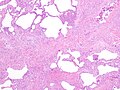
Pathological findings in usual interstitial pneumonia (UIP)
Appearance of usual interstitial pneumonia (UIP) in a surgical lung biopsy at low magnification. The tissue is stained with hematoxylin (purple dye) and eosin (pink dye) to make it visible. The pink areas in this picture represent lung fibrosis (collagen stains pink). Note the "patchwork" (quilt-like) pattern of the fibrosis.
Appearance of honeycomb change in a surgical lung biopsy at low magnification. The dilated spaces seen here are filled with mucin. Hematoxylin-eosin stain, low magnification.
A fibroblast focus in a surgical lung biopsy of UIP. Hematoxylin-eosin stain, high magnification. The white space to the left is an airspace. The pale area to the right is a fibroblast focus. It is an area of active fibroblast proliferation within the interstitium of the lung.
Differential diagnosis
The differential diagnosis includes other types of lung disease that cause similar symptoms and show similar abnormalities on chest radiographs. Some of these diseases cause fibrosis, scarring or honeycomb change. The most common considerations include:
Regardless of cause, UIP is relentlessly progressive, usually leading to respiratory failure and death without a lung transplant.[citation needed] Some patients do well for a prolonged period of time, but then deteriorate rapidly because of a superimposed acute illness (so-called "accelerated UIP"). The outlook for long-term survival is poor. In most studies, the median survival is 3 to 4 years.[citation needed] Patients with UIP in the setting of rheumatoid arthritis have a slightly better prognosis than UIP without a known cause (IPF).
↑Travis WD, King TE, Bateman ED, etal. (2002). "ATS/ERS international multidisciplinary consensus classification of idiopathic interstitial pneumonias. General principles and recommendations". American Journal of Respiratory and Critical Care Medicine. 165 (5): 277–304. doi:10.1164/ajrccm.165.2.ats01. PMID11790668.
↑Sumikawa H, etal. (2008). "Computed tomography findings in pathological usual interstitial pneumonia: relationship to survival". American Journal of Respiratory and Critical Care Medicine. 177 (4): 433–439. doi:10.1164/rccm.200611-1696OC. PMID17975197.
12345Raghu, Ganesh; Remy-Jardin, Martine; Myers, Jeffrey L.; Richeldi, Luca; Ryerson, Christopher J.; Lederer, David J.; Behr, Juergen; Cottin, Vincent; Danoff, Sonye K.; Morell, Ferran; Flaherty, Kevin R.; Wells, Athol; Martinez, Fernando J.; Azuma, Arata; Bice, Thomas J.; Bouros, Demosthenes; Brown, Kevin K.; Collard, Harold R.; Duggal, Abhijit; Galvin, Liam; Inoue, Yoshikazu; Jenkins, R. Gisli; Johkoh, Takeshi; Kazerooni, Ella A.; Kitaichi, Masanori; Knight, Shandra L.; Mansour, George; Nicholson, Andrew G.; Pipavath, Sudhakar N. J.; Buendía-Roldán, Ivette; Selman, Moisés; Travis, William D.; Walsh, Simon L. F.; Wilson, Kevin C. (2018). "Diagnosis of Idiopathic Pulmonary Fibrosis. An Official ATS/ERS/JRS/ALAT Clinical Practice Guideline". American Journal of Respiratory and Critical Care Medicine. 198 (5): e44 –e68. doi:10.1164/rccm.201807-1255ST. ISSN1073-449X.
↑Leslie, Kevin O; Wick, Mark R. (2005). Practical pulmonary pathology: a diagnostic approach. Edinburgh: Churchill Livingstone. ISBN0-443-06631-0. OCLC156861539.
This page is based on this Wikipedia article Text is available under the CC BY-SA 4.0 license; additional terms may apply. Images, videos and audio are available under their respective licenses.