| Lung nodule | |
|---|---|
 | |
| Chest X-ray showing a solitary pulmonary nodule (indicated by a black box) in the left upper lobe. | |
| Specialty | Pulmonology |
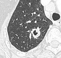
A lung nodule or pulmonary nodule is a relatively small focal density in the lung. A solitary pulmonary nodule (SPN) or coin lesion, [1] is a mass in the lung smaller than three centimeters in diameter. A pulmonary micronodule has a diameter of less than three millimetres. [2] There may also be multiple nodules.
Contents
- Causes
- Risk factors
- Diagnosis
- Definition
- CT scan
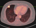
- PET scan
- Other imaging
- Histopathology
- Management
- Excision
- See also
- Footnotes
- External links
One or more lung nodules can be an incidental finding found in up to 0.2% of chest X-rays [3] and around 1% of CT scans. [4]
The nodule most commonly represents a benign tumor such as a granuloma or hamartoma, but in around 20% of cases it represents a malignant cancer, [4] especially in older adults and smokers. Conversely, 10 to 20% of patients with lung cancer are diagnosed in this way. [4] If the patient has a history of smoking or the nodule is growing, the possibility of cancer may need to be excluded through further radiological studies and interventions, possibly including surgical resection. The prognosis depends on the underlying condition.