Tuberculoma

Last updated
PET-CT of a tuberculoma PET-CT of a tuberculoma.png
PET-CT of a tuberculoma

A tuberculoma is a clinical manifestation of tuberculosis which conglomerates tubercles into a firm lump, and so can mimic cancer tumors of many types in medical imaging studies. [1] [2] They often arise within individuals in whom a primary tuberculosis infection is not well controlled. [3] When tuberculomas arise intracranially, they represent a manifestation of CNS tuberculosis. [3] Since these are evolutions of primary complex, the tuberculomas may contain caseum or calcifications.

Contents

With the passage of time, Mycobacterium tuberculosis can transform into crystals of calcium. These can affect any organ such as the brain, [4] [5] intestine, [6] [7] [8] ovaries, [9] [10] breast, [11] [12] [13] lungs, [14] [15] esophagus, [16] pancreas, [17] bones, [18] [19] and many others. Even with guideline-directed treatment they often persist for months to years. [3]

Mechanism

The exact mechanism of tuberculoma development has not been determined, although multiple theories have been proposed. It is possible that, following an initial tuberculosis infection resulting in bacteremia, a foci of granulomatous inflammation may coalesce into a caseous tuberculoma. [20] Pulmonary tuberculomas may arise due to repeated cycles of necrosis and re-encapsulation of foci, or, alternatively, the shrinkage and fusion of encapsulated densities. [21]

In regards to CNS tuberculoma, it is thought that mycobacterium tuberculosis is capable of penetrating the blood brain barrier after bacterial bacilli induce the release of cytokines by various immunologic cells, leading to an increase in barrier permeability. [22] Similar to pulmonary tuberculomas, small lesions eventually coalesce and undergo both necrosis and enlargement. [22]

Signs and symptoms

Symptoms are based on the location of the tuberculoma. Small, scattered lesions may be asymptomatic. Intracranial tuberculomas in children are often infratentorial, occurring near the cerebellum and base of the brain. In this population, symptoms such as headache, fever, focal neurologic findings and seizures have been seen [3] in addition to papilledema with or without meningitis. [20] When the size of a brainstem tuberculoma grows to the point of narrowing the fourth ventricle, obstructing hydrocephalus and its related symptoms can arise. [20] Rupture of tuberculomas adjacent to the arachnoid can lead to arachnoiditis, [23] while rupture near the subarachnoid space or ventricular system can cause meningitis. [22]

Diagnosis

Tuberculoma With Cavitation Tuberculosis - Tuberculoma with cavitation (6596009867).jpg
Tuberculoma With Cavitation

The diagnosis of tuberculoma can be challenging, as invasive testing may be required and, occasionally, concomitant malignancy may be present. [21] In children with tuberculoma, CXR is often normal despite a positive TST/IGRA. [3]

Diagnosis of brain tuberculoma can be aided with PCR of cerebrospinal fluid, but is of less utility for quickly diagnosing and treating lesions. [22] When CSF is analyzed in patients with suspected tuberculoma, high protein concentrations and cell counts are often seen. [24]

Definitive diagnosis can be made through stereotactic, CT-guided biopsy, with excision required in rare cases. Biopsy is chosen when non-invasive testing has failed to produce a diagnosis, when patients fail to respond to a treatment regimen, in cases of drug-resistant tuberculosis, and in non-compliant patients. [22]

Imaging

The appearance of a tuberculoma on imaging can vary according to the composition and age of the mass. They may appear as either non-caseating or solidly caseating lesions. [20] Initially, tuberculomas appear hypodense on computed tomography (CT) scans with significant surrounding edema. [23] [3] The "target sign" is pathognomonic for tuberculoma on CT, with a nodular ring-enhancing mass and central calcification. [24] [20] The characteristic ring-enhanced appearance is due to lack of blood supply in the central necrotic core that is visualized with injected contrast. [22] Sometimes a hypodense central area is seen instead of calcification. [25] When considering other potential intracranial masses in a differential diagnosis, such as cysticercosis, pyogenic abscess, and neoplastic lesions, tuberculoma can be identified by its larger size (>2 cm), edema, and irregular border.

Magnetic resonance imaging (MRI) is another useful imaging modality for diagnosing and characterizing of tuberculomas, especially solid caseous necrosis in which 3 zones of varying intensity are seen. [22]

Treatment

Tuberculoma is commonly treated through the HRZE drug combination (Isoniazid, Rifampin, Pyrazinamide, Ethambutol) followed by maintenance therapy. [26] Per international guidelines, 9–12 months of medical management is standard. [20] While the majority of tuberculomas resolve in 12–24 months, in patients with multiple or larger lesions prolonged treatment extending beyond two years may be required. In some patients, the release of inflammatory mediators during treatment can cause a paradoxical worsening of symptoms that is treated with anti-inflammatory medications in addition to the standard anti-tuberculosis regimen. [22]

Exceptionally large tuberculomas, those exerting a mass effect on the brain, and those which fail to respond to medical management required surgical excision. In some cases, surgical excision is necessary for diagnosis as well as treatment. [3] When intracranial pressure rises in the setting of tuberculoma, removal is considered a surgical emergency. [22]

Prognosis

Of patients with a brain tuberculoma treated with an appropriate medication regimen, almost half recover completely. Approximately 10% of those treated fail to recover and succumb to the tuberculoma. [22] Reports issued before the advent of effective anti-tuberculosis therapy showed that, when untreated, 30-50% of tuberculomas enter and remain in a stationary course. [21]

Epidemiology

Tuberculomas are most commonly seen in areas where tuberculosis is endemic. In these areas, tuberculomas can account for between 30%-50% of intracranial masses. [22] [3] India and parts of Asia are two areas where tuberculomas have been noted to be particularly prevalent. [20] They occur most often as solitary, infratentorial lesions in young children. [3] In contrast, lesions are most often supratentorial in adults. [3]

Pulmonary tuberculomas are among the most common benign nodules, with 5%-24% of all resected nodules being of tuberculous origin. [21] In areas of lower prevalence, such as the United States, they are most commonly seen in the setting of an acquired immunodeficiency. [23] Intracerebral tuberculomas, specifically, are more frequently observed in patients with an HIV infection. [20]

Related Research Articles

<span class="mw-page-title-main">Brain abscess</span> Medical condition

Brain abscess is an abscess within the brain tissue caused by inflammation and collection of infected material coming from local or remote infectious sources. The infection may also be introduced through a skull fracture following a head trauma or surgical procedures. Brain abscess is usually associated with congenital heart disease in young children. It may occur at any age but is most frequent in the third decade of life.

<span class="mw-page-title-main">Brain tumor</span> Neoplasm in the brain

A brain tumor occurs when abnormal cells form within the brain. There are two main types of tumors: malignant tumors and benign (non-cancerous) tumors. These can be further classified as primary tumors, which start within the brain, and secondary tumors, which most commonly have spread from tumors located outside the brain, known as brain metastasis tumors. All types of brain tumors may produce symptoms that vary depending on the size of the tumor and the part of the brain that is involved. Where symptoms exist, they may include headaches, seizures, problems with vision, vomiting and mental changes. Other symptoms may include difficulty walking, speaking, with sensations, or unconsciousness.

<span class="mw-page-title-main">Granuloma</span> Aggregation of macrophages in response to chronic inflammation

A granuloma is an aggregation of macrophages that forms in response to chronic inflammation. This occurs when the immune system attempts to isolate foreign substances that it is otherwise unable to eliminate. Such substances include infectious organisms including bacteria and fungi, as well as other materials such as foreign objects, keratin, and suture fragments.

<span class="mw-page-title-main">Miliary tuberculosis</span> Medical condition

Miliary tuberculosis is a form of tuberculosis that is characterized by a wide dissemination into the human body and by the tiny size of the lesions (1–5 mm). Its name comes from a distinctive pattern seen on a chest radiograph of many tiny spots distributed throughout the lung fields with the appearance similar to millet seeds—thus the term "miliary" tuberculosis. Miliary TB may infect any number of organs, including the lungs, liver, and spleen. Miliary tuberculosis is present in about 2% of all reported cases of tuberculosis and accounts for up to 20% of all extra-pulmonary tuberculosis cases.

<span class="mw-page-title-main">Pilocytic astrocytoma</span> Medical condition

Pilocytic astrocytoma is a brain tumor that occurs most commonly in children and young adults. They usually arise in the cerebellum, near the brainstem, in the hypothalamic region, or the optic chiasm, but they may occur in any area where astrocytes are present, including the cerebral hemispheres and the spinal cord. These tumors are usually slow growing and benign, corresponding to WHO malignancy grade 1.

<span class="mw-page-title-main">Craniopharyngioma</span> Medical condition

A craniopharyngioma is a rare type of brain tumor derived from pituitary gland embryonic tissue that occurs most commonly in children, but also affects adults. It may present at any age, even in the prenatal and neonatal periods, but peak incidence rates are childhood-onset at 5–14 years and adult-onset at 50–74 years. People may present with bitemporal inferior quadrantanopia leading to bitemporal hemianopsia, as the tumor may compress the optic chiasm. It has a point prevalence around two per 1,000,000. Craniopharyngiomas are distinct from Rathke's cleft tumours and intrasellar arachnoid cysts.

<span class="mw-page-title-main">Arachnoid cyst</span> Medical condition

Arachnoid cysts are cerebrospinal fluid covered by arachnoidal cells and collagen that may develop between the surface of the brain and the cranial base or on the arachnoid membrane, one of the three meningeal layers that cover the brain and the spinal cord. Primary arachnoid cysts are a congenital disorder whereas secondary arachnoid cysts are the result of head injury or trauma. Most cases of primary cysts begin during infancy; however, onset may be delayed until adolescence.

Sparganosis is a parasitic infection caused by the plerocercoid larvae of the genus Spirometra including S. mansoni, S. ranarum, S. mansonoides and S. erinacei. It was first described by Patrick Manson in 1882, and the first human case was reported by Charles Wardell Stiles from Florida in 1908. The infection is transmitted by ingestion of contaminated water, ingestion of a second intermediate host such as a frog or snake, or contact between a second intermediate host and an open wound or mucous membrane. Humans are the accidental hosts in the life cycle, while dogs, cats, and other mammals are definitive hosts. Copepods are the first intermediate hosts, and various amphibians and reptiles are second intermediate hosts.

<span class="mw-page-title-main">Hemangioblastoma</span> Medical condition

Hemangioblastomas, or haemangioblastomas, are vascular tumors of the central nervous system that originate from the vascular system, usually during middle age. Sometimes, these tumors occur in other sites such as the spinal cord and retina. They may be associated with other diseases such as polycythemia, pancreatic cysts and Von Hippel–Lindau syndrome. Hemangioblastomas are most commonly composed of stromal cells in small blood vessels and usually occur in the cerebellum, brainstem or spinal cord. They are classed as grade I tumors under the World Health Organization's classification system.

<span class="mw-page-title-main">Calciphylaxis</span> Medical condition

Calciphylaxis, also known as calcific uremic arteriolopathy (CUA) or “Grey Scale”, is a rare syndrome characterized by painful skin lesions. The pathogenesis of calciphylaxis is unclear but believed to involve calcification of the small blood vessels located within the fatty tissue and deeper layers of the skin, blood clots, and eventual death of skin cells due to lack of blood flow. It is seen mostly in people with end-stage kidney disease but can occur in the earlier stages of chronic kidney disease and rarely in people with normally functioning kidneys. Calciphylaxis is a rare but serious disease, believed to affect 1-4% of all dialysis patients. It results in chronic non-healing wounds and indicates poor prognosis, with typical life expectancy of less than one year.

<span class="mw-page-title-main">Thyroid nodule</span> Medical condition

Thyroid nodules are nodules which commonly arise within an otherwise normal thyroid gland. They may be hyperplastic or tumorous, but only a small percentage of thyroid tumors are malignant. Small, asymptomatic nodules are common, and often go unnoticed. Nodules that grow larger or produce symptoms may eventually need medical care. A goitre may have one nodule – uninodular, multiple nodules – multinodular, or be diffuse.

<span class="mw-page-title-main">Lung nodule</span> Medical condition

A lung nodule or pulmonary nodule is a relatively small focal density in the lung. A solitary pulmonary nodule (SPN) or coin lesion, is a mass in the lung smaller than three centimeters in diameter. A pulmonary micronodule has a diameter of less than three millimetres. There may also be multiple nodules.
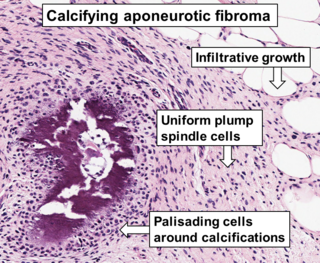
<span class="mw-page-title-main">Cerebrospinal fluid leak</span> Medical condition

A cerebrospinal fluid leak is a medical condition where the cerebrospinal fluid (CSF) surrounding the brain or spinal cord leaks out of one or more holes or tears in the dura mater. A cerebrospinal fluid leak can be either cranial or spinal, and these are two different disorders. A spinal CSF leak can be caused by one or more meningeal diverticula or CSF-venous fistulas not associated with an epidural leak.

<span class="mw-page-title-main">Spiradenoma</span> Medical condition

Spiradenomas (SA) are rare, benign cutaneous adnexal tumors that may progress to become their malignant counterparts, i.e. spiradenocarcinomas (SAC). Cutaneous adnexal tumors are a group of skin tumors consisting of tissues that have differentiated towards one of the four primary adnexal structures found in normal skin: hair follicles, sebaceous sweat glands, apocrine sweat glands, and eccrine sweat glands. SA and SAC tumors were regarded as eccrine gland tumors and termed eccrine spiradenomas and eccrine spiradenocarcinomas, respectively. However, more recent studies have found them to be hair follicle tumors and commonly term them spiradenomas and spiradenocarcinomas, respectively. Further confusing the situation, SA-like and SAC-like tumors are also 1) manifestations of the inherited disorder, CYLD cutaneous syndrome (CCS), and 2) have repeatedly been confused with an entirely different tumor, adenoid cystic carcinomas of the salivary gland. Here, SA and SAC are strictly defined as sporadic hair follicle tumors that do not include the hereditary CCS spiradenomas and heridtary spiradenocarcinoms of CCS or the adenoid cystic carcinomas.

<span class="mw-page-title-main">Malignant chondroid syringoma</span> Type of skin cancer

A malignant chondroid syringoma is a very uncommon cutaneous (skin) condition characterised by an adnexal eccrine tumour.

<span class="mw-page-title-main">Bonnet–Dechaume–Blanc syndrome</span> Medical condition

Bonnet–Dechaume–Blanc syndrome, also known as Wyburn-Mason syndrome, is a rare congenital disorder characterized by arteriovenous malformations of the brain, retina or facial nevi. The syndrome has a number of possible symptoms and can, more rarely, affect the skin, bones, kidneys, muscles, and gastrointestinal tract. When the syndrome affects the brain, people can experience severe headaches, seizures, acute stroke, meningism, and progressive neurological deficits due to acute or chronic ischaemia caused by arteriovenous shunting.

<span class="mw-page-title-main">Brain metastasis</span> Cancer that has metastasized (spread) to the brain from another location in the body

A brain metastasis is a cancer that has metastasized (spread) to the brain from another location in the body and is therefore considered a secondary brain tumor. The metastasis typically shares a cancer cell type with the original site of the cancer. Metastasis is the most common cause of brain cancer, as primary tumors that originate in the brain are less common. The most common sites of primary cancer which metastasize to the brain are lung, breast, colon, kidney, and skin cancer. Brain metastases can occur in patients months or even years after their original cancer is treated. Brain metastases have a poor prognosis for cure, but modern treatments are allowing patients to live months and sometimes years after the diagnosis.

<span class="mw-page-title-main">Computed tomography of the head</span> Cross-sectional X-rays of the head

Computed tomography of the head uses a series of X-rays in a CT scan of the head taken from many different directions; the resulting data is transformed into a series of cross sections of the brain using a computer program. CT images of the head are used to investigate and diagnose brain injuries and other neurological conditions, as well as other conditions involving the skull or sinuses; it used to guide some brain surgery procedures as well. CT scans expose the person getting them to ionizing radiation which has a risk of eventually causing cancer; some people have allergic reactions to contrast agents that are used in some CT procedures.

<span class="mw-page-title-main">Intracranial epidermoid cyst</span>

Intracranial epidermoid cysts develop in the early embryonic phases. The cysts develop when epithelial cells are confined with cells that form the brain.

<span class="mw-page-title-main">Melanocytoma</span>

A melanocytoma is a rare pigmented tumor that has been described as a variant of the melanocytic nevus and is a derivative of the neural crest. The term "melanocytoma" was introduced by Limas and Tio in 1972.

References

  1. Pitlik SD, Fainstein V, Bodey GP (May 1984). "Tuberculosis mimicking cancer--a reminder". The American Journal of Medicine. 76 (5): 822–5. doi:10.1016/0002-9343(84)90993-8. PMID   6720729.
  2. Vento S, Lanzafame M (June 2011). "Tuberculosis and cancer: a complex and dangerous liaison". The Lancet. Oncology. 12 (6): 520–2. doi:10.1016/S1470-2045(11)70105-X. PMID   21624773.
  3. 1 2 3 4 5 6 7 8 9 10 Lloyd N. Friedman; Martin Dedicoat; P. D. O. Davies, eds. (2020). Clinical tuberculosis (Sixth ed.). Boca Raton, FL. ISBN   978-1-351-24998-0. OCLC   1145905400.{{cite book}}: CS1 maint: location missing publisher (link)
  4. Dennison P, Rajakaruna G (October 2006). "Cerebral tuberculoma". Thorax. 61 (10): 922. doi:10.1136/thx.2005.054932. PMC   2104774 . PMID   17008487.
  5. Chatterjee S (October 2011). "Brain tuberculomas, tubercular meningitis, and post-tubercular hydrocephalus in children". Journal of Pediatric Neurosciences. 6 (Suppl 1): S96–S100. doi: 10.4103/1817-1745.85725 . PMC   3208909 . PMID   22069437.
  6. Herrick FC (April 1925). "Tuberculoma of the Caecum: Hyperplastic Tuberculosis". Annals of Surgery. 81 (4): 801–20. doi:10.1097/00000658-192504000-00009. PMC   1399989 . PMID   17865239.
  7. Chakravartty S, Chattopadhyay G, Ray D, Choudhury CR, Mandal S (2010). "Concomitant tuberculosis and carcinoma colon: coincidence or causal nexus?". Saudi Journal of Gastroenterology. 16 (4): 292–4. doi: 10.4103/1319-3767.70619 . PMC   2995101 . PMID   20871197.
  8. Kushwaha JK, Sonkar AA, Saraf A, Singh D, Gupta R (September 2011). "Jejunal adenocarcinoma: an elusive diagnosis". Indian Journal of Surgical Oncology. 2 (3): 197–201. doi:10.1007/s13193-011-0101-7. PMC   3272177 . PMID   22942611.
  9. Elmore RG, Li AJ (December 2007). "Peritoneal tuberculosis mimicking advanced-stage epithelial ovarian cancer". Obstetrics and Gynecology. 110 (6): 1417–9. doi:10.1097/01.AOG.0000295653.32975.4a. PMID   18055741.
  10. Rabesalama S, Mandeville K, Raherison R, Rakoto-Ratsimba H (2011). "Isolated ovarian tuberculosis mimicking ovarian carcinoma: case report and literature review". African Journal of Infectious Diseases. 5 (1): 7–10. doi:10.4314/ajid.v5i1.66508. PMC   3497843 . PMID   23878702.
  11. Baharoon S (July 2008). "Tuberculosis of the breast". Annals of Thoracic Medicine. 3 (3): 110–4. doi: 10.4103/1817-1737.41918 . PMC   2700437 . PMID   19561892.
  12. Sen M, Gorpelioglu C, Bozer M (2009). "Isolated primary breast tuberculosis: report of three cases and review of the literature". Clinics. 64 (6): 607–10. doi:10.1590/S1807-59322009000600019. PMC   2705158 . PMID   19578668.
  13. Akçay MN, Sağlam L, Polat P, Erdoğan F, Albayrak Y, Povoski SP (June 2007). "Mammary tuberculosis -- importance of recognition and differentiation from that of a breast malignancy: report of three cases and review of the literature". World Journal of Surgical Oncology. 5: 67. doi: 10.1186/1477-7819-5-67 . PMC   1910599 . PMID   17577397.
  14. Liang HY, Li XL, Yu XS, Guan P, Yin ZH, He QC, Zhou BS, et al. (December 2009). "Facts and fiction of the relationship between preexisting tuberculosis and lung cancer risk: a systematic review". International Journal of Cancer. 125 (12): 2936–44. doi: 10.1002/ijc.24636 . PMID   19521963. S2CID   21083607.
  15. Khan AN, Al-Jahdali HH, Allen CM, Irion KL, Al Ghanem S, Koteyar SS (April 2010). "The calcified lung nodule: What does it mean?". Annals of Thoracic Medicine. 5 (2): 67–79. doi: 10.4103/1817-1737.62469 . PMC   2883201 . PMID   20582171.
  16. Patnayak R, Reddy MK, Parthasarathy S, Yootla M, Reddy V, Jena A (April 2008). "Unusual presentation of esophageal tuberculosis mimicking malignancy". Saudi Journal of Gastroenterology. 14 (2): 103–4. doi: 10.4103/1319-3767.39632 . PMC   2702907 . PMID   19568514.
  17. Saluja SS, Ray S, Pal S, Kukeraja M, Srivastava DN, Sahni P, Chattopadhyay TK (June 2007). "Hepatobiliary and pancreatic tuberculosis: a two decade experience". BMC Surgery. 7 (1): 10. doi: 10.1186/1471-2482-7-10 . PMC   1925057 . PMID   17588265.
  18. Herzog A (September 2009). "Dangerous errors in the diagnosis and treatment of bony tuberculosis". Deutsches Ärzteblatt International. 106 (36): 573–7. doi:10.3238/arztebl.2009.0573. PMC   2770211 . PMID   19890413.
  19. Dhillon MS, Aggarwal S, Prabhakar S, Bachhal V (March 2012). "Tuberculosis of the foot: An osteolytic variety". Indian Journal of Orthopaedics. 46 (2): 206–11. doi: 10.4103/0019-5413.93683 . PMC   3308663 . PMID   22448060.
  20. 1 2 3 4 5 6 7 8 Gupta, Monica; Munakomi, Sunil (2022), "CNS Tuberculosis", StatPearls, Treasure Island (FL): StatPearls Publishing, PMID   36256788 , retrieved 2022-10-31
  21. 1 2 3 4 Lee, H. S.; Oh, J. Y.; Lee, J. H.; Yoo, C. G.; Lee, C. T.; Kim, Y. W.; Han, S. K.; Shim, Y. S.; Yim, J. J. (March 2004). "Response of pulmonary tuberculomas to anti-tuberculous treatment". The European Respiratory Journal. 23 (3): 452–455. doi: 10.1183/09031936.04.00087304 . ISSN   0903-1936. PMID   15065838. S2CID   16186172.
  22. 1 2 3 4 5 6 7 8 9 10 11 Perez-Malagon, Carlos David; Barrera-Rodriguez, Raul; Lopez-Gonzalez, Miguel A.; Alva-Lopez, Luis F. (December 2021). "Diagnostic and Neurological Overview of Brain Tuberculomas: A Review of Literature". Cureus. 13 (12): e20133. doi: 10.7759/cureus.20133 . ISSN   2168-8184. PMC   8648135 . PMID   34900500.
  23. 1 2 3 Martin A. Samuels; Steven K. Feske, eds. (2003). Office practice of neurology (2nd ed.). Philadelphia: Churchill Livingstone. ISBN   978-0-7020-3588-3. OCLC   324998368.
  24. 1 2 Kateryna Kon; Mahendra Rai, eds. (2018). The microbiology of central nervous system infections. London. ISBN   978-0-12-813807-6. OCLC   1023628139.{{cite book}}: CS1 maint: location missing publisher (link)
  25. Michael J. Aminoff; Scott Andrew Josephson, eds. (2021). Aminoff's neurology and general medicine (Sixth ed.). London. ISBN   978-0-12-819307-5. OCLC   1235762322.{{cite book}}: CS1 maint: location missing publisher (link)
  26. Monteiro R, Carneiro JC, Costa C, Duarte R (2013). "Cerebral tuberculomas - A clinical challenge". Respiratory Medicine Case Reports. 9: 34–7. doi:10.1016/j.rmcr.2013.04.003. PMC   3949551 . PMID   26029627.