Respiratory disease

Last updated
Respiratory disease
Emphysema low mag.jpg
Micrograph of an emphysematous lung; emphysema is a rare respiratory disease, strongly associated with smoking. H&E stain.
Specialty Pulmonology

Respiratory diseases, or lung diseases, [1] are pathological conditions affecting the organs and tissues that make gas exchange difficult in air-breathing animals. They include conditions of the respiratory tract including the trachea, bronchi, bronchioles, alveoli, pleurae, pleural cavity, the nerves and muscles of respiration. Respiratory diseases range from mild and self-limiting, such as the common cold, influenza, and pharyngitis to life-threatening diseases such as bacterial pneumonia, pulmonary embolism, tuberculosis, acute asthma, lung cancer, [2] and severe acute respiratory syndromes, such as COVID-19. [3] Respiratory diseases can be classified in many different ways, including by the organ or tissue involved, by the type and pattern of associated signs and symptoms, or by the cause of the disease.

Contents

The study of respiratory disease is known as pulmonology. A physician who specializes in respiratory disease is known as a pulmonologist, a chest medicine specialist, a respiratory medicine specialist, a respirologist or a thoracic medicine specialist.

Obstructive lung disease

Asthma, chronic bronchitis, bronchiectasis and chronic obstructive pulmonary disease (COPD) are all obstructive lung diseases characterised by airway obstruction. This limits the amount of air that is able to enter alveoli because of constriction of the bronchial tree, due to inflammation. Obstructive lung diseases are often identified because of symptoms and diagnosed with pulmonary function tests such as spirometry. Many obstructive lung diseases are managed by avoiding triggers (such as dust mites or smoking), with symptom control such as bronchodilators, and with suppression of inflammation (such as through corticosteroids) in severe cases. One common cause of COPD including emphysema, and chronic bronchitis, is tobacco smoking, and common causes of bronchiectasis include severe infections and cystic fibrosis. The definitive cause of asthma is not yet known. [4]

Restrictive lung diseases

Age-standardised disability-adjusted life year (DALY) rates from respiratory diseases by country (per 100,000 inhabitants). Respiratory diseases world map - DALY - WHO2004.svg
Age-standardised disability-adjusted life year (DALY) rates from respiratory diseases by country (per 100,000 inhabitants).

Restrictive lung diseases are a category of respiratory disease characterized by a loss of lung compliance, [5] causing incomplete lung expansion and increased lung stiffness, such as in infants with respiratory distress syndrome. Restrictive lung diseases can be divided into two categories: those caused by intrinsic factors and those caused by extrinsic factors. Restrictive lung diseases yielding from intrinsic factors occur within the lungs themselves, such as tissue death due to inflammation or toxins. Conversely, restrictive lung diseases caused by extrinsic factors result from conditions originating from outside the lungs such as neuromuscular dysfunction and irregular chest wall movements. [6]

Chronic respiratory disease

Chronic respiratory diseases (CRDs) are long-term diseases of the airways and other structures of the lung. They are characterized by a high inflammatory cell recruitment (neutrophil) and/or destructive cycle of infection, (e.g. mediated by Pseudomonas aeruginosa ). Some of the most common are asthma, chronic obstructive pulmonary disease, and acute respiratory distress syndrome. CRDs are not curable; however, various forms of treatment that help dilate major air passages and improve shortness of breath can help control symptoms and increase the quality of life. [7]

Telerehabilitation for chronic respiratory disease

The latest evidence suggests that primary pulmonary rehabilitation and maintenance rehabilitation delivered through telerehabilitation for people with chronic respiratory disease reaches outcomes similar to centre-based rehabilitation. [8] While there are no safety issues identified, the findings are based on evidence limited by a small number of studies. [8]

Respiratory tract infections

Infections can affect any part of the respiratory system. They are traditionally divided into upper respiratory tract infections and lower respiratory tract infections.[ citation needed ]

Upper respiratory tract infection

Classic steeple sign indicating croup Croup steeple sign.jpg
Classic steeple sign indicating croup

The upper airway is defined as all the structures connecting the glottis to the mouth and nose. [9] The most common upper respiratory tract infection is the common cold. However, infections of specific organs of the upper respiratory tract such as sinusitis, tonsillitis, otitis media, pharyngitis and laryngitis are also considered upper respiratory tract infections.

Epiglottitis is a bacterial infection of the larynx which causes life-threatening swelling of the epiglottis with a mortality rate of 7% in adults and 1% in children. [10] Haemophilus influenzae is still the primary cause even with vaccinations. Also Streptococcus pyogenes can cause epiglottitis. Symptoms include drooling, stridor, difficulty breathing and swallowing, and a hoarse voice. [11]

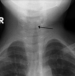
Croup (Laryngotracheobronchitis) is a viral infection of the vocal cords typically lasting five to six days. The main symptom is a barking cough and low-grade fever. On an X-ray, croup can be recognized by the "steeple sign", which is a narrowing of the trachea. It most commonly occurs in winter months in children between the ages of 3 months and 5 years. A severe form caused by bacteria is called bacterial tracheitis. [12]

Tonsillitis is swelling of the tonsils by a bacterial or viral infection. This inflammation can lead to airway obstruction. From tonsillitis can come a peritonsillar abscess which is the most common upper airway infection and occurs primarily in young adults. It causes one swelling of one of tonsils pushing the uvula to the unaffected side. [9] Diagnosis is usually made based on the presentation and examination. Symptoms generally include fever, sore throat, trouble swallowing, and sounding like they have a "hot potato" in their mouth. [13]

Lower respiratory tract infection

The most common lower respiratory tract infection is pneumonia, an infection of the lungs which is usually caused by bacteria, particularly Streptococcus pneumoniae in Western countries. Worldwide, tuberculosis is an important cause of pneumonia. Other pathogens such as viruses and fungi can cause pneumonia, for example severe acute respiratory syndrome, COVID-19 and pneumocystis pneumonia. Pneumonia may develop complications such as a lung abscess, a round cavity in the lung caused by the infection, or may spread to the pleural cavity.[ citation needed ]

Poor oral care may be a contributing factor to lower respiratory disease, as bacteria from gum disease may travel through airways and into the lungs. [14] [15]

Upper and lower respiratory tract infection

Primary ciliary dyskinesia is a genetic disorder causing the cilia to not move in a coordinated manner. This causes chronic respiratory infections, cough, and nasal congestion. This can lead to bronchiectasis, which can cause life-threatening breathing issues. [16]

Tumors

Malignant tumors

Malignant tumors of the respiratory system, particularly primary carcinomas of the lung, are a major health problem responsible for 15% of all cancer diagnoses and 30% of all cancer deaths. The majority of respiratory system cancers are attributable to smoking tobacco.[ citation needed ]

The major histological types of respiratory system cancer are:

In addition, since many cancers spread via the bloodstream and the entire cardiac output passes through the lungs, it is common for cancer metastases to occur within the lung. Breast cancer may invade directly through local spread, and through lymph node metastases. After metastasis to the liver, colon cancer frequently metastasizes to the lung. Prostate cancer, germ cell cancer and renal cell carcinoma may also metastasize to the lung.

Treatment of respiratory system cancer depends on the type of cancer. Surgical removal of part of a lung (lobectomy, segmentectomy, or wedge resection) or of an entire lung pneumonectomy), along with chemotherapy and radiotherapy, are all used. The chance of surviving lung cancer depends on the cancer stage at the time the cancer is diagnosed, and to some extent on the histology, and is only about 14–17% overall. In the case of metastases to the lung, treatment can occasionally be curative but only in certain, rare circumstances.

Benign tumors

Benign tumors are relatively rare causes of respiratory disease. Examples of benign tumors are:

Pleural cavity diseases

Pleural cavity diseases include pleural mesothelioma which are mentioned above.

A collection of fluid in the pleural cavity is known as a pleural effusion. [17] This may be due to fluid shifting from the bloodstream into the pleural cavity due to conditions such as congestive heart failure and cirrhosis. [17] It may also be due to inflammation of the pleura itself as can occur with infection, pulmonary embolus, tuberculosis, mesothelioma and other conditions. [17]

A pneumothorax is a hole in the pleura covering the lung allowing air in the lung to escape into the pleural cavity. The affected lung "collapses" like a deflated balloon. A tension pneumothorax is a particularly severe form of this condition where the air in the pleural cavity cannot escape, so the pneumothorax keeps getting bigger until it compresses the heart and blood vessels, leading to a life-threatening situation.

Pulmonary vascular disease

Pulmonary vascular diseases are conditions that affect the pulmonary circulation. Examples are: [18] [ citation needed ]

Neonatal diseases

Pulmonary diseases also impact newborns and the disorders are often unique from those that affect adults.

Infant respiratory distress syndrome most commonly occurs in less than six hours after birth in about 1% of all births in the United States. [9] The main risk factor is prematurity with the likelihood of it occurring going up to 71% in infants under 750g. [19] Other risk factors include infant of a diabetic mother (IDM), method of delivery, fetal asphyxia, genetics, prolonged rupture of membranes (PROM), maternal toxemia, chorioamnionitis, and male sex. The widely accepted pathophysiology of respiratory distress syndrome is it caused by insufficient surfactant production and immature lung and vascular development. The lack of surfactant makes the lungs atelectatic causing a ventilation to perfusion mismatch, lowered compliance, and increased air resistance. This causes hypoxia and respiratory acidosis which can lead to pulmonary hypertension. It has a ground glass appearance on an x-ray. Symptoms can include tachypnea, nasal flaring, paradoxical chest movement, grunting, and subcostal retractions. [9]

Bronchopulmonary Dysplasia is a condition that occurs after birth usually from mechanical ventilation and oxygen use. It happens almost exclusively in pre-mature infants and is characterized by the alveoli, and lung vasculature becoming inflamed and damaged. Complications from BPD can follow a patient into adulthood. As a child they may experience learning disabilities, pulmonary hypertension, and hearing problems. As an adult, there is an increased likelihood for asthma and exercise intolerance. [20]

Meconium Aspiration Syndrome occurs in full term or post-term infants who aspirate meconium. Risk factors include a diabetic mother, fetal hypoxia, precipitous delivery, and maternal high blood pressure. [21] Its diagnosis is based on meconium stained amniotic fluid at delivery and staining on the skin, nails, and umbilical cord. Aspiration can cause airway obstruction, air-trapping, pneumonia, lung inflammation, and inactivated surfactant. It presents as patchy atelectasis and hyperinflation on an x-ray with a pneumothorax of pneumomediastinum also possible. [9]

Persistent Pulmonary Hypertension of the Newborn (PPHN) is a syndrome that occurs from an abnormal transition to extra-uterine life. It is marked by an elevated pulmonary vascular resistance and vasoconstriction causing a right-to-left shunt of the blood through the foramen ovale or ductus arteriosus. [9] There are three main causes of PPHN are parenchymal diseases such as meconium aspiration syndrome, idiopathic, and hypoplastic vasculature like in a diaphragmatic hernia. It will eventually resolve in most infants. [22] This is the only syndrome that inhaled nitric oxide is approved for by the FDA. [23]

Pulmonary interstitial emphysema CXR-Pulmonary interstitial emphysema.jpg
Pulmonary interstitial emphysema

Transient Tachypnea of the Newborn is caused by the retention of alveolar fluid in the lungs. It commonly occurs in infants who are delivered via caesarean section without the onset of labor because absorption of amniotic fluid in the lungs has not yet commenced. Other risk factors are male sex, macrosomia, multiple gestations, and maternal asthma. It usually presents with tachypnea and increased work of breathing. On an x-ray diffuse infiltrates, interlobar fissures, and sometimes pleural effusions can be seen. It is a diagnosis of exclusion because of its similarity to other diseases and frequently CPAP is used to help push the lung fluid into the pulmonary vasculature. [9] [24]

Pulmonary interstitial emphysema is the condition of air escaping overdistended alveoli into the pulmonary interstitium. It is a rare disease that occurs most often in premature infants, even though it is possible to appear in adults. [25] It often presents as a slow deterioration with the need for increased ventilatory support. Chest x-ray is the standard for diagnosis where it is seen as linear or cystic translucencies extending to the edges of the lungs. [9]

Bronchiolitis is the swelling and buildup of mucus in the bronchioles. It is usually caused by respiratory syncytial virus (RSV), which is spread when an infant touches the nose or throat fluids of someone infected. [26] The virus infects the cells causing ciliary dysfunction and death. The debris, edema, and inflammation eventually leads to the symptoms. [27] It is the most common reason for admission of children under the age of one year. It can present widely from a mild respiratory infection to respiratory failure. Since there is no medication to treat the disease, it is only managed supportively with fluids and oxygen. [28]

Diagnosis

Respiratory diseases may be investigated by performing one or more of the following tests:

Epidemiology

Deaths from respiratory diseases per million persons in 2012
.mw-parser-output .div-col{margin-top:0.3em;column-width:30em}.mw-parser-output .div-col-small{font-size:90%}.mw-parser-output .div-col-rules{column-rule:1px solid #aaa}.mw-parser-output .div-col dl,.mw-parser-output .div-col ol,.mw-parser-output .div-col ul{margin-top:0}.mw-parser-output .div-col li,.mw-parser-output .div-col dd{page-break-inside:avoid;break-inside:avoid-column}
.mw-parser-output .legend{page-break-inside:avoid;break-inside:avoid-column}.mw-parser-output .legend-color{display:inline-block;min-width:1.25em;height:1.25em;line-height:1.25;margin:1px 0;text-align:center;border:1px solid black;background-color:transparent;color:black}.mw-parser-output .legend-text{}
19-125
126-169
170-196
197-225
226--75
276-308
309-365
366-440
441-593
594-1,227 Respiratory diseases world map-Deaths per million persons-WHO2012.svg
Deaths from respiratory diseases per million persons in 2012
  19–125
  126–169
  170–196
  197–225
  226-–75
  276–308
  309–365
  366–440
  441–593
  594–1,227

Respiratory disease is a common and significant cause of illness and death around the world. In the US, approximately one billion common colds occur each year. [29] A study found that in 2010, there were approximately 6.8 million emergency department visits for respiratory disorders in the U.S. for patients under the age of 18. [30] In 2012, respiratory conditions were the most frequent reasons for hospital stays among children. [31]

In the UK, approximately 1 in 7 individuals are affected by some form of chronic lung disease, most commonly chronic obstructive pulmonary disease, which includes asthma, chronic bronchitis and emphysema. [32] Respiratory diseases (including lung cancer) are responsible for over 10% of hospitalizations and over 16% of deaths in Canada. [33]

In 2011, respiratory disease with ventilator support accounted for 93.3% of ICU utilization in the United States. [34]

Related Research Articles

<span class="mw-page-title-main">Meconium aspiration syndrome</span> Medical condition affecting newborn infants

Meconium aspiration syndrome (MAS) also known as neonatal aspiration of meconium is a medical condition affecting newborn infants. It describes the spectrum of disorders and pathophysiology of newborns born in meconium-stained amniotic fluid (MSAF) and have meconium within their lungs. Therefore, MAS has a wide range of severity depending on what conditions and complications develop after parturition. Furthermore, the pathophysiology of MAS is multifactorial and extremely complex which is why it is the leading cause of morbidity and mortality in term infants.

<span class="mw-page-title-main">Lung</span> Primary organ of the respiratory system

The lungs are the central organs of the respiratory system in humans and most other animals, including some snails and a small number of fish. In mammals and most other vertebrates, two lungs are located near the backbone on either side of the heart. Their function in the respiratory system is to extract oxygen from the air and transfer it into the bloodstream, and to release carbon dioxide from the bloodstream into the atmosphere, in a process of gas exchange. The pleurae, which are thin, smooth, and moist, serve to reduce friction between the lungs and chest wall during breathing, allowing for easy and effortless movements of the lungs.

<span class="mw-page-title-main">Pneumonia</span> Inflammation of the alveoli of the lungs

Pneumonia is an inflammatory condition of the lung primarily affecting the small air sacs known as alveoli. Symptoms typically include some combination of productive or dry cough, chest pain, fever, and difficulty breathing. The severity of the condition is variable.

<span class="mw-page-title-main">Pulmonary alveolus</span> Hollow cavity found in the lungs

A pulmonary alveolus, also known as an air sac or air space, is one of millions of hollow, distensible cup-shaped cavities in the lungs where pulmonary gas exchange takes place. Oxygen is exchanged for carbon dioxide at the blood–air barrier between the alveolar air and the pulmonary capillary. Alveoli make up the functional tissue of the mammalian lungs known as the lung parenchyma, which takes up 90 percent of the total lung volume.

<span class="mw-page-title-main">Pleural cavity</span> Thin fluid-filled space between the two pulmonary pleurae (visceral and parietal) of each lung

The pleural cavity, pleural space, or interpleural space is the potential space between the pleurae of the pleural sac that surrounds each lung. A small amount of serous pleural fluid is maintained in the pleural cavity to enable lubrication between the membranes, and also to create a pressure gradient.

<span class="mw-page-title-main">Cough</span> Sudden expulsion of air from the lungs as a reflex to clear irritants

A cough is a sudden expulsion of air through the large breathing passages which can help clear them of fluids, irritants, foreign particles and microbes. As a protective reflex, coughing can be repetitive with the cough reflex following three phases: an inhalation, a forced exhalation against a closed glottis, and a violent release of air from the lungs following opening of the glottis, usually accompanied by a distinctive sound.

<span class="mw-page-title-main">Sputum</span> Mucus that is coughed up from the lower airways

Sputum is mucus that is coughed up from the lower airways. In medicine, sputum samples are usually used for a naked eye examination, microbiological investigation of respiratory infections and cytological investigations of respiratory systems. It is crucial that the specimen does not include any mucoid material from the nose or oral cavity.

<span class="mw-page-title-main">Respiratory tract</span> Organs involved in transmission of air to and from the point where gases diffuse into tissue

The respiratory tract is the subdivision of the respiratory system involved with the process of respiration in mammals. The respiratory tract is lined with respiratory epithelium as respiratory mucosa.

<span class="mw-page-title-main">Pleurisy</span> Disease of the lungs

Pleurisy, also known as pleuritis, is inflammation of the membranes that surround the lungs and line the chest cavity (pleurae). This can result in a sharp chest pain while breathing. Occasionally the pain may be a constant dull ache. Other symptoms may include shortness of breath, cough, fever, or weight loss, depending on the underlying cause. Pleurisy can be caused by a variety of conditions, including viral or bacterial infections, autoimmune disorders, and pulmonary embolism.

<span class="mw-page-title-main">Pulmonary edema</span> Fluid accumulation in the tissue and air spaces of the lungs

Pulmonary edema, also known as pulmonary congestion, is excessive fluid accumulation in the tissue or air spaces of the lungs. This leads to impaired gas exchange, most often leading to dyspnea which can progress to hypoxemia and respiratory failure. Pulmonary edema has multiple causes and is traditionally classified as cardiogenic or noncardiogenic.

<span class="mw-page-title-main">Acute respiratory distress syndrome</span> Human disease

Acute respiratory distress syndrome (ARDS) is a type of respiratory failure characterized by rapid onset of widespread inflammation in the lungs. Symptoms include shortness of breath (dyspnea), rapid breathing (tachypnea), and bluish skin coloration (cyanosis). For those who survive, a decreased quality of life is common.

<span class="mw-page-title-main">Atelectasis</span> Partial collapse of a lung causing reduced gas exchange

Atelectasis is the partial collapse or closure of a lung resulting in reduced or absent gas exchange. It is usually unilateral, affecting part or all of one lung. It is a condition where the alveoli are deflated down to little or no volume, as distinct from pulmonary consolidation, in which they are filled with liquid. It is often referred to informally as a collapsed lung, although more accurately it usually involves only a partial collapse, and that ambiguous term is also informally used for a fully collapsed lung caused by a pneumothorax.

<span class="mw-page-title-main">Pneumonitis</span> General inflammation of lung tissue

Pneumonitis describes general inflammation of lung tissue. Possible causative agents include radiation therapy of the chest, exposure to medications used during chemo-therapy, the inhalation of debris, aspiration, herbicides or fluorocarbons and some systemic diseases. If unresolved, continued inflammation can result in irreparable damage such as pulmonary fibrosis.

Community-acquired pneumonia (CAP) refers to pneumonia contracted by a person outside of the healthcare system. In contrast, hospital-acquired pneumonia (HAP) is seen in patients who have recently visited a hospital or who live in long-term care facilities. CAP is common, affecting people of all ages, and its symptoms occur as a result of oxygen-absorbing areas of the lung (alveoli) filling with fluid. This inhibits lung function, causing dyspnea, fever, chest pains and cough.

Eosinophilic pneumonia is a disease in which an eosinophil, a type of white blood cell, accumulates in the lungs. These cells cause disruption of the normal air spaces (alveoli) where oxygen is extracted from the atmosphere. Several different kinds of eosinophilic pneumonia exist and can occur in any age group. The most common symptoms include cough, fever, difficulty breathing, and sweating at night. Eosinophilic pneumonia is diagnosed by a combination of characteristic symptoms, findings on a physical examination by a health provider, and the results of blood tests and X-rays. Prognosis is excellent once most eosinophilic pneumonia is recognized and treatment with corticosteroids is begun.

Occupational lung diseases comprise a broad group of diseases, including occupational asthma, industrial bronchitis, chronic obstructive pulmonary disease (COPD), bronchiolitis obliterans, inhalation injury, interstitial lung diseases, infections, lung cancer and mesothelioma. These can be caused directly or due to immunological response to an exposure to a variety of dusts, chemicals, proteins or organisms. Occupational cases of interstitial lung disease may be misdiagnosed as COPD, idiopathic pulmonary fibrosis, or a myriad of other diseases; leading to a delay in identification of the causative agent.

Restrictive lung diseases are a category of extrapulmonary, pleural, or parenchymal respiratory diseases that restrict lung expansion, resulting in a decreased lung volume, an increased work of breathing, and inadequate ventilation and/or oxygenation. Pulmonary function test demonstrates a decrease in the forced vital capacity.

Pleural disease occurs in the pleural space, which is the thin fluid-filled area in between the two pulmonary pleurae in the human body. There are several disorders and complications that can occur within the pleural area, and the surrounding tissues in the lung.

Chronic cough is long-term coughing, sometimes defined as more than several weeks or months. Generally a cough lasting for more than eight weeks for an adult would meet the clinical definition of a chronic cough; and for children this threshold is lower. The term can be used to describe the different causes related to coughing, the three main ones being upper airway cough syndrome, asthma and gastroesophageal reflux disease. It occurs in the upper airway of the respiratory system. Generally, a cough lasts around one to two weeks; however, chronic cough can persist for an extended period of time defined as six weeks or longer. People with chronic cough often experience more than one cause present. Due to the nature of the syndrome, the treatments used are similar; however, there are a subsequent number of treatments available, and the clinical management of the patients remains a challenge. Risk factors include exposure to cigarette smoke, and exposure to pollution, especially particulates.

<span class="mw-page-title-main">Ventilation–perfusion coupling</span> Relationship between respiratory and cardiovascular processes

Ventilation-perfusion coupling is the relationship between ventilation and perfusion processes, which take place in the respiratory system and the cardiovascular system. Ventilation is the movement of gas during breathing, and perfusion is the process of pulmonary blood circulation, which delivers oxygen to body tissues. Anatomically, the lung structure, alveolar organization, and alveolar capillaries contribute to the physiological mechanism of ventilation and perfusion. Ventilation-perfusion coupling maintains a constant ventilation/perfusion ratio near 0.8 on average, while the regional variation exists within the lungs due to gravity. When the ratio gets above or below 0.8, it is considered abnormal ventilation-perfusion coupling, also known as a ventilation–perfusion mismatch. Lung diseases, cardiac shunts, and smoking can cause a ventilation-perfusion mismatch that results in significant symptoms and diseases, which can be treated through treatments like bronchodilators and oxygen therapy.

References

  1. "Lung diseases". MeSH.nlm.nih.gov. Archived from the original on 12 June 2020. Retrieved 14 August 2019.
  2. Sengupta N, Sahidullah M, Saha G (August 2016). "Lung sound classification using cepstral-based statistical features". Computers in Biology and Medicine. 75 (1): 118–29. doi:10.1016/j.compbiomed.2016.05.013. PMID   27286184.
  3. "COVID-19 and vascular disease". eBioMedicine. 58: 102966. August 2020. doi:10.1016/j.ebiom.2020.102966. PMC   7438984 . PMID   32829782.
  4. Reid PT, Innes JA (2014). "Respiratory Diseases". In Walker BR, Colledge NR, Ralston SH, Penman I (eds.). Davidson's Principles and Practice of Medicine (22nd ed.). Elsevier Health Sciences. pp. 661–730. ISBN   978-0-7020-5035-0.
  5. Sharma S (5 June 2006). Grier LR, Ouellette DR, Mosenifar Z (eds.). "Restrictive Lung Disease". Medscape. Archived from the original on 19 December 2008. Retrieved 2008-04-19.
  6. Martinez-Pitre PJ, Sabbula BR, Cascella M (2020). "Restrictive Lung Disease". StatPearls. Treasure Island (FL): StatPearls Publishing. PMID   32809715. Archived from the original on 2022-01-28. Retrieved 2020-12-04.
  7. "Chronic respiratory diseases (CRDs)". World Health Organization. Archived from the original on 2018-10-30. Retrieved 2020-10-05.
  8. 1 2 Cox NS, Dal Corso S, Hansen H, McDonald CF, Hill CJ, Zanaboni P, et al. (Cochrane Airways Group) (January 2021). "Telerehabilitation for chronic respiratory disease". The Cochrane Database of Systematic Reviews. 1 (1): CD013040. doi:10.1002/14651858.CD013040.pub2. PMC   8095032 . PMID   33511633.
  9. 1 2 3 4 5 6 7 8 Walsh, Brian (2019). Neonatal and Pediatric Respiratory Care. Elsevier.
  10. Westerhuis, Brian; Bietz, Mandi Greenway; Lindemann, Janet (2013). "Acute epiglottitis in adults: an under-recognized and life-threatening condition". South Dakota Medicine: The Journal of the South Dakota State Medical Association. 66 (8): 309–311, 313. ISSN   0038-3317. PMID   24175495. Archived from the original on 2022-09-22. Retrieved 2022-05-16.
  11. "Epiglottitis". nhs.uk. 2017-10-18. Archived from the original on 2020-12-03. Retrieved 2022-05-16.
  12. "Croup: MedlinePlus Medical Encyclopedia". medlineplus.gov. Archived from the original on 2023-02-19. Retrieved 2022-05-16.
  13. Galioto, Nicholas J. (2017-04-15). "Peritonsillar Abscess". American Family Physician. 95 (8): 501–506. ISSN   0002-838X. PMID   28409615. Archived from the original on 2022-05-16. Retrieved 2022-05-16.
  14. "Respiratory Disease & Oral Health". United Concordia Companies, Inc. Archived from the original on 2016-03-04. Retrieved 2015-01-19.
  15. Gomes-Filho IS, Passos JS, Seixas da Cruz S (December 2010). "Respiratory disease and the role of oral bacteria". Journal of Oral Microbiology. 2: 5811. doi:10.3402/jom.v2i0.5811. PMC   3084574 . PMID   21523216.
  16. "Primary ciliary dyskinesia: MedlinePlus Genetics". medlineplus.gov. Archived from the original on 2023-02-19. Retrieved 2022-05-16.
  17. 1 2 3 Karkhanis VS, Joshi JM (2012-06-22). "Pleural effusion: diagnosis, treatment, and management". Open Access Emergency Medicine. 4: 31–52. doi: 10.2147/OAEM.S29942 . PMC   4753987 . PMID   27147861.
  18. "Pulmonary Embolism". medlineplus.gov. Archived from the original on 2020-05-18. Retrieved 2022-03-22.
  19. Fanaroff, Avroy A.; Stoll, Barbara J.; Wright, Linda L.; Carlo, Waldemar A.; Ehrenkranz, Richard A.; Stark, Ann R.; Bauer, Charles R.; Donovan, Edward F.; Korones, Sheldon B.; Laptook, Abbot R.; Lemons, James A. (February 2007). "Trends in neonatal morbidity and mortality for very low birthweight infants". American Journal of Obstetrics and Gynecology. 196 (2): 147.e1–8. doi:10.1016/j.ajog.2006.09.014. ISSN   1097-6868. PMID   17306659. Archived from the original on 2022-05-06. Retrieved 2022-05-06.
  20. "Bronchopulmonary Dysplasia". www.lung.org. Archived from the original on 2023-01-11. Retrieved 2022-05-07.
  21. "Meconium aspiration syndrome: MedlinePlus Medical Encyclopedia". medlineplus.gov. Archived from the original on 2022-05-11. Retrieved 2022-05-11.
  22. Steinhorn, Robin H. (March 2010). "Neonatal Pulmonary Hypertension". Pediatric Critical Care Medicine. 11 (2 Suppl): S79–S84. doi:10.1097/PCC.0b013e3181c76cdc. ISSN   1529-7535. PMC   2843001 . PMID   20216169.
  23. "FDA" (PDF). Food and Drug Administration. Archived (PDF) from the original on 18 February 2023. Retrieved 11 May 2022.
  24. Reuter, Suzanne; Moser, Chuanpit; Baack, Michelle (2014). "Respiratory Distress in the Newborn". Pediatrics in Review. 35 (10): 417–429. doi:10.1542/pir.35-10-417. ISSN   0191-9601. PMC   4533247 . PMID   25274969.
  25. Jalota Sahota, Ruchi; Anjum, Fatima (2022), "Pulmonary Interstitial Emphysema", StatPearls, Treasure Island (FL): StatPearls Publishing, PMID   32809319, archived from the original on 2023-12-26, retrieved 2022-05-14
  26. "Bronchiolitis: MedlinePlus Medical Encyclopedia". medlineplus.gov. Archived from the original on 2023-01-30. Retrieved 2022-05-14.
  27. Justice, Nathaniel A.; Le, Jacqueline K. (2022), "Bronchiolitis", StatPearls, Treasure Island (FL): StatPearls Publishing, PMID   28722988, archived from the original on 2022-08-14, retrieved 2022-05-14
  28. Friedman, Jeremy N; Rieder, Michael J; Walton, Jennifer M (2014). "Bronchiolitis: Recommendations for diagnosis, monitoring and management of children one to 24 months of age". Paediatrics & Child Health. 19 (9): 485–491. doi:10.1093/pch/19.9.485. ISSN   1205-7088. PMC   4235450 . PMID   25414585.
  29. "National Institutes of Health – common cold". Archived from the original on 2008-10-01. Retrieved 2008-05-07.
  30. Wier LM, Yu H, Owens PL, Washington R (May 2013). "Overview of Children in the Emergency Department, 2010". HCUP Statistical Brief #157. Agency for Healthcare Research and Quality. PMID   24006551. Archived from the original on 2013-12-03. Retrieved 2013-07-17.
  31. Witt WP, Wiess AJ, Elixhauser A (December 2014). "Overview of Hospital Stays for Children in the United States, 2012". HCUP Statistical Brief #186. Rockville, MD: Agency for Healthcare Research and Quality. PMID   25695124. Archived from the original on 2018-09-24. Retrieved 2015-04-06.
  32. "What is COPD?". British Lung Foundation. 7 September 2015. Archived from the original on 20 January 2022. Retrieved 16 May 2019.
  33. "Public Health Agency of Canada – Centre for Chronic Disease Prevention and Control Chronic Respiratory Diseases". Archived from the original on 2008-04-11. Retrieved 2008-05-06.
  34. Barrett ML, Smith MW, Elizhauser A, Honigman LS, Pines JM (December 2014). "Utilization of Intensive Care Services, 2011". HCUP Statistical Brief #185. Rockville, MD: Agency for Healthcare Research and Quality. PMID   25654157. Archived from the original on 2015-04-02. Retrieved 2015-04-06.