Pleural empyema

Last updated
Pleural empyema
Other namesPyothorax, purulent pleuritis, lung empyema
Hydro pneumothorax.jpg
CT chest showing large right sided hydro-pneumothorax from pleural empyema. Arrows A: air, B: fluid
Specialty Pulmonology, cardiothoracic surgery   OOjs UI icon edit-ltr-progressive.svg
Symptoms Fever, chest pain with breathing in, cough, shortness of breath
CausesBacteria (often Strep. pneumonia)
Diagnostic method Chest X-ray, Ultrasound, CT scan, thoracentesis
Differential diagnosis Pneumonia, pleural effusion, pulmonary embolism
Treatment Supportive care, antibiotics, surgery, chest tube

Pleural empyema is a collection of pus in the pleural cavity caused by microorganisms, usually bacteria. [1] Often it happens in the context of a pneumonia, injury, or chest surgery. [1] It is one of the various kinds of pleural effusion. There are three stages: exudative, when there is an increase in pleural fluid with or without the presence of pus; fibrinopurulent, when fibrous septa form localized pus pockets; and the final organizing stage, when there is scarring of the pleura membranes with possible inability of the lung to expand. Simple pleural effusions occur in up to 40% of bacterial pneumonias. They are usually small and resolve with appropriate antibiotic therapy. If however an empyema develops additional intervention is required.

Contents

Signs and symptoms

The clinical presentation of both the adult and pediatric patient with pleural empyema depends upon several factors, including the causative micro-organism. Most cases present themselves in the setting of a pneumonia, although up to one third of patients do not have clinical signs of pneumonia and as many as 25% of cases are associated with trauma (including surgery). [2] Symptoms include fever, dry cough, sweating, difficulty breathing, and unintentional weight loss. [3] The elderly often do not have classic symptoms, but instead have anemia and exhaustion. [4]

Mechanism

When there is inflammation at the pleural space, fluid is produced at a greater level. As the disease progresses, bacteria can arrive at the fluid, which created an empyema. [5]

Diagnosis

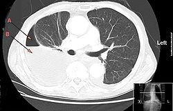
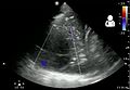
The initial investigations for suspected empyema remains chest X-ray, although it cannot differentiate an empyema from uninfected parapneumonic effusion. [6] Ultrasound must be used to confirm the presence of a pleural fluid collection and can be used to estimate the size of the effusion, differentiate between free and loculated pleural fluid and guide thoracocentesis if necessary. Chest CT and MRI do not provide additional information in most cases and should therefore not be performed routinely. [7] On a CT scan, empyema fluid most often has a radiodensity of about 0-20 Hounsfield units (HU), [8] but gets over 30 HU when becoming more thickened with time. [9]

The most often used "golden" criteria for empyema are pleural effusion with macroscopic presence of pus, a positive Gram stain or culture of pleural fluid, or a pleural fluid pH under 7.2 with normal peripheral blood pH. [10] [11] Clinical guidelines for adult patients therefore advocate diagnostic pleural fluid aspiration in patients with pleural effusion in association with sepsis or pneumonic illness. [12] Because pleural effusion in the pediatric population is almost always parapneumonic and the need for chest tube drainage can be made on clinical grounds, British guidelines for the management of pleural infection in children do not recommend diagnostic pleural fluid sampling. [7]

Blood and sputum culture has often already been performed in the setting of community acquired pneumonia needing hospitalization. It should however be noted that the micro-organism responsible for development of empyema is not necessarily the same as the organism causing the pneumonia, especially in adults. As already mentioned before, sensitivity of pleural fluid culture is generally low, often partly due to prior administration of antibiotics. It has been shown that culture yield can be increased from 44% to 69% if pleural fluid is injected into blood culture bottles (aerobic and anaerobic) immediately after aspiration. [11] Furthermore, diagnostic rates can be improved for specific pathogens using polymerase chain reaction or antigen detection, especially for Streptococcus pneumoniae, Streptococcus pyogenes and Staphylococcus aureus. In a study including 78 children with pleural empyema, the causative micro-organism could be identified using direct culture of fresh pleural fluid in 45% of patients, with an additional 28% using PCR on pleural fluid of negative cultures. Pneumococcal antigen detection in pleural fluid samples by latex agglutination can also be useful for rapid diagnosis of pneumococcal empyema. In the previously noted study, positive and negative predictive value of pneumococcal antigen detection was 95% and 90%, respectively. [13]

Treatment

Pleural fluid drainage

Proven empyema (as defined by the "golden" criteria mentioned earlier) is an indication for prompt chest tube drainage. [12] This has been shown to improve resolution of the infection and shorten hospital admission. [15] Data from a meta-analysis has shown that a pleural fluid pH of <7.2 is the most powerful indicator to predict the need for chest tube drainage in patients with non-purulent, culture negative fluid. [16]

Because of the viscous, lumpy nature of infected pleural fluid, in combination with possible septation and loculation, it has been proposed that intrapleural fibrinolytic or mucolytic therapy might improve drainage and therefore might have a positive effect on the clinical outcome. [17] Intrapleural fibrinolysis with urokinase decreased the need for surgery but there is a trend to increased serious side effects. [18]

Approximately 15 to 40 percent of people require surgical drainage of the infected pleural space because of inadequate drainage due to clogging of the chest tube or loculated empyema. [19] Patients should thus be considered for surgery if they have ongoing signs of sepsis in association with a persistent pleural collection despite drainage and antibiotics. [12]

Antibiotics

There is no readily available evidence on the route of administration and duration of antibiotics in patients with pleural empyema. Experts agree that all patients should be hospitalized and treated with antibiotics intravenously. [7] [12] The specific antimicrobial agent should be chosen based on Gram stain and culture, or on local epidemiologic data when these are not available. Anaerobic coverage must be included in all adults, and in children if aspiration is likely. Good pleural fluid and empyema penetration has been reported in adults for penicillins, ceftriaxone, metronidazole, clindamycin, vancomycin, gentamicin and ciprofloxacin. [20] [21] Aminoglycosides should typically be avoided as they have poor penetration into the pleural space. There is no clear consensus on duration of intravenous and oral therapy. Switching to oral antibiotics can be considered upon clinical and objective improvement (adequate drainage and removal of chest tube, declining CRP, temperature normalization). Oral antibiotic treatment should then be continued for another 1–4 weeks, again based on clinical, biochemical and radiological response. [7] [12]

Prognosis

All patients with empyema require outpatient follow-up with a repeat chest X-ray and inflammatory biochemistry analysis within 4 weeks following discharge. Chest radiograph returns to normal in the majority of patients by 6 months. Patients should, of course, be advised to return sooner if symptoms redevelop. Long-term sequelae of pleural empyema are rare but include bronchopleural fistula formation, recurrent empyema and pleural thickening, which may lead to functional lung impairment needing surgical decortication. [12]

Mortality in children is generally reported to be less than 3%. [7] No reliable clinical, radiological or pleural fluid characteristics accurately determine patients’ prognosis at initial presentation. [22]

Epidemiology

The incidence of pleural empyema and the prevalence of specific causative microorganisms varies depending on the source of infection (community acquired vs. hospital acquired pneumonia), the age of the patient and host immune status. Risk factors include alcoholism, drug use, HIV infection, neoplasm and pre-existent pulmonary disease. [23] Pleural empyema was found in 0.7% of 3675 patients needing hospitalization for a community acquired pneumonia in a recent Canadian single-center prospective study. [10] A multi-center study from the UK including 430 adult patients with community acquired pleural empyema found negative pleural-fluid cultures in 54% of patients, Streptococcus milleri group in 16%, Staphylococcus aureus in 12%, Streptococcus pneumoniae in 8%, other Streptococci in 7% and anaerobic bacteria in 8%. [17]

The risk of empyema in children seems to be comparable to adults. Using the United States Kids’ Inpatient Database the incidence is calculated to be around 1.5% in children hospitalized for community acquired pneumonia, [24] although percentages up to 30% have been reported in individual hospitals, [25] a difference which may be explained by an transient endemic of highly invasive serotype or overdiagnosis of small parapneumonic effusions. The distribution of causative organisms does differ greatly from that in adults: in an analysis of 78 children with community acquired pleural empyema, no micro-organism was found in 27% of patients, Streptococcus pneumoniae in 51%, Streptococcus pyogenes in 9% and Staphylococcus aureus in 8%. [13]

Although pneumococcal vaccination dramatically decreased the incidence of pneumonia in children, it did not have this effect on the incidence of complicated pneumonia. It has been shown that the incidence of empyema in children was already on the rise at the end of the 20th century, and that the widespread use of pneumococcal vaccination did not slow down this trend. [26] This might in part be explained by a change in prevalence of (more invasive) pneumococcal serotypes, some of which are not covered by the vaccine, as well a rise in incidence of pneumonia caused by other streptococci and staphylococci. [27]

Related Research Articles

<span class="mw-page-title-main">Pneumonia</span> Inflammation of the alveoli of the lungs

Pneumonia is an inflammatory condition of the lung primarily affecting the small air sacs known as alveoli. Symptoms typically include some combination of productive or dry cough, chest pain, fever, and difficulty breathing. The severity of the condition is variable.

<span class="mw-page-title-main">Acute bronchitis</span> Medical condition

Acute bronchitis, also known as a chest cold, is short-term bronchitis – inflammation of the bronchi of the lungs. The most common symptom is a cough. Other symptoms include coughing up mucus, wheezing, shortness of breath, fever, and chest discomfort. The infection may last from a few to ten days. The cough may persist for several weeks afterward with the total duration of symptoms usually around three weeks. Some have symptoms for up to six weeks.

<span class="mw-page-title-main">Pleurisy</span> Disease of the lungs

Pleurisy, also known as pleuritis, is inflammation of the membranes that surround the lungs and line the chest cavity (pleurae). This can result in a sharp chest pain while breathing. Occasionally the pain may be a constant dull ache. Other symptoms may include shortness of breath, cough, fever, or weight loss, depending on the underlying cause. Pleurisy can be caused by a variety of conditions, including viral or bacterial infections, autoimmune disorders, and pulmonary embolism.

<span class="mw-page-title-main">Pleural effusion</span> Accumulation of excess fluid in the pleural cavity

A pleural effusion is accumulation of excessive fluid in the pleural space, the potential space that surrounds each lung. Under normal conditions, pleural fluid is secreted by the parietal pleural capillaries at a rate of 0.6 millilitre per kilogram weight per hour, and is cleared by lymphatic absorption leaving behind only 5–15 millilitres of fluid, which helps to maintain a functional vacuum between the parietal and visceral pleurae. Excess fluid within the pleural space can impair inspiration by upsetting the functional vacuum and hydrostatically increasing the resistance against lung expansion, resulting in a fully or partially collapsed lung.

<i>Streptococcus pneumoniae</i> Species of bacterium

Streptococcus pneumoniae, or pneumococcus, is a Gram-positive, spherical bacteria, alpha-hemolytic member of the genus Streptococcus. They are usually found in pairs (diplococci) and do not form spores and are non motile. As a significant human pathogenic bacterium S. pneumoniae was recognized as a major cause of pneumonia in the late 19th century, and is the subject of many humoral immunity studies.

<span class="mw-page-title-main">Chest tube</span> Type of surgical drain

A chest tube is a surgical drain that is inserted through the chest wall and into the pleural space or the mediastinum in order to remove clinically undesired substances such as air (pneumothorax), excess fluid, blood (hemothorax), chyle (chylothorax) or pus (empyema) from the intrathoracic space. An intrapleural chest tube is also known as a Bülau drain or an intercostal catheter (ICC), and can either be a thin, flexible silicone tube, or a larger, semi-rigid, fenestrated plastic tube, which often involves a flutter valve or underwater seal.

<span class="mw-page-title-main">Aspiration pneumonia</span> Medical condition

Aspiration pneumonia is a type of lung infection that is due to a relatively large amount of material from the stomach or mouth entering the lungs. Signs and symptoms often include fever and cough of relatively rapid onset. Complications may include lung abscess, acute respiratory distress syndrome, empyema, and parapneumonic effusion. Some include chemical induced inflammation of the lungs as a subtype, which occurs from acidic but non-infectious stomach contents entering the lungs.

<span class="mw-page-title-main">Hemothorax</span> Blood accumulation in the pleural cavity

A hemothorax is an accumulation of blood within the pleural cavity. The symptoms of a hemothorax may include chest pain and difficulty breathing, while the clinical signs may include reduced breath sounds on the affected side and a rapid heart rate. Hemothoraces are usually caused by an injury, but they may occur spontaneously due to cancer invading the pleural cavity, as a result of a blood clotting disorder, as an unusual manifestation of endometriosis, in response to Pneumothorax, or rarely in association with other conditions.

<span class="mw-page-title-main">Lung abscess</span> Medical condition

Lung abscess is a type of liquefactive necrosis of the lung tissue and formation of cavities containing necrotic debris or fluid caused by microbial infection.

<span class="mw-page-title-main">Thoracentesis</span> Medical procedure

Thoracentesis, also known as thoracocentesis, pleural tap, needle thoracostomy, or needle decompression, is an invasive medical procedure to remove fluid or air from the pleural space for diagnostic or therapeutic purposes. A cannula, or hollow needle, is carefully introduced into the thorax, generally after administration of local anesthesia. The procedure was first performed by Morrill Wyman in 1850 and then described by Henry Ingersoll Bowditch in 1852.

Community-acquired pneumonia (CAP) refers to pneumonia contracted by a person outside of the healthcare system. In contrast, hospital-acquired pneumonia (HAP) is seen in patients who have recently visited a hospital or who live in long-term care facilities. CAP is common, affecting people of all ages, and its symptoms occur as a result of oxygen-absorbing areas of the lung (alveoli) filling with fluid. This inhibits lung function, causing dyspnea, fever, chest pains and cough.

Pneumococcal pneumonia is a type of bacterial pneumonia that is caused by Streptococcus pneumoniae (pneumococcus). It is the most common bacterial pneumonia found in adults, the most common type of community-acquired pneumonia, and one of the common types of pneumococcal infection. The estimated number of Americans with pneumococcal pneumonia is 900,000 annually, with almost 400,000 cases hospitalized and fatalities accounting for 5-7% of these cases.

<span class="mw-page-title-main">Pericardial effusion</span> Medical condition

A pericardial effusion is an abnormal accumulation of fluid in the pericardial cavity. The pericardium is a two-part membrane surrounding the heart: the outer fibrous connective membrane and an inner two-layered serous membrane. The two layers of the serous membrane enclose the pericardial cavity between them. This pericardial space contains a small amount of pericardial fluid, normally 15-50 mL in volume. The pericardium, specifically the pericardial fluid provides lubrication, maintains the anatomic position of the heart in the chest, and also serves as a barrier to protect the heart from infection and inflammation in adjacent tissues and organs.

<span class="mw-page-title-main">Hospital-acquired pneumonia</span>

Hospital-acquired pneumonia (HAP) or nosocomial pneumonia refers to any pneumonia contracted by a patient in a hospital at least 48–72 hours after being admitted. It is thus distinguished from community-acquired pneumonia. It is usually caused by a bacterial infection, rather than a virus.

<span class="mw-page-title-main">Parapneumonic effusion</span> Medical condition

A parapneumonic effusion is a type of pleural effusion that arises as a result of a pneumonia, lung abscess, or bronchiectasis. There are three types of parapneumonic effusions: uncomplicated effusions, complicated effusions, and empyema. Uncomplicated effusions generally respond well to appropriate antibiotic treatment.

Malignant pleural effusion is a condition in which cancer causes an abnormal amount of fluid to collect between the thin layers of tissue (pleura) lining the outside of the lung and the wall of the chest cavity. Lung cancer and breast cancer account for about 50-65% of malignant pleural effusions. Other common causes include pleural mesothelioma and lymphoma.

Pneumococcal infection is an infection caused by the bacterium Streptococcus pneumoniae.

Austrian syndrome, also known as Osler's triad, is a medical condition that was named after Robert Austrian in 1957. The presentation of the condition consists of pneumonia, endocarditis, and meningitis, all caused by Streptococcus pneumoniae. It is associated with alcoholism due to hyposplenism and can be seen in males between the ages of 40 and 60 years old. Robert Austrian was not the first one to describe the condition, but Richard Heschl or William Osler were not able to link the signs to the bacteria because microbiology was not yet developed.

<span class="mw-page-title-main">Eloesser flap</span> Surgical procedure

The Eloesser flap is a surgical procedure developed by Dr. Leo Eloesser in 1935 at the San Francisco General Hospital. It was originally intended to aid with drainage of tuberculous empyemas, since at the time there were no effective medications to treat tuberculosis. The procedure was used extensively until the development of effective chemotherapy for tuberculosis in the late 1940s and early 1950s. It is still used occasionally for chronic empyemas.

Necrotizing pneumonia (NP), also known as cavitary pneumonia or cavitatory necrosis, is a rare but severe complication of lung parenchymal infection. In necrotizing pneumonia, there is a substantial liquefaction following death of the lung tissue, which may lead to gangrene formation in the lung. In most cases patients with NP have fever, cough and bad breath, and those with more indolent infections have weight loss. Often patients clinically present with acute respiratory failure. The most common pathogens responsible for NP are Streptococcus pneumonia, Staphylococcus aureus, Klebsiella pneumoniae. Diagnosis is usually done by chest imaging, e.g. chest X-ray, CT scan. Among these CT scan is the most sensitive test which shows loss of lung architecture and multiple small thin walled cavities. Often cultures from bronchoalveolar lavage and blood may be done for identification of the causative organism(s). It is primarily managed by supportive care along with appropriate antibiotics. However, if patient develops severe complications like sepsis or fails to medical therapy, surgical resection is a reasonable option for saving life.

References

  1. 1 2 Redden MD, Chin TY, van Driel ML (March 2017). "Surgical versus non-surgical management for pleural empyema". Cochrane Database Syst Rev. 2017 (3): CD010651. doi:10.1002/14651858.CD010651.pub2. ISSN   1469-493X. PMC   6464687 . PMID   28304084.
  2. Fernández-Cotarelo MJ, López-Medrano F, San Juan R, Díaz-Pedroche C, Lizasoain M, Chaves F, Aguado JM (March 2007). "Protean manifestations of pleural empyema caused by Streptococcus pneumoniae in adults". Eur. J. Intern. Med. 18 (2): 141–5. doi:10.1016/j.ejim.2006.09.017. PMID   17338967.
  3. "Empyema: MedlinePlus Medical Encyclopedia". medlineplus.gov. Retrieved 2021-06-19.
  4. Feller-Kopman, David; Light, Richard (2018-02-21). "Pleural Disease". New England Journal of Medicine. 378 (8): 740–751. doi:10.1056/NEJMra1403503. PMID   29466146. S2CID   40061070.
  5. Garvia, Veronica; Paul, Manju (2021), "Empyema", StatPearls, Treasure Island (FL): StatPearls Publishing, PMID   29083780 , retrieved 2021-06-19
  6. King S, Thomson A (2002). "Radiological perspectives in empyema". British Medical Bulletin. 61: 203–14. doi: 10.1093/bmb/61.1.203 . PMID   11997307.
  7. 1 2 3 4 5 Balfour-Lynn IM, Abrahamson E, Cohen G, Hartley J, King S, Parikh D, Spencer D, Thomson AH, Urquhart D (2005). "BTS guidelines for the management of pleural infection in children". Thorax. 60 (Suppl 1): 1–21. doi:10.1136/thx.2004.030676. PMC   1766040 . PMID   15681514.
  8. Yildiz S, Cece H, Turksoy O (October 2010). "Discriminative role of CT in exudative and transudative pleural effusions". AJR Am J Roentgenol. 195 (4): W305, author reply W306. doi:10.2214/AJR.10.4437. ISSN   0361-803X. PMID   20858794.
  9. Moshe Schein, John C. Marshall (2013). Source Control: A Guide to the Management of Surgical Infections. Springer Science & Business Media. ISBN   9783642559143.
  10. 1 2 Ahmed R, Marri T, Huang J (2006). "Thoracic empyema in patients with community-acquired pneumonia". American Journal of Medicine. 119 (10): 877–83. doi:10.1016/j.amjmed.2006.03.042. PMID   17000220.
  11. 1 2 Ferrer A, Osset J, Alegre J, Suriñach JM, Crespo E, Fernández de Sevilla T, Fernández F (April 1999). "Prospective clinical and microbiological study of pleural effusions". Eur. J. Clin. Microbiol. Infect. Dis. 18 (4): 237–41. doi:10.1007/s100960050270. PMID   10385010. S2CID   6550696.
  12. 1 2 3 4 5 6 Davies H, Davies R, Davies C (2010). "Management of pleural infection in adults: British Thoracic Society Pleural Disease Guideline 2010". Thorax. 65 (Suppl 2): 41–53. doi: 10.1136/thx.2010.137000 . PMID   20696693.
  13. 1 2 Le Monnier A, Carbonnelle E, Zahar JR, Le Bourgeois M, Abachin E, Quesne G, Varon E, Descamps P, De Blic J, Scheinmann P, Berche P, Ferroni A (2006). "Microbiological diagnosis of empyema in children: comparative evaluations by culture, polymerase chain reaction, and pneumococcal antigen detection in pleural fluids". Clinical Infectious Diseases. 42 (8): 1135–40. doi: 10.1086/502680 . PMID   16575731.
  14. 1 2 3 4 "UOTW #28 - Ultrasound of the Week". Ultrasound of the Week. 3 December 2014. Retrieved 27 May 2017.
  15. Sasse S, Nguyen TK, Mulligan M, Wang NS, Mahutte CK, Light RW (June 1997). "The effects of early chest tube placement on empyema resolution". Chest. 111 (6): 1679–83. doi:10.1378/chest.111.6.1679. PMID   9187193. S2CID   7554750.
  16. Heffner JE, Brown LK, Barbieri C, DeLeo JM (June 1995). "Pleural fluid chemical analysis in parapneumonic effusions. A meta-analysis". Am. J. Respir. Crit. Care Med. 151 (6): 1700–8. doi:10.1164/ajrccm.151.6.7767510. PMID   7767510.
  17. 1 2 Maskell NA, Davies CW, Nunn AJ, Hedley EL, Gleeson FV, Miller R, Gabe R, Rees GL, Peto TE, Woodhead MA, Lane DJ, Darbyshire JH, Davies RJ (2005). "U.K. controlled trial of intrapleural streptokinase for pleural infection". New England Journal of Medicine. 352 (9): 865–74. doi: 10.1056/nejmoa042473 . PMID   15745977.
  18. Nie W, Liu Y, Ye J, Shi L, Shao F, Ying K, Zhang R (July 2014). "Efficacy of intrapleural instillation of fibrinolytics for treating pleural empyema and parapneumonic effusion: a meta-analysis of randomized control trials". The Clinical Respiratory Journal. 8 (3): 281–91. doi:10.1111/crj.12068. PMID   24428897. S2CID   30223456.
  19. Ferguson AD, Prescott RJ, Selkon JB, Watson D, Swinburn CR (April 1996). "The clinical course and management of thoracic empyema". QJM. 89 (4): 285–9. doi: 10.1093/qjmed/89.4.285 . PMID   8733515.
  20. Teixeira LR, Sasse SA, Villarino MA, Nguyen T, Mulligan ME, Light RW (June 2000). "Antibiotic levels in empyemic pleural fluid". Chest. 117 (6): 1734–9. doi:10.1378/chest.117.6.1734. PMID   10858410.
  21. Umut S, Demir T, Akkan G, Keskiner N, Yilmaz V, Yildirim N, Sipahioğlu B, Hasan A, Barlas A, Sözer K (April 1993). "Penetration of ciprofloxacin into pleural fluid". J Chemother. 5 (2): 110–2. doi:10.1080/1120009X.1993.11739217. PMID   8515292.
  22. Davies CW, Kearney SE, Gleeson FV, Davies RJ (November 1999). "Predictors of outcome and long-term survival in patients with pleural infection". Am. J. Respir. Crit. Care Med. 160 (5 Pt 1): 1682–7. doi:10.1164/ajrccm.160.5.9903002. PMID   10556140.
  23. Alfageme I, Muñoz F, Peña N, Umbría S (March 1993). "Empyema of the thorax in adults. Etiology, microbiologic findings, and management". Chest. 103 (3): 839–43. doi:10.1378/chest.103.3.839. PMID   8449078. S2CID   23185011.
  24. Li S, Tancredi D (2010). "Empyema hospitalizations increased in US children despite pneumococcal conjugate vaccine". Pediatrics. 125 (1): 26–33. doi:10.1542/peds.2009-0184. PMID   19948570. S2CID   33176058.
  25. Byington CL, Spencer LY, Johnson TA, Pavia AT, Allen D, Mason EO, Kaplan S, Carroll KC, Daly JA, Christenson JC, Samore MH (2002). "An epidemiological investigation of a sustained high rate of pediatric parapneumonic empyema: risk factors and microbiological associations". Clinical Infectious Diseases. 34 (4): 434–40. doi: 10.1086/338460 . PMID   11797168.
  26. Grijalva CG, Nuorti JP, Zhu Y, Griffin MR (2010). "Increasing incidence of empyema complicating childhood community-acquired pneumonia in the United States". Clinical Infectious Diseases. 50 (6): 805–13. doi:10.1086/650573. PMC   4696869 . PMID   20166818.
  27. Obando I, Camacho-Lovillo MS, Porras A, Gandía-González MA, Molinos A, Vazquez-Barba I, Morillo-Gutierrez B, Neth OW, Tarrago D (August 2012). "Sustained high prevalence of pneumococcal serotype 1 in paediatric parapneumonic empyema in southern Spain from 2005 to 2009". Clin. Microbiol. Infect. 18 (8): 763–8. doi: 10.1111/j.1469-0691.2011.03632.x . PMID   21910779.