C-reactive protein

Last updated
CRP
PDB 1b09 EBI.jpg
Available structures
PDB Ortholog search: PDBe RCSB
Identifiers
Aliases CRP , PTX1, C-reactive protein, pentraxin-related, C-Reactive Protein
External IDs OMIM: 123260 MGI: 88512 HomoloGene: 128039 GeneCards: CRP
Orthologs
SpeciesHumanMouse
Entrez
Ensembl
UniProt
RefSeq (mRNA)

NM_000567
NM_001329057
NM_001329058
NM_001382703

NM_007768

RefSeq (protein)

NP_000558
NP_001315986
NP_001315987
NP_001369632

NP_031794

Location (UCSC) Chr 1: 159.71 – 159.71 Mb Chr 1: 172.53 – 172.66 Mb
PubMed search [3] [4]
Wikidata
View/Edit Human View/Edit Mouse

C-reactive protein (CRP) is an annular (ring-shaped) pentameric protein found in blood plasma, whose circulating concentrations rise in response to inflammation. It is an acute-phase protein of hepatic origin that increases following interleukin-6 secretion by macrophages and T cells. Its physiological role is to bind to lysophosphatidylcholine expressed on the surface of dead or dying cells (and some types of bacteria) in order to activate the complement system via C1q. [5]

Contents

CRP is synthesized by the liver [6] in response to factors released by macrophages, T cells and fat cells (adipocytes). [7] It is a member of the pentraxin family of proteins. [6] It is not related to C-peptide (insulin) or protein C (blood coagulation). C-reactive protein was the first pattern recognition receptor (PRR) to be identified. [8]

History and etymology

Discovered by Tillett and Francis in 1930, [9] it was initially thought that CRP might be a pathogenic secretion since it was elevated in a variety of illnesses, including cancer. [6] The later discovery of hepatic synthesis (made in the liver) demonstrated that it is a native protein. [10] [11] [12] Initially, CRP was measured using the quellung reaction which gave a positive or a negative result. More precise methods nowadays use dynamic light scattering after reaction with CRP-specific antibodies. [13]

CRP was so named because it was first identified as a substance in the serum of patients with acute inflammation that reacted with the cell wall polysaccharide (C-polysaccharide) of pneumococcus. [14]

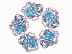
Genetics and structure

It is a member of the small pentraxins family (also known as short pentraxins). [15] The polypeptide encoded by this gene has 224 amino acids. [16] The full-length polypeptide is not present in the body in significant quantities due to signal peptide, which is removed by signal peptidase before translation is completed. The complete protein, composed of five monomers, has a total mass of approximately 120,000 Da. In serum, it assembles into stable pentameric structure with a discoid shape. [17]

Function

CRP binds to the phosphocholine expressed on the surface of bacterial cells such as pneumococcus bacteria. This activates the complement system, promoting phagocytosis by macrophages, which clears necrotic and apoptotic cells and bacteria. [18] [13] With this mechanism, CRP also binds to ischemic/hypoxic cells, which could regenerate with more time. However, the binding of CRP causes them to be disposed of prematurely. [19] [20] CRP is a prehistoric antibody and binds to the Fc-gamma receptor IIa, to which antibodies also bind. [21] In addition, CRP activates the classical complement pathway via C1q binding. [22] [23] CRP thus forms immune complexes in the same way as IgG antibodies.

This so-called acute phase response occurs as a result of increasing concentrations of interleukin-6 (IL-6), which is produced by macrophages [6] as well as adipocytes [7] in response to a wide range of acute and chronic inflammatory conditions such as bacterial, viral, or fungal infections; rheumatic and other inflammatory diseases; malignancy; and tissue injury and necrosis. These conditions cause release of IL-6 and other cytokines that trigger the synthesis of CRP and fibrinogen by the liver.

CRP binds to phosphocholine on micro-organisms. It is thought to assist in complement binding to foreign and damaged cells and enhances phagocytosis by macrophages (opsonin-mediated phagocytosis), which express a receptor for CRP. It plays a role in innate immunity as an early defense system against infections. [13]

Serum levels

C-reactive protein
PurposeDetection of inflammation in body. [24]
Test ofThe amount of CRP in the blood. [24]

Measurement methods

Traditional CRP measurement only detected CRP in the range of 10 to 1,000 mg/L, whereas high sensitivity CRP (hs-CRP) detects CRP in the range of 0.5 to 10 mg/L. [25] hs-CRP can detect cardiovascular disease risk when in excess of 3 mg/L, whereas below 1 mg/L would be low risk. [26] Traditional CRP measurement is faster and less costly than hs-CRP, and can be adequate for some applications, such as monitoring hemodialysis patients. [27]

Normal

In healthy adults, the normal concentrations of CRP varies between 0.8 mg/L and 3.0 mg/L. However, some healthy adults show elevated CRP at 10 mg/L. CRP concentrations also increase with age, possibly due to subclinical conditions. There are also no seasonal variations of CRP concentrations. Gene polymorphism of interleukin-1 family, interleukin 6, and polymorphic GT repeat of the CRP gene do affect the usual CRP concentrations when a person does not have any medical illnesses. [6]

Acute inflammation

When there is a stimulus, the CRP level can increase 10,000-fold from less than 50 μg/L to more than 500 mg/L. Its concentration can increase to 5 mg/L by 6 hours and peak at 48 hours. The plasma half-life of CRP is 19 hours, and is constant in all medical conditions. [28] Therefore, the only factor that affects the blood CRP concentration is its production rate, which increases with inflammation, infection, trauma, necrosis, malignancy, and allergic reactions.[ citation needed ] Other inflammatory mediators that can increase CRP are TGF beta 1, and tumor necrosis factor alpha. In acute inflammation, CRP can increase as much as 50 to 100 mg/L within 4 to 6 hours in mild to moderate inflammation or an insult such as skin infection, cystitis, or bronchitis [ clarification needed ]. It can double every 8 hours and reaches its peak at 36 to 50 hours following injury or inflammation. CRP between 100 and 500 mg/L is considered highly predictive of inflammation due to bacterial infection. Once inflammation subsides, CRP level falls quickly because of its relatively short half-life. [13]

Metabolic inflammation

CRP concentrations between 2 and 10 mg/L are considered as metabolic inflammation: metabolic pathways that cause arteriosclerosis [29] and type II diabetes mellitus [30]

Clinical significance

Diagnostic use

CRP is used mainly as an inflammation marker. Apart from liver failure, there are few known factors that interfere with CRP production. [6] Interferon alpha inhibits CRP production from liver cells which may explain the relatively low levels of CRP found during viral infections compared to bacterial infections [31] [32]

Measuring and charting CRP values can prove useful in determining disease progress or the effectiveness of treatments. ELISA, immunoturbidimetry, nephelometry, radial immunodiffusion [33] [26] [ clarification needed ]

Normal levels increase with aging. [34] Higher levels are found in late pregnant women, mild inflammation and viral infections (10–40 mg/L), active inflammation, bacterial infection (40–200 mg/L), severe bacterial infections and burns (>200 mg/L). [35]

CRP cut-off levels indicating bacterial from non-bacterial illness can vary due to co-morbidities such as malaria, HIV and malnutrition and the stage of disease presentation. [36]

CRP is a more sensitive and accurate reflection of the acute phase response than the ESR [37] (erythrocyte sedimentation rate). ESR may be normal while CRP is elevated. CRP returns to normal more quickly than ESR in response to therapy.[ citation needed ]

Cardiovascular disease

Recent research suggests that patients with elevated basal levels of CRP are at an increased risk of diabetes, [38] [39] hypertension and cardiovascular disease. A study of over 700 nurses showed that those in the highest quartile of trans fat consumption had blood levels of CRP that were 73% higher than those in the lowest quartile. [40] Although one group of researchers indicated that CRP may be only a moderate risk factor for cardiovascular disease, [41] this study (known as the Reykjavik Study) was found to have some problems for this type of analysis related to the characteristics of the population studied, and there was an extremely long follow-up time, which may have attenuated the association between CRP and future outcomes. [42] Others have shown that CRP can exacerbate ischemic necrosis in a complement-dependent fashion and that CRP inhibition can be a safe and effective therapy for myocardial and cerebral infarcts; this has been demonstrated in animal models and humans. [43] [44] [45]

It has been hypothesized that patients with high CRP levels might benefit from use of statins. This is based on the JUPITER trial that found that elevated CRP levels without hyperlipidemia benefited. Statins were selected because they have been proven to reduce levels of CRP. [6] [46] Studies comparing effect of various statins in hs-CRP revealed similar effects of different statins. [47] [48] A subsequent trial however failed to find that CRP was useful for determining statin benefit. [49]

In a meta-analysis of 20 studies involving 1,466 patients with coronary artery disease, CRP levels were found to be reduced after exercise interventions. Among those studies, higher CRP concentrations or poorer lipid profiles before beginning exercise were associated with greater reductions in CRP. [50]

To clarify whether CRP is a bystander or active participant in atherogenesis, a 2008 study compared people with various genetic CRP variants. Those with a high CRP due to genetic variation had no increased risk of cardiovascular disease compared to those with a normal or low CRP. [51] A study published in 2011 shows that CRP is associated with lipid responses to low-fat and high-polyunsaturated fat diets. [52]

Coronary heart disease risk

Arterial damage results from white blood cell invasion and inflammation within the wall. CRP is a general marker for inflammation and infection, so it can be used as a very rough proxy for heart disease risk. Since many things can cause elevated CRP, this is not a very specific prognostic indicator. [53] [54] Nevertheless, a level above 2.4 mg/L has been associated with a doubled risk of a coronary event compared to levels below 1 mg/L; [6] however, the study group in this case consisted of patients who had been diagnosed with unstable angina pectoris; whether elevated CRP has any predictive value of acute coronary events in the general population of all age ranges remains unclear. Currently, C-reactive protein is not recommended as a cardiovascular disease screening test for average-risk adults without symptoms. [55]

The American Heart Association and U.S. Centers for Disease Control and Prevention have defined risk groups as follows: [56] [26]

But hs-CRP is not to be used alone and should be combined with elevated levels of cholesterol, LDL-C, triglycerides, and glucose level. Smoking, hypertension and diabetes also increase the risk level of cardiovascular disease.

Fibrosis and inflammation

Scleroderma, polymyositis, and dermatomyositis elicit little or no CRP response.[ citation needed ] CRP levels also tend not to be elevated in systemic lupus erythematosus (SLE) unless serositis or synovitis is present. Elevations of CRP in the absence of clinically significant inflammation can occur in kidney failure. CRP level is an independent risk factor for atherosclerotic disease. Patients with high CRP concentrations are more likely to develop stroke, myocardial infarction, and severe peripheral vascular disease. [57] Elevated level of CRP can also be observed in inflammatory bowel disease (IBD), including Crohn's disease and ulcerative colitis. [37] [58]

High levels of CRP has been associated to point mutation Cys130Arg in the APOE gene, coding for apolipoprotein E, establishing a link between lipid values and inflammatory markers modulation. [59] [ unreliable medical source? ] [58]

Cancer

The role of inflammation in cancer is not well understood. Some organs of the body show greater risk of cancer when they are chronically inflamed. [60] While there is an association between increased levels of C-reactive protein and risk of developing cancer, there is no association between genetic polymorphisms influencing circulating levels of CRP and cancer risk. [61]

In a 2004 prospective cohort study on colon cancer risk associated with CRP levels, people with colon cancer had higher average CRP concentrations than people without colon cancer. [62] It can be noted that the average CRP levels in both groups were well within the range of CRP levels usually found in healthy people. However, these findings may suggest that low inflammation level can be associated with a lower risk of colon cancer, concurring with previous studies that indicate anti-inflammatory drugs could lower colon cancer risk. [63]

Obstructive sleep apnea

C-reactive protein (CRP), a marker of systemic inflammation, is also increased in obstructive sleep apnea (OSA). CRP and interleukin-6 (IL-6) levels were significantly higher in patients with OSA compared to obese control subjects. [64] Patients with OSA have higher plasma CRP concentrations that increased corresponding to the severity of their apnea-hypopnea index score. Treatment of OSA with CPAP (continuous positive airway pressure) significantly alleviated the effect of OSA on CRP and IL-6 levels. [64]

Rheumatoid arthritis

In the context of rheumatoid arthritis (RA), CRP is one of the acute phase reactants, whose assessment is defined as part of the joint 2010 ACR/EULAR classification criteria for RA with abnormal levels accounting for a single point within the criteria [65] Higher levels of CRP are associated with more severe disease and a higher likelihood of radiographic progression. Rheumatoid arthritis associated antibodies together with 14-3-3η YWHAH have been reported to complement CRP in predicting clinical and radiographic outcomes in patients with recent onset inflammatory polyarthritis. [66] Elevated levels of CRP appear to be associated with common comorbidities including cardiovascular disease, metabolic syndrome, diabetes and interstitial lung (pulmonary) disease. Mechanistically, CRP also appears to influence osteoclast activity leading to bone resorption and also stimulates RANKL expression in peripheral blood monocytes. [67]

It has previously been speculated that single-nucleotide polymorphisms in the CRP gene may affect clinical decision-making based on CRP in rheumatoid arthritis, e.g. DAS28 (Disease Activity Score 28 joints). A recent study showed that CRP genotype and haplotype were only marginally associated with serum CRP levels and without any association to the DAS28 score. [68] Thus, that DAS28, which is the core parameter for inflammatory activity in RA, can be used for clinical decision-making without adjustment for CRP gene variants.[ citation needed ]

Viral infections

Increased blood CRP levels were higher in people with avian flu H7N9 compared to those with H1N1 (more common) influenza, [69] with a review reporting that severe H1N1 influenza had elevated CRP. [70] In 2020, people infected with COVID-19 in Wuhan, China, had elevated CRP. [71] [72] [73]

Additional images

Related Research Articles

<span class="mw-page-title-main">Rheumatoid arthritis</span> Type of autoimmune arthritis

Rheumatoid arthritis (RA) is a long-term autoimmune disorder that primarily affects joints. It typically results in warm, swollen, and painful joints. Pain and stiffness often worsen following rest. Most commonly, the wrist and hands are involved, with the same joints typically involved on both sides of the body. The disease may also affect other parts of the body, including skin, eyes, lungs, heart, nerves, and blood. This may result in a low red blood cell count, inflammation around the lungs, and inflammation around the heart. Fever and low energy may also be present. Often, symptoms come on gradually over weeks to months.

<span class="mw-page-title-main">Ulcerative colitis</span> Inflammatory bowel disease that causes ulcers in the colon

Ulcerative colitis (UC) is a type of inflammatory bowel disease (IBD). It is a long-term condition that results in inflammation and ulcers of the colon and rectum. The primary symptoms of active disease are abdominal pain and diarrhea mixed with blood (hematochezia). Weight loss, fever, and anemia may also occur. Often, symptoms come on slowly and can range from mild to severe. Symptoms typically occur intermittently with periods of no symptoms between flares. Complications may include abnormal dilation of the colon (megacolon), inflammation of the eye, joints, or liver, and colon cancer.

<span class="mw-page-title-main">Inflammation</span> Physical effects resulting from activation of the immune system

Inflammation is part of the biological response of body tissues to harmful stimuli, such as pathogens, damaged cells, or irritants. It is a protective response involving immune cells, blood vessels, and molecular mediators. The function of inflammation is to eliminate the initial cause of cell injury, clear out damaged cells and tissues, and initiate tissue repair.

<span class="mw-page-title-main">Autoimmunity</span> Immune response against an organisms own healthy cells

In immunology, autoimmunity is the system of immune responses of an organism against its own healthy cells, tissues and other normal body constituents. Any disease resulting from this type of immune response is termed an "autoimmune disease". Prominent examples include celiac disease, diabetes mellitus type 1, Henoch–Schönlein purpura (HSP), systemic lupus erythematosus (SLE), Sjögren syndrome, eosinophilic granulomatosis with polyangiitis, Hashimoto's thyroiditis, Graves' disease, idiopathic thrombocytopenic purpura, Addison's disease, rheumatoid arthritis (RA), ankylosing spondylitis, polymyositis (PM), dermatomyositis (DM), and multiple sclerosis (MS). Autoimmune diseases are very often treated with steroids.

<span class="mw-page-title-main">Erythrocyte sedimentation rate</span> Physiological quantity

The erythrocyte sedimentation rate is the rate at which red blood cells in anticoagulated whole blood descend in a standardized tube over a period of one hour. It is a common hematology test, and is a non-specific measure of inflammation. To perform the test, anticoagulated blood is traditionally placed in an upright tube, known as a Westergren tube, and the distance which the red blood cells fall is measured and reported in millimetres at the end of one hour.

<span class="mw-page-title-main">Procalcitonin</span> Precursor of the peptide hormone calcitonin

Procalcitonin (PCT) is a peptide precursor of the hormone calcitonin, the latter being involved with calcium homeostasis. It arises once preprocalcitonin is cleaved by endopeptidase. It was first identified by Leonard J. Deftos and Bernard A. Roos in the 1970s. It is composed of 116 amino acids and is produced by parafollicular cells of the thyroid and by the neuroendocrine cells of the lung and the intestine.

<span class="mw-page-title-main">Paul Ridker</span> American epidemiologist and academic

Paul M. Ridker is a cardiovascular epidemiologist and biomedical researcher. He is currently the Eugene Braunwald Professor of Medicine at Harvard University and Brigham and Women's Hospital, where he directs the Center for Cardiovascular Disease Prevention. Ridker also holds an appointment as Professor in the Department of Epidemiology at the Harvard T.H. Chan School of Public Health.

<span class="mw-page-title-main">Pentraxins</span> Protein family

Pentraxins (PTX), also known as pentaxins, are an evolutionary conserved family of proteins characterised by containing a pentraxin protein domain. Proteins of the pentraxin family are involved in acute immunological responses. They are a class of pattern recognition receptors (PRRs). They are a superfamily of multifunctional conserved proteins, some of which are components of the humoral arm of innate immunity and behave as functional ancestors of antibodies (Abs). They are known as classical acute phase proteins (APP), known for over a century.

<span class="mw-page-title-main">Cystatin C</span>

Cystatin C or cystatin 3, a protein encoded by the CST3 gene, is mainly used as a biomarker of kidney function. Recently, it has been studied for its role in predicting new-onset or deteriorating cardiovascular disease. It also seems to play a role in brain disorders involving amyloid, such as Alzheimer's disease. In humans, all cells with a nucleus produce cystatin C as a chain of 120 amino acids. It is found in virtually all tissues and body fluids. It is a potent inhibitor of lysosomal proteinases and probably one of the most important extracellular inhibitors of cysteine proteases. Cystatin C belongs to the type 2 cystatin gene family.

<span class="mw-page-title-main">PTX3</span>

Pentraxin-related protein PTX3 also known as TNF-inducible gene 14 protein (TSG-14) is a protein that in humans is encoded by the PTX3 gene.

Chronic systemic inflammation (SI) is the result of release of pro-inflammatory cytokines from immune-related cells and the chronic activation of the innate immune system. It can contribute to the development or progression of certain conditions such as cardiovascular disease, cancer, diabetes mellitus, chronic kidney disease, non-alcoholic fatty liver disease, autoimmune and neurodegenerative disorders, and coronary heart disease.

<span class="mw-page-title-main">Autoimmune disease</span> Disorders of adaptive immune system

An autoimmune disease is a condition that results from an anomalous response of the adaptive immune system, wherein it mistakenly targets and attacks healthy, functioning parts of the body as if they were foreign organisms. It is estimated that there are more than 80 recognized autoimmune diseases, with recent scientific evidence suggesting the existence of potentially more than 100 distinct conditions. Nearly any body part can be involved.

The JUPITER trial was a clinical trial aimed at evaluating whether statins reduce heart attacks and strokes in people with normal cholesterol levels.

Cryoglobulinemic vasculitis is a form of inflammation affecting the blood vessels caused by the deposition of abnormal proteins called cryoglobulins. These immunoglobulin proteins are soluble at normal body temperatures, but become insoluble below 37 °C (98.6 °F) and subsequently may aggregate within smaller blood vessels. Inflammation within these obstructed blood vessels is due to the deposition of complement proteins which activate inflammatory pathways.

<span class="mw-page-title-main">Elevated alkaline phosphatase</span> Medical condition

Elevated alkaline phosphatase occurs when levels of alkaline phosphatase (ALP) exceed the reference range. This group of enzymes has a low substrate specificity and catalyzes the hydrolysis of phosphate esters in a basic environment. The major function of alkaline phosphatase is transporting chemicals across cell membranes. Alkaline phosphatases are present in many human tissues, including bone, intestine, kidney, liver, placenta and white blood cells. Damage to these tissues causes the release of ALP into the bloodstream. Elevated levels can be detected through a blood test. Elevated alkaline phosphate is associated with certain medical conditions or syndromes. It serves as a significant indicator for certain medical conditions, diseases and syndromes.

A silent stroke is a stroke that does not have any outward symptoms associated with stroke, and the patient is typically unaware they have suffered a stroke. Despite not causing identifiable symptoms, a silent stroke still causes damage to the brain and places the patient at increased risk for both transient ischemic attack and major stroke in the future. In a broad study in 1998, more than 11 million people were estimated to have experienced a stroke in the United States. Approximately 770,000 of these strokes were symptomatic and 11 million were first-ever silent MRI infarcts or hemorrhages. Silent strokes typically cause lesions which are detected via the use of neuroimaging such as MRI. The risk of silent stroke increases with age but may also affect younger adults. Women appear to be at increased risk for silent stroke, with hypertension and current cigarette smoking being amongst the predisposing factors.

<span class="mw-page-title-main">Secukinumab</span> Monoclonal antibody against IL-17

Secukinumab, sold under the brand name Cosentyx among others, is a human IgG1κ monoclonal antibody used for the treatment of psoriasis, ankylosing spondylitis, and psoriatic arthritis. It binds to the protein interleukin (IL)-17A and is marketed by Novartis.

Major depression is often associated or correlated with immune function dysregulation, and the two are thought to share similar physiological pathways and risk factors. Primarily seen through increased inflammation, this relationship is bidirectional with depression often resulting in increased immune response and illness resulting in prolonged sadness and lack of activity. This association is seen both long-term and short-term, with the presence of one often being accompanied by the other and both inflammation and depression often being co-morbid with other conditions.

The BaleDoneen Method is a risk assessment and treatment protocol aimed at preventing heart attack and stroke. The method also seeks to prevent or reduce the effects of type 2 diabetes. The method was developed by Bradley Field Bale and Amy Doneen.

<span class="mw-page-title-main">Inflammaging</span> Chronic low-grade inflammation that develops with advanced age

Inflammaging is a chronic, sterile, low-grade inflammation that develops with advanced age, in the absence of overt infection, and may contribute to clinical manifestations of other age-related pathologies. Inflammaging is thought to be caused by a loss of control over systemic inflammation resulting in chronic overstimulation of the innate immune system. Inflammaging is a significant risk factor in mortality and morbidity in aged individuals.

References

  1. 1 2 3 GRCh38: Ensembl release 89: ENSG00000132693 - Ensembl, May 2017
  2. 1 2 3 GRCm38: Ensembl release 89: ENSMUSG00000037942 - Ensembl, May 2017
  3. "Human PubMed Reference:". National Center for Biotechnology Information, U.S. National Library of Medicine.
  4. "Mouse PubMed Reference:". National Center for Biotechnology Information, U.S. National Library of Medicine.
  5. Thompson D, Pepys MB, Wood SP (February 1999). "The physiological structure of human C-reactive protein and its complex with phosphocholine". Structure. 7 (2): 169–177. doi: 10.1016/S0969-2126(99)80023-9 . PMID   10368284.
  6. 1 2 3 4 5 6 7 8 Pepys MB, Hirschfield GM (June 2003). "C-reactive protein: a critical update". The Journal of Clinical Investigation. 111 (12): 1805–1812. doi:10.1172/JCI18921. PMC   161431 . PMID   12813013.
  7. 1 2 Lau DC, Dhillon B, Yan H, Szmitko PE, Verma S (May 2005). "Adipokines: molecular links between obesity and atheroslcerosis". American Journal of Physiology. Heart and Circulatory Physiology. 288 (5): H2031–H2041. doi:10.1152/ajpheart.01058.2004. PMID   15653761.
  8. Mantovani A, Garlanda C, Doni A, Bottazzi B (January 2008). "Pentraxins in innate immunity: from C-reactive protein to the long pentraxin PTX3". Journal of Clinical Immunology. 28 (1): 1–13. doi:10.1007/s10875-007-9126-7. PMID   17828584. S2CID   20300531.
  9. Tillett WS, Francis T (September 1930). "Serological Reactions in Pneumonia with a Non-Protein Somatic Fraction of Pneumococcus". The Journal of Experimental Medicine. 52 (4): 561–571. doi:10.1084/jem.52.4.561. PMC   2131884 . PMID   19869788.
  10. Kennelly PJ, Murray RF, Rodwell VW, Botham KM (2009). Harper's illustrated biochemistry. McGraw-Hill Medical. ISBN   978-0-07-162591-3.
  11. Pincus MR, McPherson RA, Henry JB (2007). Henry's clinical diagnosis and management by laboratory methods. Saunders Elsevier. ISBN   978-1-4160-0287-1.
  12. Ratey JJ, Noskin GA, Braun R, Hanley EN Jr, McInnes IB, Ruddy S (2008). Kelley's Textbook of Rheumatology: 2-Volume Set, Expert Consult: Online and Print (Textbook of Rheumatology (Kelley's)(2 Vol)). Philadelphia: Saunders. ISBN   978-1-4160-3285-4.
  13. 1 2 3 4 Bray C, Bell LN, Liang H, Haykal R, Kaiksow F, Mazza JJ, Yale SH (December 2016). "Erythrocyte Sedimentation Rate and C-reactive Protein Measurements and Their Relevance in Clinical Medicine" (PDF). WMJ. 115 (6): 317–321. PMID   29094869.
  14. Mold C, Nakayama S, Holzer TJ, Gewurz H, Du Clos TW (November 1981). "C-reactive protein is protective against Streptococcus pneumoniae infection in mice". The Journal of Experimental Medicine. 154 (5): 1703–1708. doi:10.1084/jem.154.5.1703. PMC   2186532 . PMID   7299351.
  15. "Gene group: Short pentraxins". HUGO Gene Nomenclature Committee.
  16. "#CAA39671". NCBI Entrez Protein.
  17. "Human C-reactive protein complexed with phosphocholine". Protein Data Bank in Europe.
  18. Enocsson H, Karlsson J, Li HY, Wu Y, Kushner I, Wetterö J, Sjöwall C (December 2021). "The Complex Role of C-Reactive Protein in Systemic Lupus Erythematosus". Journal of Clinical Medicine. 10 (24): 5837. doi: 10.3390/jcm10245837 . PMC   8708507 . PMID   34945133.
  19. Sheriff A, Kayser S, Brunner P, Vogt B (2021). "C-Reactive Protein Triggers Cell Death in Ischemic Cells". Frontiers in Immunology. 12: 630430. doi: 10.3389/fimmu.2021.630430 . PMC   7934421 . PMID   33679775.
  20. Sheriff A, Kunze R, Brunner P, Vogt B (August 2023). "Being Eaten Alive: How Energy-Deprived Cells Are Disposed of, Mediated by C-Reactive Protein-Including a Treatment Option". Biomedicines. 11 (8): 2279. doi: 10.3390/biomedicines11082279 . PMC   10452736 . PMID   37626775.
  21. Sheriff A (June 2022). "Special Issue "C-Reactive Protein and Cardiovascular Disease: Clinical Aspects"". Journal of Clinical Medicine. 11 (13): 3610. doi: 10.3390/jcm11133610 . PMC   9267697 . PMID   35806892.
  22. Buerke M, Sheriff A, Garlichs CD (April 2022). "[CRP apheresis in acute myocardial infarction and COVID-19]". Medizinische Klinik, Intensivmedizin und Notfallmedizin (in German). 117 (3): 191–199. doi:10.1007/s00063-022-00911-x. PMC   8951661 . PMID   35333926.
  23. Sproston NR, Ashworth JJ (2018). "Role of C-Reactive Protein at Sites of Inflammation and Infection". Frontiers in Immunology. 9: 754. doi: 10.3389/fimmu.2018.00754 . PMC   5908901 . PMID   29706967.
  24. 1 2 "C-Reactive Protein (CRP)". Lab Tests Online. Retrieved 2019-12-23.
  25. Knight ML (February 18, 2015). "The Application of High-Sensitivity C-Reactive Protein in Clinical Practice: A 2015 Update". Cardiovascular. U.S. Pharmacist . Retrieved 2020-12-28.
  26. 1 2 3 Pearson TA, Mensah GA, Alexander RW, Anderson JL, Cannon RO, Criqui M, et al. (January 2003). "Markers of inflammation and cardiovascular disease: application to clinical and public health practice: A statement for healthcare professionals from the Centers for Disease Control and Prevention and the American Heart Association". Circulation. 107 (3): 499–511. doi: 10.1161/01.cir.0000052939.59093.45 . PMID   12551878.
  27. Helal I, Zerelli L, Krid M, ElYounsi F, Ben Maiz H, Zouari B, et al. (May 2012). "Comparison of C-reactive protein and high-sensitivity C-reactive protein levels in patients on hemodialysis" (PDF). Saudi Journal of Kidney Diseases and Transplantation. 23 (3): 477–483. PMID   22569431. Archived from the original (PDF) on 2021-12-09. Retrieved 2020-12-28.
  28. Vigushin DM, Pepys MB, Hawkins PN (April 1993). "Metabolic and scintigraphic studies of radioiodinated human C-reactive protein in health and disease". The Journal of Clinical Investigation. 91 (4): 1351–1357. doi:10.1172/JCI116336. PMC   288106 . PMID   8473487.
  29. Nilsson J (August 2005). "CRP--marker or maker of cardiovascular disease?". Arteriosclerosis, Thrombosis, and Vascular Biology. 25 (8): 1527–1528. doi: 10.1161/01.ATV.0000174796.81443.3f . PMID   16055753.
  30. Wang X, Bao W, Liu J, Ouyang YY, Wang D, Rong S, et al. (January 2013). "Inflammatory markers and risk of type 2 diabetes: a systematic review and meta-analysis". Diabetes Care. 36 (1): 166–175. doi:10.2337/dc12-0702. PMC   3526249 . PMID   23264288.
  31. Enocsson H, Sjöwall C, Skogh T, Eloranta ML, Rönnblom L, Wetterö J (December 2009). "Interferon-alpha mediates suppression of C-reactive protein: explanation for muted C-reactive protein response in lupus flares?". Arthritis and Rheumatism. 60 (12): 3755–3760. doi: 10.1002/art.25042 . PMID   19950271.
  32. Enocsson H, Gullstrand B, Eloranta ML, Wetterö J, Leonard D, Rönnblom L, et al. (2020). "C-Reactive Protein Levels in Systemic Lupus Erythematosus Are Modulated by the Interferon Gene Signature and CRP Gene Polymorphism rs1205". Frontiers in Immunology. 11: 622326. doi: 10.3389/fimmu.2020.622326 . PMC   7876312 . PMID   33584722.
  33. Grützmeier S, von Schenck H (March 1989). "Four immunochemical methods for measuring C-reactive protein in plasma compared". Clinical Chemistry. 35 (3): 461–463. doi: 10.1093/clinchem/35.3.461 . PMID   2493344.
  34. Thomas, Lothar, Labor und Diagnose. TH-Books, Frankfurt, 2008, p. 1010
  35. Chew KS (April 2012). "What's new in Emergencies Trauma and Shock? C-reactive protein as a potential clinical biomarker for influenza infection: More questions than answers". Journal of Emergencies, Trauma, and Shock. 5 (2): 115–117. doi: 10.4103/0974-2700.96477 . PMC   3391832 . PMID   22787338.
  36. Dittrich S, Tadesse BT, Moussy F, Chua A, Zorzet A, Tängdén T, et al. (2016-08-25). Yansouni C (ed.). "Target Product Profile for a Diagnostic Assay to Differentiate between Bacterial and Non-Bacterial Infections and Reduce Antimicrobial Overuse in Resource-Limited Settings: An Expert Consensus". PLOS ONE. 11 (8): e0161721. Bibcode:2016PLoSO..1161721D. doi: 10.1371/journal.pone.0161721 . PMC   4999186 . PMID   27559728.
  37. 1 2 Liu S, Ren J, Xia Q, Wu X, Han G, Ren H, et al. (December 2013). "Preliminary case-control study to evaluate diagnostic values of C-reactive protein and erythrocyte sedimentation rate in differentiating active Crohn's disease from intestinal lymphoma, intestinal tuberculosis and Behcet's syndrome". The American Journal of the Medical Sciences. 346 (6): 467–472. doi:10.1097/MAJ.0b013e3182959a18. PMID   23689052. S2CID   5173681.
  38. Pradhan AD, Manson JE, Rifai N, Buring JE, Ridker PM (July 2001). "C-reactive protein, interleukin 6, and risk of developing type 2 diabetes mellitus". JAMA. 286 (3): 327–334. doi: 10.1001/jama.286.3.327 . PMID   11466099.
  39. Dehghan A, Kardys I, de Maat MP, Uitterlinden AG, Sijbrands EJ, Bootsma AH, et al. (March 2007). "Genetic variation, C-reactive protein levels, and incidence of diabetes". Diabetes. 56 (3): 872–878. doi: 10.2337/db06-0922 . PMID   17327459.
  40. Lopez-Garcia E, Schulze MB, Meigs JB, Manson JE, Rifai N, Stampfer MJ, et al. (March 2005). "Consumption of trans fatty acids is related to plasma biomarkers of inflammation and endothelial dysfunction". The Journal of Nutrition. 135 (3): 562–566. doi: 10.1093/jn/135.3.562 . PMID   15735094.
  41. Danesh J, Wheeler JG, Hirschfield GM, Eda S, Eiriksdottir G, Rumley A, et al. (April 2004). "C-reactive protein and other circulating markers of inflammation in the prediction of coronary heart disease". The New England Journal of Medicine. 350 (14): 1387–1397. doi: 10.1056/NEJMoa032804 . PMID   15070788.
  42. Koenig, Wolfgang (2006). "C-reactive protein - a critical cardiovascular risk marker". CRPhealth.com.
  43. Pepys MB, Hirschfield GM, Tennent GA, Gallimore JR, Kahan MC, Bellotti V, et al. (April 2006). "Targeting C-reactive protein for the treatment of cardiovascular disease". Nature. 440 (7088): 1217–1221. Bibcode:2006Natur.440.1217P. doi:10.1038/nature04672. PMID   16642000. S2CID   4324584.
  44. Ding Z, Wei Y, Peng J, Wang S, Chen G, Sun J (October 2023). "The Potential Role of C-Reactive Protein in Metabolic-Dysfunction-Associated Fatty Liver Disease and Aging". Biomedicines. 11 (10): 2711. doi: 10.3390/biomedicines11102711 . PMC   10603830 . PMID   37893085.
  45. Torzewski J, Brunner P, Ries W, Garlichs CD, Kayser S, Heigl F, Sheriff A (March 2022). "Targeting C-Reactive Protein by Selective Apheresis in Humans: Pros and Cons". Journal of Clinical Medicine. 11 (7): 1771. doi: 10.3390/jcm11071771 . PMC   8999816 . PMID   35407379.
  46. Ridker PM, Danielson E, Fonseca FA, Genest J, Gotto AM, Kastelein JJ, et al. (November 2008). "Rosuvastatin to prevent vascular events in men and women with elevated C-reactive protein". The New England Journal of Medicine. 359 (21): 2195–2207. doi: 10.1056/NEJMoa0807646 . PMID   18997196.
  47. Sindhu S, Singh HK, Salman MT, Fatima J, Verma VK (October 2011). "Effects of atorvastatin and rosuvastatin on high-sensitivity C-reactive protein and lipid profile in obese type 2 diabetes mellitus patients". Journal of Pharmacology & Pharmacotherapeutics. 2 (4): 261–265. doi: 10.4103/0976-500X.85954 . PMC   3198521 . PMID   22025854.
  48. Jialal I, Stein D, Balis D, Grundy SM, Adams-Huet B, Devaraj S (April 2001). "Effect of hydroxymethyl glutaryl coenzyme a reductase inhibitor therapy on high sensitive C-reactive protein levels". Circulation. 103 (15): 1933–1935. doi: 10.1161/01.CIR.103.15.1933 . PMID   11306519.
  49. Jonathan E, Derrick B, Emma L, Sarah P, John D, Jane A, Rory C, et al. (Heart Protection Study Collaborative Group) (February 2011). "C-reactive protein concentration and the vascular benefits of statin therapy: an analysis of 20,536 patients in the Heart Protection Study". Lancet. 377 (9764): 469–476. doi:10.1016/S0140-6736(10)62174-5. PMC   3042687 . PMID   21277016.
  50. Swardfager W, Herrmann N, Cornish S, Mazereeuw G, Marzolini S, Sham L, Lanctôt KL (April 2012). "Exercise intervention and inflammatory markers in coronary artery disease: a meta-analysis". American Heart Journal. 163 (4): 666–76.e1–3. doi:10.1016/j.ahj.2011.12.017. PMID   22520533.
  51. Zacho J, Tybjaerg-Hansen A, Jensen JS, Grande P, Sillesen H, Nordestgaard BG (October 2008). "Genetically elevated C-reactive protein and ischemic vascular disease". The New England Journal of Medicine. 359 (18): 1897–1908. doi: 10.1056/NEJMoa0707402 . PMID   18971492.
  52. St-Onge MP, Zhang S, Darnell B, Allison DB (April 2009). "Baseline serum C-reactive protein is associated with lipid responses to low-fat and high-polyunsaturated fat diets". The Journal of Nutrition. 139 (4): 680–683. doi:10.3945/jn.108.098251. PMC   2666362 . PMID   19297430.
  53. Lloyd-Jones DM, Liu K, Tian L, Greenland P (July 2006). "Narrative review: Assessment of C-reactive protein in risk prediction for cardiovascular disease". Annals of Internal Medicine. 145 (1): 35–42. doi: 10.7326/0003-4819-145-1-200607040-00129 . PMID   16818927.
  54. Bower JK, Lazo M, Juraschek SP, Selvin E (October 2012). "Within-person variability in high-sensitivity C-reactive protein". Archives of Internal Medicine. 172 (19): 1519–1521. doi:10.1001/archinternmed.2012.3712. PMC   3613132 . PMID   22945505.
  55. Goldman L (2011). Goldman's Cecil Medicine (24th ed.). Philadelphia: Elsevier Saunders. pp.  54. ISBN   978-1437727883.
  56. "hs-CRP" . Retrieved June 3, 2013.
  57. Clearfield MB (September 2005). "C-reactive protein: a new risk assessment tool for cardiovascular disease". The Journal of the American Osteopathic Association. 105 (9): 409–416. PMID   16239491. Archived from the original on 2012-01-10. Retrieved 2013-02-10.
  58. 1 2 Vermeire, Séverine; Van Assche, Gert; Rutgeerts, Paul (September 2004). "C-Reactive Protein as a Marker for Inflammatory Bowel Disease". Inflammatory Bowel Diseases. 10 (5): 661–665. doi:10.1097/00054725-200409000-00026. ISSN   1078-0998.
  59. Sidore C, Busonero F, Maschio A, Porcu E, Naitza S, Zoledziewska M, et al. (November 2015). "Genome sequencing elucidates Sardinian genetic architecture and augments association analyses for lipid and blood inflammatory markers". Nature Genetics. 47 (11): 1272–1281. doi:10.1038/ng.3368. PMC   4627508 . PMID   26366554.
  60. Lu H, Ouyang W, Huang C (April 2006). "Inflammation, a key event in cancer development". Molecular Cancer Research. 4 (4): 221–233. doi: 10.1158/1541-7786.MCR-05-0261 . PMID   16603636.
  61. Allin KH, Nordestgaard BG (2011). "Elevated C-reactive protein in the diagnosis, prognosis, and cause of cancer". Critical Reviews in Clinical Laboratory Sciences. 48 (4): 155–170. doi:10.3109/10408363.2011.599831. PMID   22035340. S2CID   40322991.
  62. Erlinger TP, Platz EA, Rifai N, Helzlsouer KJ (February 2004). "C-reactive protein and the risk of incident colorectal cancer". JAMA. 291 (5): 585–590. doi: 10.1001/jama.291.5.585 . PMID   14762037.
  63. Baron JA, Cole BF, Sandler RS, Haile RW, Ahnen D, Bresalier R, et al. (March 2003). "A randomized trial of aspirin to prevent colorectal adenomas". The New England Journal of Medicine. 348 (10): 891–899. doi: 10.1056/NEJMoa021735 . PMID   12621133.
  64. 1 2 Latina JM, Estes NA, Garlitski AC (2013). "The Relationship between Obstructive Sleep Apnea and Atrial Fibrillation: A Complex Interplay". Pulmonary Medicine. 2013: 621736. doi: 10.1155/2013/621736 . PMC   3600315 . PMID   23533751.
  65. Kay J, Upchurch KS. ACR/EULAR 2010 rheumatoid arthritis classification criteria. Rheumatology (Oxford). 2012 Dec;51 Suppl 6:vi5-9. doi: 10.1093/rheumatology/kes279. PMID 23221588.
  66. Carrier N, Marotta A, de Brum-Fernandes AJ, Liang P, Masetto A, Ménard HA, et al. (February 2016). "Serum levels of 14-3-3η protein supplement C-reactive protein and rheumatoid arthritis-associated antibodies to predict clinical and radiographic outcomes in a prospective cohort of patients with recent-onset inflammatory polyarthritis". Arthritis Research & Therapy. 18 (37): 37. doi: 10.1186/s13075-016-0935-z . PMC   4736641 . PMID   26832367. S2CID   1926353.
  67. Pope JE, Choy EH (February 2021). "C-reactive protein and implications in rheumatoid arthritis and associated comorbidities". Seminars in Arthritis and Rheumatism. 51 (1): 219–229. doi: 10.1016/j.semarthrit.2020.11.005 . PMID   33385862. S2CID   230108148.
  68. Ammitzbøll CG, Steffensen R, Bøgsted M, Hørslev-Petersen K, Hetland ML, Junker P, et al. (October 2014). "CRP genotype and haplotype associations with serum C-reactive protein level and DAS28 in untreated early rheumatoid arthritis patients". Arthritis Research & Therapy. 16 (5): 475. doi: 10.1186/s13075-014-0475-3 . PMC   4247621 . PMID   25359432.
  69. Wu W, Shi D, Fang D, Guo F, Guo J, Huang F, et al. (March 2016). "A new perspective on C-reactive protein in H7N9 infections". International Journal of Infectious Diseases. 44: 31–36. doi: 10.1016/j.ijid.2016.01.009 . PMID   26809124.
  70. Vasileva D, Badawi A (January 2019). "C-reactive protein as a biomarker of severe H1N1 influenza". Inflammation Research. 68 (1): 39–46. doi:10.1007/s00011-018-1188-x. PMC   6314979 . PMID   30288556.
  71. Wang D, Hu B, Hu C, Zhu F, Liu X, Zhang J, et al. (March 2020). "Clinical Characteristics of 138 Hospitalized Patients With 2019 Novel Coronavirus-Infected Pneumonia in Wuhan, China". JAMA. 323 (11): 1061–1069. doi: 10.1001/jama.2020.1585 . PMC   7042881 . PMID   32031570.
  72. Chen N, Zhou M, Dong X, Qu J, Gong F, Han Y, et al. (February 2020). "Epidemiological and clinical characteristics of 99 cases of 2019 novel coronavirus pneumonia in Wuhan, China: a descriptive study". Lancet. 395 (10223): 507–513. doi: 10.1016/S0140-6736(20)30211-7 . PMC   7135076 . PMID   32007143.
  73. Zhang J, Zhou L, Yang Y, Peng W, Wang W, Chen X (March 2020). "Therapeutic and triage strategies for 2019 novel coronavirus disease in fever clinics". The Lancet. Respiratory Medicine. 8 (3): e11–e12. doi: 10.1016/S2213-2600(20)30071-0 . PMC   7159020 . PMID   32061335.