Interleukin-1 family

Last updated
Interleukin-1 / 18
2ILA.png
Crystal structure of IL-1a ( PDB: 2ILA ).
Identifiers
SymbolIL1
Pfam PF00340
InterPro IPR000975
PROSITE PDOC00226
SCOP2 1i1b / SCOPe / SUPFAM
Available protein structures:
Pfam   structures / ECOD  
PDB RCSB PDB; PDBe; PDBj
PDBsum structure summary

The Interleukin-1 family (IL-1 family) is a group of 11 cytokines that plays a central role in the regulation of immune and inflammatory responses to infections or sterile insults.

Contents

Discovery

Discovery of these cytokines began with studies on the pathogenesis of fever. The studies were performed by Eli Menkin and Paul Beeson in 1943–1948 on the fever-producing properties of proteins released from rabbit peritoneal exudate cells. These studies were followed by contributions of several investigators, who were primarily interested in the link between fever and infection/inflammation. [1] The basis for the term "interleukin" was to streamline the growing number of biological properties attributed to soluble factors from macrophages and lymphocytes. IL-1 was the name given to the macrophage product, whereas IL-2 was used to define the lymphocyte product. At the time of the assignment of these names, there was no amino acid sequence analysis known and the terms were used to define biological properties.

In 1985 two distinct, but distantly related complementary DNAs encoding proteins sharing human IL-1 activity were reported to be isolated from a macrophage cDNA library, thus defining two individual members of the IL-1 family – IL-1α and IL-1β. [2] [3] [4]

The Interleukin-1 superfamily

IL-1 family is a group of 11 cytokines, which induces a complex network of proinflammatory cytokines and via expression of integrins on leukocytes and endothelial cells, regulates and initiates inflammatory responses. [5] [6]

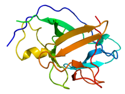
IL-1α and IL-1β are the most studied members because they were discovered first and because they possess strong proinflammatory effects. They have a natural antagonist IL-1Ra (IL-1 receptor antagonist). All three of them include a beta trefoil fold and bind IL-1 receptor (IL-1R) and activate signaling via MyD88 adaptor, which is described in the Signaling section of this page. IL-1Ra regulates IL-1α and IL-1β proinflammatory activity by competing with them for binding sites of the receptor. [5] [7] [8]

Nine IL-1 superfamily members occur in a single cluster on human chromosome two; sequence and chromosomal anatomy evidence suggest these formed through a series of gene duplications of a proto-IL-1β ligand. [9] In this way, IL-1β, IL-1α, IL-36α, IL-36β, IL-36γ, IL-36RA, IL-37, IL-38, and IL-1RA are very likely ancestral family members sharing a common lineage. [9] However, IL-18 and IL-33 are on different chromosomes and there is insufficient sequence or chromosomal anatomy evidence to suggest they share common ancestry with the other IL-1 superfamily members. IL-33 and IL-18 have been included into the IL-1 superfamily due to structural similarities, overlap in function and the receptors involved in their signalling. [9] [10] [11]

Synthesis

All of the members of IL-1 family, except IL-1Ra, are first synthesized as a precursor protein, which means it is synthesized as a long form of a protein which has to be proteolytically cleaved to a shorter, active molecule, which is generally called a mature protein. IL-1 family precursors do not have a clear signal peptide for processing and secretion and none of them are found in the Golgi; they belong to so-called leaderless secretory protein group. The similar feature of IL-1α and IL-33 is that their precursor forms can bind to their respective receptor and can activate signal transduction. But this is not a common feature for all IL-1 family members, since IL-1β and IL-18 precursor forms do not bind their receptors and require proteolytic cleavage by either intracellular caspase-1 or extracellular neutrophilic proteases. [5]

Nomenclature

The interleukin-1 superfamily has 11 members, which have similar gene structure, although originally it contained only four members IL-1α, IL-1β, IL-1Ra and IL-18. After discovery of another 5 members the updated nomenclature was generally accepted which included all members of IL-1 cytokine family. The old IL-1 members were renamed to IL-1F1, IL-1F2, IL-1F3 and IL-1F4. [12]

But according to new trends in nomenclature, the old names of IL-1 family returned. In 2010, laboratories all around the world agreed that IL-1α, IL-1β, IL-1Ra and IL-18 are more familiar to the general scientific knowledge. According to that, they suggested that IL-1F6, IL-1F8 and IL-1F9 should get new names IL-36α, IL-36β and IL-36γ, even though they are encoded by distinct genes, they use the same receptor complex IL-1Rrp2 and coreceptor IL-1RAcP and deliver almost identical signals. The nomenclature also proposes that IL-1F5 should be renamed to IL-36Ra, because it works as an antagonist to IL-36α, IL-36β and IL-36γ similar to how IL-1Ra works for IL-1α and IL-1β. Another revision was the renaming of IL-1F7 to IL-37 because this suppressing cytokine has many splicing variants, they should be called IL-37a, IL-37b and so on. For IL-1F10 there is a reserved name, IL-38. [13]

NameFamily nameReceptorCoreceptorPropertyChromosomal location
IL-1α IL-1F1 IL-1RI IL-1RAcP Proinflammatory 2q14.1
IL-1β IL-1F2IL-1RIIL-1RAcPProinflammatory2q14.1
IL-1Ra IL-1F3IL-1RINAAntagonist for IL-1α, IL-1β2q14.1
IL-18 IL-1F4 IL-18Rα IL-18Rβ Proinflammatory11q23.1
IL-36Ra IL-1F5 IL-1Rrp2 NAAntagonist for IL-36α, IL-36β, IL-36γ2q14.1
IL-36α IL-1F6IL-1Rrp2 IL-1RAcP Proinflammatory2q14.1
IL-37 IL-1F7UnknownUnknown Anti-inflammatory 2q14.1
IL-36β IL-1F8IL-1Rrp2IL-1RAcPProinflammatory2q14.1
IL-36γ IL-1F9IL-1Rrp2IL-1RAcPProinflammatory2q14.1
IL-38 IL-1F10IL-1Rrp2UnknownAnti-inflammatory2q14.1
IL-33 IL-1F11 ST2 IL-1RAcP Th2 responses, proinflammatory9p24.1

[5] [14] [15]

Signaling

IL-1α and IL-1β bind to the same receptor molecule, which is called type I IL-1 receptor (IL-1RI). There is a third ligand of this receptor – the Interleukin 1 receptor antagonist (IL-1Ra), which does not activate downstream signaling, so it acts as an inhibitor of IL-1α and IL-1β signaling by competing with them for binding sites of the receptor. [5] [16]

IL-1α or IL-1β bind first to the first extracellular chain of IL-1RI, that recruits the IL-1 receptor accessory protein (IL-1RAcP), which serves as a coreceptor and is necessary for signal transduction and it is also needed for activation of IL-1RI by IL-18 and IL-33. [16]

After the formation of receptor heterodimeric complex which is assembled by IL-1α or IL-1β, IL-1RI and IL-1RAcP, two intracellular adaptor proteins are assembled by conserved cytosolic regions called Toll- and IL-1R-like (TIR) domains. They are called the myeloid differentiation primary response gene 88 (MYD88) and interleukin-1 receptor-activated protein kinase (IRAK) 4. Phosphorylation of IRAK4 is followed by phosphorylation of IRAK1, IRAK2 and tumor necrosis factor receptor-associated factor (TRAF) 6. TRAF6 is a ubiquitin E3 ligase, that in association with ubiquitin-conjugating enzyme (ubiquitin E2 ligase) complex attaches K63-linked polyubiquitin chains to some of IL-1signaling intermediates, for instance TGF-β-activated protein kinase (TAK-1). That facilitates the association of TAK-1 with TRAF6 and with MEKK3. [16] These signaling pathways lead to activation of many transcription factors, such as NF-κB, AP-1, c-Jun N-terminal kinase (JNK) and p38 MAPK. [16] [17]

IL-1α precursor and mature IL-1β lack a signal peptide which should direct them into the endoplasmic/Golgi-dependent secretion pathway and they are secreted by an unconventional protein secretion pathway, of which the mechanism and regulation are not known. [18]

Biological activity

IL-1 is intensely produced by tissue macrophages, monocytes, fibroblasts, and dendritic cells, but is also expressed by B lymphocytes, NK cells, microglia, and epithelial cells. They form an important part of the inflammatory response of the body against infection. These cytokines increase the expression of adhesion factors on endothelial cells to enable transmigration (also called diapedesis) of immunocompetent cells, such as phagocytes, lymphocytes and others, to sites of infection. They also affect the activity of the hypothalamus, the thermoregulatory center, which leads to a rise in body temperature (fever). That is why IL-1 is called an endogenous pyrogen. Besides fever, IL-1 also causes hyperalgesia (increased pain sensitivity), vasodilation and hypotension. [18]

IL-1α

IL-1α is a “dual-function cytokine”, which means it plays a role in the nucleus by affecting transcription, as well as its extracellular receptor-mediated effects as a classical cytokine. IL-33 also belongs in this group. [19]

IL-1α is synthesized as a precursor protein and it is constitutively stored in the cytoplasm of cells of mesenchymal origin and in epithelial cells. In contrast, monocytes and macrophages do not contain preformed IL-1α precursors, but instead rely on de novo synthesis. The IL-1α precursor is processed to its mature 17-kDa form by a Ca2+-activated protease, calpain. Processing liberates the 16-kDa N-terminal propiece cleavage product (ppIL-1α), which contains a nuclear localization sequence (NLS), and translocates to the nucleus, functioning as a transcription factor. The precursor form of IL-1α, which has both the N-terminal and C-terminal receptor interacting domains, acts as a damage-associated molecular pattern (DAMP) molecule. DAMPs, also known as alarmins, are recognized by innate immunity cells by pattern recognition receptors (PRRs) and function as danger signals for the immune system. In short, DAMPs are released from stressed cells, which undergo necrosis or pyroptosis and their intracellular components are released into extracellular space. Because of misfolding and other oxidative changes of these molecules in the context of altered pH, they are recognized by the innate immune system as molecules that should not be in extracellular space. Cell stress could be due to infection, injury, ischemia, hypoxia, acidosis and complement lysis. The IL-33 precursor molecule acts in a similar way as a DAMP molecule. [19]

Inflammatory responses in the absence of infection (such as ischemia) are only dependent on IL-1α signaling via the Interleukin-1 receptor (IL-1R), rather than TLRs signaling. IL-1α also stimulates transcription and secretion of IL-1β from monocytes, so the initiator of immune responses is likely IL-1α precursor by induction of neutrophil infiltration. IL-1β seems to be an amplifier of inflammation by recruitment of macrophages in the context of sterile inflammation. [19] [20] [21]

IL-1β

IL-1β is synthesized as a precursor form protein only after stimulation, in contrast to IL-1α. Its expression is induced by transcription factor NF-κB after exposure of innate immune cells to alarmins. This occurs, for instance, after exposure of macrophages and dendritic cells to lipopolysaccharide (LPS), which binds to TLR4 and acts as pathogen-associated molecular pattern, which is another group of alarmins. [18] [21]

The synthesis of IL-1β precursor (and IL-18) is induced by stimulation of innate immune cells by Toll-like receptors (TLRs) or RIG-like receptors (RLRs), but to gain the ability to bind to IL-1 receptor, the IL-1β precursor has to be cleaved by a cysteine protease called caspase-1. Caspase-1 needs to be activated by a formation called the inflammasome which is mediated by cytoplasmic pattern recognition receptor signaling. So, the secretion of IL-1β needs these two steps and activation of different receptors to be activated. Under special circumstances IL-1β can be processed also by other proteases, like during high neutrophilic inflammation. [18] [22]

IL-18 is also synthesized as a precursor which is cleaved by caspase-1. [18]

There are indications that IL-1, not least IL-1beta, is of importance for regulation energy metabolism. For instance, Rothwell and coworkers reported evidence that Leptin actions on food intake and body temperature are mediated by IL-1 at the level of the CNS (Luheshi GN, Gardner JD, Rushforth DA, Loudon AS, Rothwell NJ: Leptin actions on food intake and body temperature are mediated by IL-1. Proc Natl Acad Sci U S A 96:7047–7052, 1999). Moreover, lack of IL-1RI–mediated biological activity in IL-1 receptor knockout mice causes mature-onset obesity (Garcia M, Wernstedt I, Berndtsson A, Enge M, Bell M, Hultgren O, Horn M, Ahren B, Enerbäck S, Ohlsson C, Wallenius V, Jansson J-O. 2006. Mature onset obesity in interleukin-1 receptor I (IL-1RI) knockout mice. Diabetes, 55:1205-1213). A similar mature onset obesity has also been observed in IL-6 knockout mice (Wallenius V, Wallenius K, Ahrén B, Rudling M, Dickson SL, Ohlsson C, Jansson J-O. 2002 Interleukin-6 deficient mice develop mature-onset obesity. Nature Medicine 8:75-79). There are fewer reports on the effects on obesity by TNFalpha, the third classic proinflammatory cytokine, although Spiegelman and co-workers found that it has profound affects on glucose metabolism Gokhan S Hotamisligil, Narinder S Shargill, Bruce M. Spiegelman. Adipose expression of tumor necrosis factor-alpha: direct role in obesity-linked insulin resistance. Science 01 Jan 1993: Vol. 259, Issue 5091, pp. 87–91DOI: 10.1126/science.7678183).

IL-1ra

IL-1ra is produced by monocytes, macrophages, neutrophils, fibroblasts, epithelial cells, Sertoli cells, microglia. IL-1ra is synthesized as a preprotein containing a classical 25 amino acid long signal sequence that allows secretion via the endoplasmic reticulum / Golgi apparatus. Mouse, rat and rabbit IL-1ra show 77, 75, and 78% sequence homology to human IL-1ra. [23] L-1ra shows approximately 30% homology to IL-1β at the protein level. Several forms of IL-1ra have been identified: the 17 kDa form, called sIL-1ra (s = soluble) or also IL-1ra1. It contains the classical signal sequence and is a secreted form of IL-1ra. [24] The other 2 forms, commonly referred to as icIL-1ra or IL-1ra2 and IL-1ra3, do not have a signal sequence, are not secreted, and remain strictly interacellular. [25] The soluble form is produced by hepatocytes and regulated by pro-inflammatory cytokines (IL1-β and a combination of IL1-β and IL-6) and other acute phase proteins. The intracellular form was found in fibroblasts, monocytes, neutrophils, keratinocytes and bronchial epithelial cells. IL-1ra is an important regulator of IL-1-induced expression and physiological responses elicited by IL-1. IL-1ra functions as a competitive inhibitor of IL-1 receptor in vivo and in vitro. It counteracts the effects of both IL-1α and IL-1β. Upon binding of IL-1ra, the IL-1 receptor does not transmit a signal to the cell. IL-1ra inhibits the release of both IL-1α and IL-1β, IL-2 secretion, cell surface IL-2 receptor expression. It blocks the stimulation of prostaglandin E2 synthesis in synovial cells and thymocyte proliferation. It also inhibits the release of leukotriene B4 from monocytes after stimulation with bacterial lipopolysaccharides. It blocks insulin release from isolated pancreatic cells.

Polymorphism of this gene is associated with an increased risk of osteoporotic fractures. [26] IL-1ra antagonist deficiency (DIRA) is a rare congenital disease. Affected children experience severe skin and bone inflammation, other organs such as the lungs may be affected. [27] IL-1ra is used in the treatment of rheumatoid arthritis. It is commercially produced as a recombinant form of IL-1ra and is called anakinra.

IL-18

IL-18 is known as a factor that induces the production of interferon gamma (IFN-γ). [28] It is a pro-inflammatory cytokine that shares similar biological effects to IL-12 and structural forms with the IL-1 family. Together with IL-12 it mediates cellular immunity. It binds to the IL-18Rα receptor. It is produced by monocytes, macrophages, osteoblasts, keratinocytes. It is synthesized as an inactive precursor that is proteolytically cleaved to the active 18 kDa form. [29] IL-18 stimulates IFN-γ production by T cells and NK cells. It acts either independently or synergizes with IL-12, which may lead to rapid activation of the monocyte / macrophage system. [30] The combination of this cytokine and IL-12 inhibits IL-4 dependent production of IgE and IgG1 and, in turn, promotes IgG2 production by B cells. [31] In addition to these physiological functions, IL-18 is involved in several serious inflammatory reactions. The amount of IL-18 receptor mRNA in the endometrium as well as the ratio of the amount of binding protein to interleukin is demonstrably increased in patients with endomyosis compared to individuals without endomyosis. [32] IL-18 is also amplified in Hashimoto's thyroiditis. [33] This interleukin has been shown to increase β amyloid production in neurons in Alzheimer's disease. [34]

IL-33

IL-33 is synthesized as a 31-kDa precursor form and binds the ST2 receptor and IL-1RAcP coreceptor, which stimulates signaling that activates transcription factors as NF-κB and ERK, p38 and JNK MAPKs. The signaling can be triggered by a precursor form of IL-33 in the same way as IL-1α precursor activates signaling through the IL-1 receptor. On the other hand, the mature forms IL-3395-270, IL-3399-270 and IL-33109-270, which are processed from a precursor by serine proteases cathepsin G and elastase, are even more potent activators of inflammatory responses. In contrast with IL-1, processing by caspases, like caspase-1, results in IL-33 inactivation. [35] [36] [37]

IL-33 is a dual function cytokine. Besides its chromatin-associated function, it is constitutively expressed in healthy endothelial cells, because it acts as DAMPs after its release to extracellular space of cells in the context of immunologic not-silent cell death (necrosis or pyroptosis), and drives cytokine production in natural helper cells, nuocytes, Th2 lymphocytes, mast cells, basophils, eosinophils, invariant natural killer and natural killer T cells. It is involved in allergic and parasite-induced inflammatory responses. [35] [36]

IL-36α

IL-36α is expressed in spleen, lymph nodes, tonsils, bone marrow, B-cells. This member is unique in that it is additionally synthesized by T lymphocytes. It is most related to IL-37 and IL-36β. [38]

IL-36β

IL-36β is expressed in the tonsils, bone marrow, heart, placenta, lung, testes, intestine, monocytes and B-lymphocytes. It is most similar to IL-36α (IL-1F6). Two alternative transcripts encoding the same protein have been described. [39]

IL-36γ

IL-36γ is most produced by keratinocytes. It activates NF-κB via interleukin 1 receptor-like 2 (IL-1Rrp2) and is specifically inhibited by IL-36ra. [40] Its production increases after IL-1β and TNF-α stimulation, but not after IL-18 or IFN-γ stimulation. IL-36γ plays an important role in skin immunity and inflammation. Expression is increased during chronic contact hypersensitivity, herpes simplex virus infection [41] and psoriasis. [38]

IL-36ra

IL-36ra is highly expressed by keratinocytes, in psoriatic skin, placenta, uterus, brain, kidneys, monocytes, B-lymphocytes and dendritic cells. IL-36ra is 155 amino acids long and lacks a signal sequence. IL-36ra shares with IL-1ra 52% homology in the amino acid sequence. IL-36ra acts as a non-specific inhibitor of inflammation and innate immunity. It inhibits IL-36α induced NF-κB activation. [42]

IL-37

IL-37 is expressed in most tissues. It is the first member of the IL-1 family to form homodimers. [43] IL-37 non-specifically inhibits the inflammatory response and innate immunity. IL-1F7 has also been found in the nucleus where it can function as a nuclear factor. This cytokine may bind or may itself be a ligand of the IL-18 receptor (IL18R1 / IL-1Rrp). It binds to the interleukin 18 binding protein (IL18BP), forming a complex with the beta subunit of the IL-18 receptor (IL-1F4), thereby inhibiting its activity. 5 alternative transcripts encoding different IL-37 isoforms have been described. [44]

IL-38

IL-38 is expressed in the skin as well as in the tonsils. It regulates both innate and adaptive immunity. It binds to the soluble IL-1RI receptor. Two alternative transcripts encoding the same protein have been described. [45]

Cytokine-induced effector cytokine production

IL-33 has a role in so called cytokine-induced effector cytokine production, which means that a production of effector cytokines by differentiated T helper lymphocytes is cytokine dependent and can happen without antigen stimulation by T-cell receptor of these cells. IL-33 in combination with some STAT5 activators, such as IL-2, IL-7 or TSLP, up-regulates expression of its own receptor on already differentiated Th2 lymphocytes, because naive T helper cells nor Th1 nor Th17 populations do not have ST2 receptors. This up-regulation works as a positive feedback which causes even more strong activation of IL-33 dependent-signaling pathways in the lymphocyte. This up-regulation is directly controlled by GATA3 transcription factor. IL-33 combined with IL-2, IL-7 or TSLP also stimulates cell proliferation. The effector cytokine which is secreted from IL-33- and STAT5 activator-stimulated Th2 cells is IL-13, which is NF-κB dependent. IL-13 is very similar to IL-4 in amino acid sequence and structure. They also used the same type II IL-4 receptor to activate STAT6. [37]

Similar functions have IL-1 to Th17 cells and IL-18 to Th1 lymphocytes. IL-1 combined with some STAT3 activators, such as IL-6, IL-21 or IL-23, which are important for Th17 lymphocytes differentiation, have similar positive feedback in Th17 cells just like IL-33 and STAT5 activators have in Th2 cells. They highly up-regulate expression of IL-1 receptor and RORγt on the surface of stimulated Th17 lymphocytes. The effector cytokines mediated by this signalization are IL-17A, IL-4 and IL-6. IL-18 with IL-12, which is a STAT4 activator, have similar effects on Th1 cells by up-regulating expression of IL-18R1 receptor and T-bet. [37] [46]

IL-1 in disease and its clinical significance

IL-1 has a major role in neuroinflammation. [47] During inflammation, there are increased levels of TNF and IL-1 in the brain, [48] [49] and their presence may cause the breakdown of the blood-brain barrier. [48] Polymorphisms in IL-1 genes have been found to contribute to genetic susceptibility to some cancers, [50] ankylosing spondylitis, [51] and Graves' disease. [52]

In terms of clinical use, because of its characterization as a hematopoietic factor, IL-1 was given to patients after bone marrow transplantation to improve the engraftment. But soon[ when? ] it was discovered that the patients were experiencing symptoms of systemic inflammation. Pharmacological blockade of these receptors was then sought in order to relieve symptoms. The endogenous IL-1 receptor antagonist (IL-1Ra), also known as anakinra, was tried in clinical trials to lessen systemic inflammation, but did not demonstrate a statistically significant difference from placebo. [5]

Nowadays, the blockade of IL-1 activity (especially IL-1β) is a standard therapy for patients with autoimmune diseases or lymphomas. Anakinra (IL-1Ra) is FDA-approved as a therapy for patients with rheumatoid arthritis, [53] because it reduces symptoms and slows joint destruction of this inflammatory disease. It has also been prescribed to patients with indolent or smoldering myeloma with a high risk of progression to multiple myeloma. In combination with other medication, IL-1Ra provides a significant increase in the number of years of progression-free disease in its recipients. The benefits of this treatment are the natural structure and no toxicity or gastrointestinal disturbances. [5]

Related Research Articles

<span class="mw-page-title-main">Cytokine</span> Broad and loose category of small proteins important in cell signaling

Cytokines are a broad and loose category of small proteins important in cell signaling. Due to their size, cytokines cannot cross the lipid bilayer of cells to enter the cytoplasm and therefore typically exert their functions by interacting with specific cytokine receptors on the target cell surface. Cytokines have been shown to be involved in autocrine, paracrine and endocrine signaling as immunomodulating agents.

Interleukins (ILs) are a group of cytokines that are expressed and secreted by white blood cells (leukocytes) as well as some other body cells. The human genome encodes more than 50 interleukins and related proteins.

<span class="mw-page-title-main">Interleukin 1-alpha</span> Protein-coding gene in the species Homo sapiens

Interleukin-1 alpha also known as hematopoietin 1 is a cytokine of the interleukin 1 family that in humans is encoded by the IL1A gene. In general, Interleukin 1 is responsible for the production of inflammation, as well as the promotion of fever and sepsis. IL-1α inhibitors are being developed to interrupt those processes and treat diseases.

<span class="mw-page-title-main">Interleukin 1 beta</span> Mammalian protein found in Homo sapiens

Interleukin-1 beta (IL-1β) also known as leukocytic pyrogen, leukocytic endogenous mediator, mononuclear cell factor, lymphocyte activating factor and other names, is a cytokine protein that in humans is encoded by the IL1B gene. There are two genes for interleukin-1 (IL-1): IL-1 alpha and IL-1 beta. IL-1β precursor is cleaved by cytosolic caspase 1 to form mature IL-1β.

<span class="mw-page-title-main">Macrophage inflammatory protein</span> Protein family

Macrophage Inflammatory Proteins (MIP) belong to the family of chemotactic cytokines known as chemokines. In humans, there are two major forms, MIP-1α and MIP-1β, renamed CCL3 and CCL4 respectively, since 2000. However, other names are sometimes encountered in older literature, such as LD78α, AT 464.1 and GOS19-1 for human CCL3 and AT 744, Act-2, LAG-1, HC21 and G-26 for human CCL4. Other macrophage inflammatory proteins include MIP-2, MIP-3 and MIP-5.

<span class="mw-page-title-main">Caspase 1</span> Protein-coding gene in the species Homo sapiens

Caspase-1/Interleukin-1 converting enzyme (ICE) is an evolutionarily conserved enzyme that proteolytically cleaves other proteins, such as the precursors of the inflammatory cytokines interleukin 1β and interleukin 18 as well as the pyroptosis inducer Gasdermin D, into active mature peptides. It plays a central role in cell immunity as an inflammatory response initiator. Once activated through formation of an inflammasome complex, it initiates a proinflammatory response through the cleavage and thus activation of the two inflammatory cytokines, interleukin 1β (IL-1β) and interleukin 18 (IL-18) as well as pyroptosis, a programmed lytic cell death pathway, through cleavage of Gasdermin D. The two inflammatory cytokines activated by Caspase-1 are excreted from the cell to further induce the inflammatory response in neighboring cells.

<span class="mw-page-title-main">Integrin alpha M</span> Mammalian protein found in Homo sapiens

Integrin alpha M (ITGAM) is one protein subunit that forms heterodimeric integrin alpha-M beta-2 (αMβ2) molecule, also known as macrophage-1 antigen (Mac-1) or complement receptor 3 (CR3). ITGAM is also known as CR3A, and cluster of differentiation molecule 11B (CD11B). The second chain of αMβ2 is the common integrin β2 subunit known as CD18, and integrin αMβ2 thus belongs to the β2 subfamily integrins.

<span class="mw-page-title-main">Interleukin 15</span> Cytokine with structural similarity to Interleukin-2

Interleukin-15 (IL-15) is a protein that in humans is encoded by the IL15 gene. IL-15 is an inflammatory cytokine with structural similarity to Interleukin-2 (IL-2). Like IL-2, IL-15 binds to and signals through a complex composed of IL-2/IL-15 receptor beta chain (CD122) and the common gamma chain. IL-15 is secreted by mononuclear phagocytes following infection by virus(es). This cytokine induces the proliferation of natural killer cells, i.e. cells of the innate immune system whose principal role is to kill virally infected cells.

<span class="mw-page-title-main">Interleukin 31</span>

Interleukin-31 (IL-31) is a protein that in humans is encoded by the IL31 gene that resides on chromosome 12. IL-31 is an inflammatory cytokine that helps trigger cell-mediated immunity against pathogens. It has also been identified as a major player in a number of chronic inflammatory diseases, including atopic dermatitis.

<span class="mw-page-title-main">Interleukin 26</span>

Interleukin-26 (IL-26) is a protein that in humans is encoded by the IL26 gene.

<span class="mw-page-title-main">Interleukin 17</span> Group of proteins

Interleukin 17 family is a family of pro-inflammatory cystine knot cytokines. They are produced by a group of T helper cell known as T helper 17 cell in response to their stimulation with IL-23. Originally, Th17 was identified in 1993 by Rouvier et al. who isolated IL17A transcript from a rodent T-cell hybridoma. The protein encoded by IL17A is a founding member of IL-17 family. IL17A protein exhibits a high homology with a viral IL-17-like protein encoded in the genome of T-lymphotropic rhadinovirus Herpesvirus saimiri. In rodents, IL-17A is often referred to as CTLA8.

<span class="mw-page-title-main">Interleukin 19</span> Protein-coding gene in the species Homo sapiens

Interleukin 19 (IL-19) is an immunosuppressive protein that belongs to the IL-10 cytokine subfamily.

Chemokine ligands 4 previously known as macrophage inflammatory protein (MIP-1β), is a protein which in humans is encoded by the CCL4 gene. CCL4 belongs to a cluster of genes located on 17q11-q21 of the chromosomal region. Identification and localization of the gene on the chromosome 17 was in 1990 although the discovery of MIP-1 was initiated in 1988 with the purification of a protein doublet corresponding to inflammatory activity from supernatant of endotoxin-stimulated murine macrophages. At that time, it was also named as "macrophage inflammatory protein-1" (MIP-1) due to its inflammatory properties.

<span class="mw-page-title-main">Interleukin 37</span> Protein-coding gene in the species Homo sapiens

Interleukin 37 (IL-37), also known as Interleukin-1 family member 7 (IL-1F7), is an anti-inflammatory cytokine important for the downregulation of pro-inflammatory cytokine production as well as the suppression of tumor cell growth.

<span class="mw-page-title-main">SIGIRR</span> Protein-coding gene in the species Homo sapiens

Single Ig IL-1-related receptor (SIGIRR), also called Toll/Interleukin-1 receptor 8 (TIR8) or Interleukin-1 receptor 8 (IL-1R8), is transmembrane protein encoded by gene SIGIRR, which modulate inflammation, immune response, and tumorigenesis of colonic epithelial cells.

Interleukin-28 receptor is a type II cytokine receptor found largely in epithelial cells. It binds type 3 interferons, interleukin-28 A, Interleukin-28B, interleukin 29 and interferon lambda 4. It consists of an α chain and shares a common β subunit with the interleukin-10 receptor. Binding to the interleukin-28 receptor, which is restricted to select cell types, is important for fighting infection. Binding of the type 3 interferons to the receptor results in activation of the JAK/STAT signaling pathway.

The IL-10 family is a family of interleukins.

Members of the very wide interleukin-1 receptor (IL-1R) family are characterized by extracellular immunoglobulin-like domains and intracellular Toll/Interleukin-1R (TIR) domain. It is a group of structurally homologous proteins, conserved throughout the species as it was identified from plants to mammals. Proteins of this family play important role in host defence, injury and stress. There are four main groups of TIR domain-containing proteins in animals; Toll-like receptors, Interleukin-1 receptor (IL-1R), cytosolic adaptor proteins and insect and nematode Toll. Each of these groups is involved mainly in host defence; Toll receptors are also involved in embryogenesis.

<span class="mw-page-title-main">CLEC6A</span> Protein-coding gene in humans

Dectin-2 or C-type lectin domain containing 6A is a protein that in humans is encoded by the CLEC6A gene. Dectin-2 is a member of the C-type lectin/C-type lectin-like domain (CTL/CTLD) superfamily. The encoded protein is a type II transmembrane protein with an extracellular carbohydrate recognition domain. It functions as a pattern recognition receptor recognizing α-mannans and as such plays an important role in innate immune response to fungi. Expression is found on macrophages and dendritic cells. It can also be found at low levels in Langerhans cells and peripheral blood monocytes, where expression levels could be increased upon induction of inflammation.

Autoinflammatory diseases (AIDs) are a group of rare disorders caused by dysfunction of the innate immune system. They are characterized by periodic or chronic systemic inflammation, usually without the involvement of adaptive immunity.

References

  1. Dinarello CA (2015). "The history of fever, leukocytic pyrogen and interleukin-1". Temperature. 2 (1): 8–16. doi:10.1080/23328940.2015.1017086. PMC   4843879 . PMID   27226996.
  2. March CJ, Mosley B, Larsen A, Cerretti DP, Braedt G, Price V, et al. (Aug 1985). "Cloning, sequence and expression of two distinct human interleukin-1 complementary DNAs". Nature. 315 (6021): 641–7. Bibcode:1985Natur.315..641M. doi:10.1038/315641a0. PMID   2989698. S2CID   4240002.
  3. Auron PE, Webb AC, Rosenwasser LJ, Mucci SF, Rich A, Wolff SM, Dinarello CA (December 1984). "Nucleotide sequence of human monocyte interleukin 1 precursor cDNA". Proceedings of the National Academy of Sciences of the United States of America. 81 (24): 7907–11. Bibcode:1984PNAS...81.7907A. doi: 10.1073/pnas.81.24.7907 . PMC   392262 . PMID   6083565.
  4. Dinarello CA (December 1994). "The interleukin-1 family: 10 years of discovery". FASEB Journal. 8 (15): 1314–25. doi: 10.1096/fasebj.8.15.8001745 . PMID   8001745. S2CID   10404996.
  5. 1 2 3 4 5 6 7 Dinarello CA (April 2011). "Interleukin-1 in the pathogenesis and treatment of inflammatory diseases". Blood. 117 (14): 3720–32. doi:10.1182/blood-2010-07-273417. PMC   3083294 . PMID   21304099.
  6. Macleod, T; Berekmeri, A; Bridgewood, C; Stacey, M; McGonagle, D; Wittmann, M (2021). "The Immunological Impact of IL-1 Family Cytokines on the Epidermal Barrier". Frontiers in Immunology. 12: 808012. doi: 10.3389/fimmu.2021.808012 . PMC   8733307 . PMID   35003136.
  7. Murzin AG, Lesk AM, Chothia C (January 1992). "beta-Trefoil fold. Patterns of structure and sequence in the Kunitz inhibitors interleukins-1 beta and 1 alpha and fibroblast growth factors". Journal of Molecular Biology. 223 (2): 531–43. doi:10.1016/0022-2836(92)90668-A. PMID   1738162.
  8. Gosavi S, Whitford PC, Jennings PA, Onuchic JN (July 2008). "Extracting function from a beta-trefoil folding motif". Proceedings of the National Academy of Sciences of the United States of America. 105 (30): 10384–9. Bibcode:2008PNAS..10510384G. doi: 10.1073/pnas.0801343105 . PMC   2492465 . PMID   18650393.
  9. 1 2 3 Rivers-Auty J, Daniels MJ, Colliver I, Robertson DL, Brough D (March 2018). "Redefining the ancestral origins of the interleukin-1 superfamily". Nature Communications. 9 (1): 1156. Bibcode:2018NatCo...9.1156R. doi:10.1038/s41467-018-03362-1. PMC   5861070 . PMID   29559685.
  10. Schmitz J, Owyang A, Oldham E, Song Y, Murphy E, McClanahan TK, et al. (November 2005). "IL-33, an interleukin-1-like cytokine that signals via the IL-1 receptor-related protein ST2 and induces T helper type 2-associated cytokines". Immunity. 23 (5): 479–90. doi: 10.1016/j.immuni.2005.09.015 . PMID   16286016.
  11. Dao T, Ohashi K, Kayano T, Kurimoto M, Okamura H (November 1996). "Interferon-gamma-inducing factor, a novel cytokine, enhances Fas ligand-mediated cytotoxicity of murine T helper 1 cells". Cellular Immunology. 173 (2): 230–5. doi:10.1006/cimm.1996.0272. PMID   8912881.
  12. Sims JE, Nicklin MJ, Bazan JF, Barton JL, Busfield SJ, Ford JE, et al. (October 2001). "A new nomenclature for IL-1-family genes". Trends in Immunology. 22 (10): 536–7. doi:10.1016/S1471-4906(01)02040-3. PMID   11574262.
  13. Dinarello C, Arend W, Sims J, Smith D, Blumberg H, O'Neill L, et al. (November 2010). "IL-1 family nomenclature". Nature Immunology. 11 (11): 973. doi:10.1038/ni1110-973. PMC   4174560 . PMID   20959797.
  14. van de Veerdonk FL, Stoeckman AK, Wu G, Boeckermann AN, Azam T, Netea MG, et al. (February 2012). "IL-38 binds to the IL-36 receptor and has biological effects on immune cells similar to IL-36 receptor antagonist". Proceedings of the National Academy of Sciences of the United States of America. 109 (8): 3001–5. Bibcode:2012PNAS..109.3001V. doi: 10.1073/pnas.1121534109 . PMC   3286950 . PMID   22315422.
  15. "Interleukins and interleukin receptors - HUGO Gene Nomenclature Committee". www.genenames.org. Archived from the original on 15 July 2017. Retrieved 3 May 2018.
  16. 1 2 3 4 Weber A, Wasiliew P, Kracht M (January 2010). "Interleukin-1 (IL-1) pathway". Science Signaling. 3 (105): cm1. doi:10.1126/scisignal.3105cm1. PMID   20086235. S2CID   10388683.
  17. 1. Simi A, Tsakiri N, Wang P, Rothwell NJ. Interleukin-1 and inflammatory neurodegeneration. Biochemical Society Transactions. 2007 Oct 1;35(5):1122–6.
  18. 1 2 3 4 5 Contassot E, Beer HD, French LE (May 2012). "Interleukin-1, inflammasomes, autoinflammation and the skin". Swiss Medical Weekly. 142: w13590. doi: 10.4414/smw.2012.13590 . PMID   22653747.
  19. 1 2 3 Cohen I, Rider P, Carmi Y, Braiman A, Dotan S, White MR, et al. (February 2010). "Differential release of chromatin-bound IL-1alpha discriminates between necrotic and apoptotic cell death by the ability to induce sterile inflammation". Proceedings of the National Academy of Sciences of the United States of America. 107 (6): 2574–9. Bibcode:2010PNAS..107.2574C. doi: 10.1073/pnas.0915018107 . PMC   2823886 . PMID   20133797.
  20. Rider P, Carmi Y, Guttman O, Braiman A, Cohen I, Voronov E, et al. (November 2011). "IL-1α and IL-1β recruit different myeloid cells and promote different stages of sterile inflammation". Journal of Immunology. 187 (9): 4835–43. doi: 10.4049/jimmunol.1102048 . PMID   21930960.
  21. 1 2 Matzinger P (May 2012). "The evolution of the danger theory. Interview by Lauren Constable, Commissioning Editor". Expert Review of Clinical Immunology. 8 (4): 311–7. doi:10.1586/eci.12.21. PMC   4803042 . PMID   22607177.
  22. Sahoo M, Ceballos-Olvera I, del Barrio L, Re F (2011). "Role of the inflammasome, IL-1β, and IL-18 in bacterial infections". TheScientificWorldJournal. 11: 2037–50. doi: 10.1100/2011/212680 . PMC   3217589 . PMID   22125454.
  23. Cominelli F, Bortolami M, Pizarro TT, Monsacchi L, Ferretti M, Brewer MT, et al. (March 1994). "Rabbit interleukin-1 receptor antagonist. Cloning, expression, functional characterization, and regulation during intestinal inflammation". The Journal of Biological Chemistry. 269 (9): 6962–71. doi: 10.1016/S0021-9258(17)37468-9 . PMID   7509813.
  24. Muzio M, Polentarutti N, Sironi M, Poli G, De Gioia L, Introna M, et al. (August 1995). "Cloning and characterization of a new isoform of the interleukin 1 receptor antagonist". The Journal of Experimental Medicine. 182 (2): 623–8. doi:10.1084/jem.182.2.623. PMC   2192137 . PMID   7629520.
  25. Muzio M, Polentarutti N, Facchetti F, Peri G, Doni A, Sironi M, et al. (March 1999). "Characterization of type II intracellular IL-1 receptor antagonist (IL-1ra3): a depot IL-1ra". European Journal of Immunology. 29 (3): 781–8. doi: 10.1002/(SICI)1521-4141(199903)29:03<781::AID-IMMU781>3.0.CO;2-0 . PMID   10092080.
  26. Langdahl BL, Løkke E, Carstens M, Stenkjaer LL, Eriksen EF (March 2000). "Osteoporotic fractures are associated with an 86-base pair repeat polymorphism in the interleukin-1--receptor antagonist gene but not with polymorphisms in the interleukin-1beta gene". Journal of Bone and Mineral Research. 15 (3): 402–14. doi: 10.1359/jbmr.2000.15.3.402 . PMID   10750554.
  27. Aksentijevich I, Masters SL, Ferguson PJ, Dancey P, Frenkel J, van Royen-Kerkhoff A, et al. (June 2009). "An autoinflammatory disease with deficiency of the interleukin-1-receptor antagonist". The New England Journal of Medicine. 360 (23): 2426–37. doi:10.1056/NEJMoa0807865. PMC   2876877 . PMID   19494218.
  28. Okamura H, Tsutsi H, Komatsu T, Yutsudo M, Hakura A, Tanimoto T, et al. (November 1995). "Cloning of a new cytokine that induces IFN-gamma production by T cells". Nature. 378 (6552): 88–91. Bibcode:1995Natur.378...88O. doi:10.1038/378088a0. PMID   7477296. S2CID   4323405.
  29. Ushio S, Namba M, Okura T, Hattori K, Nukada Y, Akita K, et al. (June 1996). "Cloning of the cDNA for human IFN-gamma-inducing factor, expression in Escherichia coli, and studies on the biologic activities of the protein". Journal of Immunology. 156 (11): 4274–9. doi:10.4049/jimmunol.156.11.4274. PMID   8666798. S2CID   30629282.
  30. Billiau A (1996). "Interferon-γ: Biology and Role in Pathogenesis". Interferon-gamma: biology and role in pathogenesis. Advances in Immunology. Vol. 62. pp. 61–130. doi:10.1016/s0065-2776(08)60428-9. ISBN   9780120224623. PMID   8781267.
  31. Yoshimoto T, Okamura H, Tagawa YI, Iwakura Y, Nakanishi K (April 1997). "Interleukin 18 together with interleukin 12 inhibits IgE production by induction of interferon-gamma production from activated B cells". Proceedings of the National Academy of Sciences of the United States of America. 94 (8): 3948–53. Bibcode:1997PNAS...94.3948Y. doi: 10.1073/pnas.94.8.3948 . PMC   20548 . PMID   9108085.
  32. Huang HY, Yu HT, Chan SH, Lee CL, Wang HS, Soong YK (June 2010). "Eutopic endometrial interleukin-18 system mRNA and protein expression at the level of endometrial-myometrial interface in adenomyosis patients". Fertility and Sterility. 94 (1): 33–9. doi: 10.1016/j.fertnstert.2009.01.132 . PMID   19394601.
  33. Liu Z, Wang H, Xiao W, Wang C, Liu G, Hong T (October 2010). "Thyrocyte interleukin-18 expression is up-regulated by interferon-γ and may contribute to thyroid destruction in Hashimoto's thyroiditis". International Journal of Experimental Pathology. 91 (5): 420–5. doi:10.1111/j.1365-2613.2010.00715.x. PMC   3003839 . PMID   20586818.
  34. Sutinen EM, Pirttilä T, Anderson G, Salminen A, Ojala JO (August 2012). "Pro-inflammatory interleukin-18 increases Alzheimer's disease-associated amyloid-β production in human neuron-like cells". Journal of Neuroinflammation. 9 (1): 199. doi: 10.1186/1742-2094-9-199 . PMC   3458954 . PMID   22898493.
  35. 1 2 Lefrançais E, Roga S, Gautier V, Gonzalez-de-Peredo A, Monsarrat B, Girard JP, Cayrol C (January 2012). "IL-33 is processed into mature bioactive forms by neutrophil elastase and cathepsin G". Proceedings of the National Academy of Sciences of the United States of America. 109 (5): 1673–8. Bibcode:2012PNAS..109.1673L. doi: 10.1073/pnas.1115884109 . PMC   3277172 . PMID   22307629.
  36. 1 2 Cayrol C, Girard JP (June 2009). "The IL-1-like cytokine IL-33 is inactivated after maturation by caspase-1". Proceedings of the National Academy of Sciences of the United States of America. 106 (22): 9021–6. Bibcode:2009PNAS..106.9021C. doi: 10.1073/pnas.0812690106 . PMC   2690027 . PMID   19439663.
  37. 1 2 3 Guo L, Wei G, Zhu J, Liao W, Leonard WJ, Zhao K, Paul W (August 2009). "IL-1 family members and STAT activators induce cytokine production by Th2, Th17, and Th1 cells". Proceedings of the National Academy of Sciences of the United States of America. 106 (32): 13463–8. Bibcode:2009PNAS..10613463G. doi: 10.1073/pnas.0906988106 . PMC   2726336 . PMID   19666510.
  38. 1 2 Smith DE, Renshaw BR, Ketchem RR, Kubin M, Garka KE, Sims JE (January 2000). "Four new members expand the interleukin-1 superfamily". The Journal of Biological Chemistry. 275 (2): 1169–75. doi: 10.1074/jbc.275.2.1169 . PMID   10625660.
  39. "IL36B interleukin 36 beta [Homo sapiens (human)] - Gene - NCBI". www.ncbi.nlm.nih.gov. Retrieved 2019-08-31.
  40. Debets R, Timans JC, Homey B, Zurawski S, Sana TR, Lo S, et al. (August 2001). "Two novel IL-1 family members, IL-1 delta and IL-1 epsilon, function as an antagonist and agonist of NF-kappa B activation through the orphan IL-1 receptor-related protein 2". Journal of Immunology. 167 (3): 1440–6. doi: 10.4049/jimmunol.167.3.1440 . PMID   11466363.
  41. Kumar S, McDonnell PC, Lehr R, Tierney L, Tzimas MN, Griswold DE, et al. (April 2000). "Identification and initial characterization of four novel members of the interleukin-1 family". The Journal of Biological Chemistry. 275 (14): 10308–14. doi: 10.1074/jbc.275.14.10308 . PMID   10744718.
  42. "Interleukin-1 Family: Ligands & Receptors". www.rndsystems.com. Retrieved 2019-08-31.
  43. Kumar S, Hanning CR, Brigham-Burke MR, Rieman DJ, Lehr R, Khandekar S, et al. (April 2002). "Interleukin-1F7B (IL-1H4/IL-1F7) is processed by caspase-1 and mature IL-1F7B binds to the IL-18 receptor but does not induce IFN-gamma production". Cytokine. 18 (2): 61–71. doi:10.1006/cyto.2002.0873. PMID   12096920.
  44. "IL37 interleukin 37 [Homo sapiens (human)] - Gene - NCBI". www.ncbi.nlm.nih.gov. Retrieved 2019-08-31.
  45. "IL1F10 interleukin 1 family member 10 [Homo sapiens (human)] - Gene - NCBI". www.ncbi.nlm.nih.gov. Retrieved 2019-08-31.
  46. Ben-Sasson SZ, Hu-Li J, Quiel J, Cauchetaux S, Ratner M, Shapira I, et al. (April 2009). "IL-1 acts directly on CD4 T cells to enhance their antigen-driven expansion and differentiation". Proceedings of the National Academy of Sciences of the United States of America. 106 (17): 7119–24. Bibcode:2009PNAS..106.7119B. doi: 10.1073/pnas.0902745106 . PMC   2678417 . PMID   19359475.
  47. Moynagh PN (September 2005). "The interleukin-1 signalling pathway in astrocytes: a key contributor to inflammation in the brain". Journal of Anatomy. 207 (3): 265–9. doi:10.1111/j.1469-7580.2005.00445.x. PMC   1571539 . PMID   16185251.
  48. 1 2 Hofman FM, von Hanwehr RI, Dinarello CA, Mizel SB, Hinton D, Merrill JE (May 1986). "Immunoregulatory molecules and IL 2 receptors identified in multiple sclerosis brain". Journal of Immunology. 136 (9): 3239–45. doi: 10.4049/jimmunol.136.9.3239 . PMID   3082983. S2CID   39751767.
  49. Zhu H, Wang Z, Yu J, Yang X, He F, Liu Z, Che F, Chen X, Ren H, Hong M, Wang J (March 2019). "Role and mechanisms of cytokines in the secondary brain injury after intracerebral hemorrhage". Prog. Neurobiol. 178: 101610. doi:10.1016/j.pneurobio.2019.03.003. PMID   30923023. S2CID   85495400.
  50. Durães C, Muñoz X, Bonet C, García N, Venceslá A, Carneiro F, et al. (September 2014). "Genetic variants in the IL1A gene region contribute to intestinal-type gastric carcinoma susceptibility in European populations". International Journal of Cancer. 135 (6): 1343–55. doi:10.1002/ijc.28776. PMID   24615437. S2CID   667280.
  51. Timms AE, Crane AM, Sims AM, Cordell HJ, Bradbury LA, Abbott A, et al. (October 2004). "The interleukin 1 gene cluster contains a major susceptibility locus for ankylosing spondylitis". American Journal of Human Genetics. 75 (4): 587–95. doi:10.1086/424695. PMC   1182046 . PMID   15309690.
  52. Liu N, Li X, Liu C, Zhao Y, Cui B, Ning G (April 2010). "The association of interleukin-1alpha and interleukin-1beta polymorphisms with the risk of Graves' disease in a case-control study and meta-analysis". Human Immunology. 71 (4): 397–401. doi:10.1016/j.humimm.2010.01.023. PMID   20116409.
  53. "Anakinra". DrugBank Version 4.1. Archived from the original on 2014-01-29. Retrieved Jan 29, 2014.